Validating Molecular Dynamics Ensembles with NMR Data: A Comprehensive Guide for Biomedical Researchers
This article provides a comprehensive overview of the synergistic integration of Nuclear Magnetic Resonance (NMR) spectroscopy and Molecular Dynamics (MD) simulations for determining accurate, dynamic conformational ensembles of proteins, including...
Validating Molecular Dynamics Ensembles with NMR Data: A Comprehensive Guide for Biomedical Researchers
Abstract
This article provides a comprehensive overview of the synergistic integration of Nuclear Magnetic Resonance (NMR) spectroscopy and Molecular Dynamics (MD) simulations for determining accurate, dynamic conformational ensembles of proteins, including challenging intrinsically disordered proteins (IDPs). It covers foundational principles, current methodological workflows like maximum entropy reweighting, and practical guidance for troubleshooting common pitfalls in force field selection and sampling. The content also explores advanced validation strategies and comparative analyses to achieve force-field independent ensembles, highlighting applications in drug discovery and the study of complex biological systems within a cellular context. This resource is tailored for researchers, scientists, and drug development professionals seeking to leverage integrative approaches for robust structural biology insights.
The Synergy of NMR and MD: Establishing Dynamic Structural Biology
Traditional structural biology has long operated under the paradigm that proteins adopt single, well-defined three-dimensional structures. However, this static view fails to capture the intrinsic dynamism essential for biological function. The conformational ensemble paradigm has emerged as a more accurate representation, conceptualizing proteins as dynamic systems populating multiple interconverting states in solution. This shift is particularly crucial for understanding intrinsically disordered proteins (IDPs) and flexible regions in multidomain proteins, where structural heterogeneity defines functional mechanisms [1] [2]. Nuclear Magnetic Resonance (NMR) spectroscopy serves as a powerful technique for characterizing these ensembles, providing atomic-level insights into dynamics across multiple timescales. This guide compares contemporary computational strategies for deriving conformational ensembles, focusing on their integration with NMR data for validation and refinement.
Methodologies for Ensemble Generation and Validation
Molecular Dynamics Simulations
Molecular Dynamics (MD) simulations generate conformational ensembles by numerically solving Newton's equations of motion for all atoms in a system, providing atomically detailed trajectories. The accuracy of these ensembles is highly dependent on the force field employed. Recent improvements have yielded force fields like a99SB-disp, CHARMM36m, and AMBER ff99SB-ILDN, which show improved performance for both folded and disordered proteins [2] [3]. Standard MD protocols involve solvating the protein in an explicit water box, energy minimization, equilibration, and finally production runs. While MD can in principle provide a true dynamical ensemble, limitations in sampling and force field accuracy mean resulting ensembles often require validation against experimental data [3].
Integrative Approaches: Combining Computation and Experiment
Integrative methods combine computational sampling with experimental data to generate accurate ensembles. Two primary philosophies exist: ensemble restraining and ensemble reweighting.
- Ensemble Restraining: Experimental data are incorporated as restraints during the simulation itself. For example, NMR-derived order parameters (S²) can be used in ensemble-averaged restraining simulations, ensuring the evolving ensemble conforms to experimental dynamics [1].
- Ensemble Reweighting: A large pool of conformations is generated first (e.g., via MD), and statistical weights are subsequently adjusted to achieve optimal agreement with experimental data. Maximum Entropy Reweighting is a key technique that minimally perturbs the initial computational weights to match experiments, preventing overfitting [4] [2].
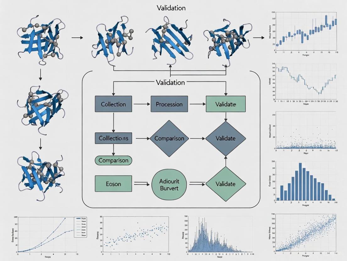
Diagram: Integrative Ensemble Workflow. A generalized workflow for determining conformational ensembles by integrating computational sampling (e.g., MD) with experimental data via reweighting.
The Conformational Filter for Specific Conformations
For systems with suspected distinct states (e.g., "open" and "closed"), a conformational filter can identify which states dominate in solution. This involves generating candidate ensembles (e.g., from MD) and comparing back-calculated NMR relaxation parameters with experimental values. The ensemble whose back-calculated data best matches the experiment is considered the most accurate representation of the solution state [5].
Quantitative Comparison of Methodologies
The table below summarizes the core methodologies, their key features, and primary applications, providing a comparative overview for researchers.
Table 1: Comparison of Core Methodologies for Conformational Ensemble Determination
| Method | Key Features | Experimental Data Used | Primary Applications | Key Advantages |
|---|---|---|---|---|
| Molecular Dynamics (MD) [2] [3] | Generates dynamics trajectories using physics-based force fields. | Used for validation, not generation. | Studying folding, conformational changes, and molecular interactions. | Provides atomic detail and time-resolved dynamics. |
| Maximum Entropy Reweighting [4] [2] | Minimally adjusts weights of a pre-generated ensemble (e.g., from MD) to fit data. | NMR (CS, RDCs, Relaxation), SAXS. | Refining ensembles of IDPs and flexible proteins. | Minimally biased; combines force field accuracy with experimental data. |
| Ensemble Restraining [1] | Incorporates experimental data as restraints during simulation. | NMR S² order parameters, NOEs. | Refining ensembles of globular proteins and IDPs. | Ensures simulation conforms to experimental data throughout. |
| Conformational Filter [5] | Selects among discrete candidate ensembles based on best fit to data. | NMR relaxation parameters (R₁, R₂, NOE, ηxy). | Identifying predominant conformational states in solution. | Unambiguous identification of true conformational states. |
Performance Benchmarking: Force Fields and Integrative Approaches
The choice of force field and the strategy for integrating experimental data significantly impact the quality of the resulting ensemble. The following table benchmarks different approaches based on recent studies.
Table 2: Performance Benchmarking of Force Fields and Integrative Methods
| Force Field / Method | Test System(s) | Agreement with NMR Data | Agreement with SAXS Data | Key Findings |
|---|---|---|---|---|
| a99SB-disp [2] | Aβ40, drkN SH3, ACTR, PaaA2, α-synuclein | Good to excellent for chemical shifts and J-couplings. | Good | One of the top performers in initial agreement with experiment; reweighting further improves agreement. |
| CHARMM36m [2] | Aβ40, drkN SH3, ACTR, PaaA2, α-synuclein | Good to excellent for chemical shifts and J-couplings. | Good | Strong performance, particularly for IDPs; reweighted ensembles often converge with a99SB-disp. |
| MaxEnt Reweighting [2] | Aβ40, drkN SH3, ACTR, PaaA2, α-synuclein | Excellent after reweighting (χ² ≈ 1). | Excellent after reweighting | Effectively produces accurate, force-field-independent ensembles when initial sampling is reasonable. |
| Conformational Filter [5] | Dengue protease NS2B/NS3pro | Correctly identified "closed" conformation as dominant. | N/A | Proven effective at rejecting artifactual conformations induced by crystal packing. |
Essential Research Reagents and Computational Tools
Successful ensemble generation relies on a suite of software tools and computational resources. The following table details key "research reagents" in the computational scientist's toolkit.
Table 3: The Scientist's Toolkit: Key Software and Resources
| Tool / Resource | Type | Function | Applicability |
|---|---|---|---|
| GROMACS [6] [3] | MD Software Package | Performs high-performance MD simulations. | General-purpose MD for folded and disordered proteins. |
| DeePMD-kit [6] | Machine Learning Potential | ML-accelerated potential for efficient, accurate dynamics. | Accelerating ab initio accuracy MD for specific chemical spaces. |
| XPLOR-NIH [1] | NMR Structure Determination | Structure calculation with ensemble restraining capabilities. | Refining ensembles against NMR data. |
| Flexible-meccano [4] | Conformer Generator | Efficiently generates conformational ensembles of IDPs. | Creating prior ensembles of disordered proteins for reweighting. |
| Bayesian/Maximum Entropy (BME) [4] [2] | Reweighting Software | Reweights ensembles to match experimental data. | Integrative modeling with NMR and SAXS data. |
| IR-NMR Multimodal Dataset [6] | Synthetic Spectral Dataset | Provides computed IR and NMR spectra for ~177K molecules. | Benchmarking AI models for spectral prediction and structure elucidation. |
Experimental Protocols for Ensemble Validation
NMR Relaxation Measurements for Dynamics
Purpose: To probe fast (ps-ns) backbone dynamics and extract the Generalized Order Parameter (S²), which quantifies spatial confinement of bond vectors. Workflow:
- Data Collection: Measure longitudinal (R₁), transverse (R₂) relaxation rates, and heteronuclear {¹H}-¹⁵N NOEs for backbone amides [7].
- Model-Free Analysis: Analyze relaxation data using the Model-Free approach to derive S² and internal correlation time (τₑ) for each residue [1] [7].
- Validation: S² values can be directly back-calculated from an MD ensemble by analyzing the angular fluctuations of N-H vectors over the trajectory and compared to experimental values [5] [7].
Protocol for Maximum Entropy Reweighting
Purpose: To refine an initial, computationally generated ensemble (e.g., from MD) against experimental data without introducing undue bias. Workflow [4] [2]:
- Generate Prior Ensemble: Run long-timescale MD simulations with a state-of-the-art force field (e.g., a99SB-disp, CHARMM36m) to create a structural pool.
- Calculate Observables: Use forward models to predict experimental observables (e.g., NMR chemical shifts, J-couplings, SAXS profile) for every structure in the ensemble.
- Define Objective Function: Minimize a pseudo-free energy function,
L = (m/2)χ²_red - θS_rel, which balances agreement with experiment (χ²red) and minimal deviation from the prior distribution (relative entropy, Srel). - Optimize Weights: Iteratively adjust the statistical weights of each conformation to minimize
L. The effective ensemble size is controlled via the Kish ratio to prevent overfitting. - Cross-Validate: Validate the final reweighted ensemble against experimental data not used in the reweighting process.
Diagram: Path to Force-Field Independent Ensembles. When initial ensembles from different force fields are reasonably accurate, Maximum Entropy reweighting can make them converge to a highly similar, force-field-independent solution ensemble [2].
Protocol for a Conformational Filter
Purpose: To unambiguously identify which conformational state (e.g., "open" or "closed") dominates in solution [5]. Workflow:
- Generate Candidate Ensembles: Use free MD simulations or homology modeling to generate representative structural models for each proposed conformational state.
- Run Restrained MD: Perform MD simulations (e.g., 1 μs) incorporating available experimental restraints (e.g., NOE distances, dihedral angles) for each candidate.
- Cluster and Select Models: Perform cluster analysis on the resulting trajectories to select representative models for each conformational family.
- Back-calculate and Compare: Calculate NMR relaxation parameters (backbone and methyl) for each ensemble and compare quantitatively with experimental values.
- Identify Best Fit: The conformational ensemble whose back-calculated data shows the best statistical agreement with experiment is identified as the true solution ensemble.
The paradigm of conformational ensembles represents a fundamental advancement in structural biology, moving beyond static snapshots to embrace the dynamic nature of proteins. As demonstrated, no single method reigns supreme. MD simulations provide dynamical context, while integrative approaches like Maximum Entropy reweighting and conformational filtering leverage experimental data to achieve accuracy and specificity. The convergence of reweighted ensembles from different force fields suggests that determining accurate, force-field independent atomic-resolution models of conformational ensembles is an achievable goal [2]. This progress, powered by the synergy between computational power, algorithmic innovation, and sophisticated NMR experiments, provides a more realistic framework for understanding biological function and guiding drug discovery, especially for highly dynamic targets.
The Unique Strengths of NMR for Probing Dynamics and Averaged States
A central challenge in modern structural biology, particularly in drug development, is moving beyond static snapshots to understand the dynamic conformational ensembles that underlie protein function. Among available analytical techniques, Nuclear Magnetic Resonance (NMR) spectroscopy is uniquely powerful for probing both the structure and dynamics of biomolecules in solution at atomic resolution. This guide objectively compares NMR's performance against other structural methods and details its critical role in validating molecular dynamics (MD) ensembles.
Theoretical Foundation: Why NMR is Uniquely Suited for Dynamics
NMR spectroscopy is fundamentally different from many other analytical techniques because its parameters are directly computable from a molecule's electronic structure using quantum mechanics [8]. The chemical shifts and J-couplings observed in an NMR spectrum are not empirical markers; they are physical manifestations of the magnetic environment of each nucleus, which can be accurately predicted using quantum chemical methods like Density Functional Theory (DFT) [8]. This direct link to first principles makes NMR intrinsically more computable than techniques like mass spectrometry (MS) or chromatography, where predictive modeling of fragmentation patterns or retention behavior often remains empirical [8].
Furthermore, NMR is an ensemble-averaging technique. Unlike X-ray crystallography, which typically produces a single, static structure often constrained by crystal packing, NMR captures the physical properties of biomolecules averaged across all populated conformations over time [7]. This allows it to inherently represent the dynamic and flexible nature of proteins, especially intrinsically disordered proteins (IDPs) that lack a fixed three-dimensional structure [2]. NMR provides site-specific information across a wide range of timescales, from fast picosecond-nanosecond backbone motions to slower microsecond-millisecond conformational exchanges, which are critical for understanding allosteric mechanisms and binding events [9].
Performance Comparison: NMR vs. Alternative Techniques
The table below summarizes a quantitative comparison of NMR's capabilities against other major structural biology techniques.
Table 1: Quantitative Comparison of NMR with Other Structural Techniques
| Technique | Sample State | Atomic Resolution | Timescale Sensitivity | Key Dynamic Parameters | Key Limitations |
|---|---|---|---|---|---|
| NMR Spectroscopy | Solution (near-native) | Yes | Picoseconds - Seconds | Chemical Shifts, J-couplings, Relaxation rates (R1, R2), NOEs, Residual Dipolar Couplings (RDCs), Order Parameters (S²) [7] [9] | Molecular weight limit, requires isotope labeling, low sensitivity [7] |
| X-ray Crystallography | Crystalline solid | Yes | Static (except via temperature factors) | Crystallographic B-factors (motional rigidity) | Requires high-quality crystals, may not represent solution state [7] |
| Cryo-Electron Microscopy (Cryo-EM) | Vitrified solution | Near-atomic to Atomic | Static | — | Can struggle with highly flexible or heterogeneous systems [7] |
| Infrared (IR) Spectroscopy | Various | No | Femtoseconds - Picoseconds | Bond vibration frequencies (functional group focus) [10] | Lacks atomic resolution for full structure, fingerprint region hard to interpret [10] |
Complementary Data from IR Spectroscopy
While not a direct competitor for structure determination, IR spectroscopy provides complementary information based on bond vibrations. A 2025 study demonstrated that combining proton NMR and IR data significantly outperforms either technique alone for automated structure verification, especially for distinguishing challenging isomer pairs [10]. At a true positive rate of 90%, the unsolved pairs were reduced to 0–15% using the combination, compared to 27–49% using individual techniques [10]. This highlights NMR's strength as part of a multi-technique approach.
Experimental Protocols for Probing Dynamics
NMR offers a diverse toolkit of experiments to characterize different aspects of molecular dynamics. The core workflow for studying protein dynamics and validating MD ensembles is shown below.
Diagram Title: Workflow for Integrating NMR and MD Simulations
Protocol 1: Backbone Dynamics via Relaxation Measurements
This methodology characterizes fast internal motions on the picosecond-to-nanosecond timescale [9].
- Sample Preparation: A uniformly ¹⁵N-labeled protein sample is essential. The protein must be soluble and stable at concentrations typically between 0.1 - 1.0 mM in a suitable buffer [9].
- Data Acquisition: A series of two-dimensional ¹H-¹⁵N correlation spectra are acquired to measure:
- Data Analysis: The relaxation parameters are interpreted using the Model-Free (MF) approach, which yields the generalized order parameter (S²) and the effective correlation time (τₑ). The order parameter S² quantifies the spatial restriction of bond vector motion, ranging from 0 (completely disordered) to 1 (completely rigid) [7] [9].
- Integration with MD: Order parameters are back-calculated from the MD trajectory and compared to experimental values. Segments of the MD trajectory that are inconsistent with the experimental dynamics can be identified and selected against [7].
Protocol 2: Cross-Correlated Relaxation for Enhanced Validation
This method provides a more robust measurement by being less sensitive to slow conformational exchange that can bias R2 rates [7].
- Sample Preparation: Same as Protocol 1 (¹⁵N-labeled protein).
- Data Acquisition: Measurement of cross-correlated relaxation (ηxy) rates [7].
- Data Analysis: ηxy rates are used alongside R1 and NOE data to select MD trajectory segments that best align with experimental observables. This approach helps identify biologically relevant conformational ensembles without the confounding effects of slow exchange [7].
Protocol 3: Integrative Ensemble Determination with Maximum Entropy Reweighting
This advanced protocol is used for determining accurate atomic-resolution conformational ensembles of challenging systems like IDPs [2].
- Prerequisites: Long-timescale, all-atom MD simulations (e.g., 30 µs) and extensive experimental data, including NMR chemical shifts, relaxation data, and often Small-Angle X-Ray Scattering (SAXS) [2].
- Method: A maximum entropy reweighting procedure is applied. This method finds the minimal perturbation to the statistical weights of the MD-derived conformational ensemble required to achieve agreement with the experimental data [2].
- Key Metric: The Kish ratio (K) is used to monitor the statistical robustness of the reweighted ensemble, ensuring that the final ensemble does not overfit the data and retains a sufficient number of conformations (e.g., K=0.10 for ~3000 structures from an initial 30,000) [2].
- Outcome: A force-field independent conformational ensemble that represents the most accurate approximation of the protein's solution-state dynamics [2].
The Timescales of Molecular Motion Accessible by NMR
The following diagram illustrates the wide range of dynamic processes that NMR can characterize, correlating them with specific NMR experiments.
Diagram Title: NMR Accessible Timescales of Motion
The Scientist's Toolkit: Essential Research Reagents and Solutions
The table below details key materials and software essential for conducting the NMR experiments and analyses described in this guide.
Table 2: Essential Research Reagents and Computational Tools
| Item Name | Function / Description | Application in NMR/MD Workflow |
|---|---|---|
| ¹⁵N-labeled Protein | Protein expressed with the stable nitrogen-15 isotope, enabling detection of the protein backbone. | Required for all multidimensional NMR experiments probing backbone dynamics and assignments [9]. |
| Cryoprobes | NMR probeheads cooled to reduce electronic noise, significantly increasing sensitivity. | Enables study of proteins at lower concentrations or of higher molecular weight [8]. |
| Molecular Dynamics Software | Software packages like GROMACS, AMBER, or NAMD for running MD simulations. | Generates initial atomic-resolution conformational ensembles for integration with NMR data [7] [2]. |
| Maximum Entropy Reweighting Code | Custom or published scripts (e.g., from GitHub repositories) for integrative modeling. | Refines initial MD ensembles to achieve optimal agreement with experimental NMR data [2]. |
| AlphaFold2 | AI-based protein structure prediction tool. | Provides high-quality starting structural models for MD simulations, especially for folded domains [7]. |
| Density Functional Theory (DFT) Software | Quantum chemical calculation software (e.g., Gaussian, ORCA). | Predicts NMR chemical shifts and J-couplings from first principles for structural validation [8] [10]. |
Molecular dynamics (MD) simulations provide unparalleled insight into the atomic-scale motions that govern biological processes, from protein folding and drug binding to allosteric regulation. The core value of MD lies in its sampling power—the ability to explore the conformational landscape of a biomolecular system. However, a central challenge persists: the timescale problem, where biologically relevant events often occur over microseconds to seconds, while all-atom simulations may be limited to nanoseconds or microseconds [11]. This limitation has driven the development of sophisticated enhanced sampling techniques and multiscale modeling strategies to expand the scope of MD simulations while maintaining atomic fidelity.
The validation of these sampled ensembles, particularly through experimental techniques like Nuclear Magnetic Resonance spectroscopy, forms a critical pillar of modern structural biology. NMR provides ensemble-averaged, site-specific structural and dynamic parameters that serve as essential benchmarks for assessing the quality and reality of MD-derived conformational ensembles [12]. This guide compares the performance of different MD simulation approaches in sampling biomolecular dynamics, focusing on their integration with NMR validation to achieve experimentally-grounded understanding.
Comparing MD Sampling Approaches: Techniques and Applications
Table 1: Key Enhanced Sampling Techniques in Molecular Dynamics
| Technique | Sampling Principle | Typical System Size | Effective Timescale | Key Applications | Validation Methods |
|---|---|---|---|---|---|
| GaMD (Gaussian-accelerated MD) | Adds harmonic boost potential to smoothen energy surface | Medium-large (proteins, ligands) | Microseconds-milliseconds | Ligand binding pathways, conformational transitions | NMR chemical shifts, SAXS [11] |
| REST (Replica Exchange with Solute Tempering) | Parallel simulations at different temperatures | Small-medium (peptides, small proteins) | Nanoseconds-microseconds | IDP conformational ensembles, peptide folding | NMR J-couplings, NOEs [11] |
| Metadynamics | Uses history-dependent bias to escape energy minima | Small-medium (enzyme active sites, binding pockets) | Microseconds-seconds | Protein-ligand binding, allosteric mechanism | NMR relaxation, chemical shifts [11] |
| Coarse-grained MD (MARTINI) | Reduces system complexity by grouping atoms | Very large (membranes, protein complexes) | Microseconds-milliseconds | Membrane remodeling, protein association | NMR lipid interactions, FRET [11] [13] |
| Markov State Models (MSM) | Constructs kinetic network from multiple short simulations | Small-very large | Milliseconds-seconds | Protein folding, large conformational changes | NMR relaxation, hydrogen exchange [11] |
Table 2: Performance Comparison of MD Sampling for Different Biological Systems
| Biological System | Sampling Challenge | Recommended Approaches | Achievable Resolution | NMR Validation Metrics |
|---|---|---|---|---|
| Intrinsically Disordered Proteins (IDPs) | Rugged, shallow energy landscape | REST, Metadynamics, integrative modeling | Atomic detail of transient structures | Chemical shifts, PRE, RDCs [11] [12] |
| GPCRs and Membrane Proteins | Slow dynamics, lipid interactions | GaMD, Coarse-grained MD, conventional MD | Allosteric sites, activation mechanisms | Chemical shifts, relaxation [14] |
| Protein-Ligand Binding | Rare binding/unbinding events | GaMD, Metadynamics, Markov State Models | Binding pathways, intermediate states | Chemical shifts, NOEs, RDCs [11] |
| Amorphous Pharmaceuticals | Dynamic disorder, glass transitions | Conventional MD, machine learning potentials | Molecular conformations, interactions | Chemical shifts, relaxation [15] |
| Protein Folding | High energy barriers, multiple pathways | REST, Markov State Models, specialized hardware | Secondary structure formation | Chemical shifts, J-couplings, RDCs [16] |
Experimental Protocols: Integrating MD Sampling with NMR Validation
Protocol 1: Validating Dynamic Conformational Ensembles with NMR Relaxation
Application: Characterizing the functional dynamics of folded proteins, such as the extracellular region of Streptococcus pneumoniae PsrSp [17].
MD Methodology:
- Perform free MD simulations starting from AlphaFold-generated structures or experimental structures
- Conduct long-scale simulations (hundreds of nanoseconds to microseconds) using modern force fields
- Extract trajectory segments with stable RMSD plateaus for analysis
- Back-calculate NMR relaxation parameters (R₁, R₂, NOE, ηxy) directly from MD trajectories
NMR Methodology:
- Measure 15N cross-correlated relaxation rates using optimized pulse programs
- Determine experimental order parameters (S²) from relaxation data
- Acquire site-specific chemical shifts and residual dipolar couplings
Integration Approach: Identify MD trajectory segments consistent with experimental relaxation data through statistical comparison. Segments showing strong agreement with back-calculated parameters represent valid conformational ensembles [17].
Protocol 2: Characterizing Amorphous Drug Forms
Application: Understanding molecular behavior in amorphous pharmaceuticals like irbesartan [15].
MD Methodology:
- Build simulation boxes with 100+ randomly oriented drug molecules
- Perform equilibration cycles (compression at 500 K, 1000 bar) followed by production runs (200+ ns)
- Sample snapshots regularly (every 400 ps) for ensemble analysis
- Employ machine learning predictors (ShiftML2) to calculate NMR chemical shifts from MD snapshots
NMR Methodology:
- Acquire experimental 13C, 15N, and 1H chemical shifts for amorphous forms
- Measure NMR relaxation to probe molecular dynamics
- Analyze linewidths and shift distributions for dynamic information
Integration Approach: Compare averaged predicted shifts from MD ensembles with experimental NMR data. Use differences to refine force fields and validate the representation of transient interactions like hydrogen bonding [15].
Protocol 3: Ensemble Modeling of Intrinsically Disordered Proteins
Application: Determining conformational ensembles of IDPs, which lack stable tertiary structure [12].
MD Methodology:
- Conduct all-atom MD simulations with explicit solvent
- Apply enhanced sampling techniques (REST, metadynamics) for sufficient conformational sampling
- Generate thousands of conformations representing the ensemble
NMR Methodology:
- Measure chemical shifts, residual dipolar couplings, and paramagnetic relaxation enhancements
- Collect J-couplings and NOE data where possible
Integration Approach: Use NMR data as restraints in MD simulations or to reweight ensemble populations. Statistical reweighting techniques ensure the final ensemble matches experimental observations while maintaining physical realism [12].
Visualization of Methodologies
Integrative MD-NMR Workflow for Conformational Sampling
Figure 1: This workflow illustrates the synergistic integration of molecular dynamics sampling approaches with experimental NMR validation to generate accurate conformational ensembles, highlighting the cyclical nature of computational and experimental integration.
Research Reagent Solutions: Essential Tools for MD-NMR Studies
Table 3: Essential Research Reagents and Tools for MD-NMR Integration
| Tool/Resource | Type | Primary Function | Application Examples |
|---|---|---|---|
| GROMACS | Software | High-performance MD simulation package | Simulating protein dynamics, membrane systems [15] |
| ShiftML2 | Machine Learning Model | Predicting chemical shifts from structures | Validating MD ensembles against NMR data [15] |
| GPCRmd | Database/Platform | Sharing and analyzing GPCR MD simulations | Large-scale dynamics of membrane proteins [14] |
| AMBER/GAFF | Force Field | Parameterizing organic molecules | Pharmaceutical compounds, drug-like molecules [15] |
| MARTINI | Coarse-grained Force Field | Simulating large systems over longer timescales | Membrane remodeling, protein-lipid interactions [11] [13] |
| CPMG Relaxation Dispersion | NMR Technique | Probing millisecond-timescale dynamics | Validating rare event sampling in MD [18] |
| Residual Dipolar Couplings | NMR Measurement | Determining molecular orientations in aligned media | Validating structural ensembles from MD [17] |
The sampling power of MD simulations has expanded dramatically through enhanced sampling algorithms, coarse-grained models, and machine learning acceleration. However, the true validation of this sampling occurs through integration with experimental techniques, particularly NMR spectroscopy, which provides site-specific, dynamic information across multiple timescales. The future of the field lies in developing more sophisticated integrative approaches that leverage the complementary strengths of computation and experiment—using MD to provide atomic detail and continuous trajectories, while employing NMR to ground these ensembles in experimental reality. As force fields continue to improve and sampling algorithms become more efficient, this synergy will enable researchers to tackle increasingly complex biological questions, from drug binding mechanisms to the dynamics of disordered proteins, with greater confidence and atomic-level insight.
Molecular dynamics (MD) simulations are an indispensable tool in computational structural biology, providing atomic-level insights into the motions and interactions of proteins, nucleic acids, and lipids. These simulations empower research in drug discovery and basic science by visualizing processes that are difficult to observe experimentally [19] [20]. However, the predictive power and scalability of MD are constrained by three persistent challenges: the accuracy of the underlying force fields, the limited sampling of conformational space, and the data sparsity problem associated with storing and analyzing massive simulation trajectories [21] [22] [23]. These challenges are particularly acute when MD ensembles are used in conjunction with experimental data like Nuclear Magnetic Resonance (NMR) for validation, as the fidelity of the simulation directly impacts the ability to interpret experimental observables [19] [8]. This guide objectively compares contemporary solutions addressing these bottlenecks, providing researchers with a clear overview of the current landscape.
Force Field Accuracy
The accuracy of a molecular mechanics (MM) force field, which calculates the potential energy of a system, is foundational to any MD simulation. Traditional force fields rely on fixed parameters assigned from a finite set of atom types, which can struggle to capture the full complexity of diverse molecular systems, especially those outside the well-characterized regions of chemical space [21].
Comparative Analysis of Modern Force Field Approaches
The table below compares the performance of traditional, machine-learned, and specialized force fields based on recent studies.
Table 1: Comparison of Modern Force Field Approaches and Their Performance
| Force Field Approach | Key Methodology | Reported Performance / Advantages | Limitations / Challenges |
|---|---|---|---|
| Traditional MM (e.g., AMBER, CHARMM) [21] | Pre-defined parameters based on atom type lookup tables. | Established, highly efficient, and widely validated for standard biomolecules [21]. | Accuracy trade-off; limited transferability to novel molecules like peptide radicals [21]. |
| Machine-Learned (Grappa) [21] | Graph neural network predicts MM parameters directly from molecular structure. | Outperforms traditional MM and other machine-learned fields (Espaloma) on small molecules, peptides, and RNA; matches AMBER FF19SB on dihedrals without corrective maps (CMAPs); transferable to proteins and viruses [21]. | Currently predicts only bonded interactions; nonbonded parameters are taken from established force fields [21]. |
| Specialized (BLipidFF) [24] | Quantum mechanics (QM)-based parameterization for specific bacterial lipids. | For α-mycolic acid, captures unique membrane rigidity and diffusion rates, showing excellent agreement with fluorescence spectroscopy and FRAP experiments, outperforming general fields (GAFF, CGenFF) [24]. | Development is resource-intensive and specific to a class of molecules (e.g., mycobacterial lipids) [24]. |
Experimental Protocol: Development of a Specialized Force Field (BLipidFF)
The creation of the BLipidFF force field for mycobacterial lipids exemplifies a rigorous, QM-driven parameterization protocol [24]:
- Atom Type Definition: Atoms are categorized based on location and chemical environment (e.g.,
cTfor tail carbon,oGfor glycosidic oxygen), resulting in 18 distinct atom types to capture unique lipid features [24]. - Charge Calculation: A "divide-and-conquer" strategy is employed:
- Large lipid molecules are divided into smaller segments.
- Each segment undergoes geometry optimization at the B3LYP/def2SVP level of theory.
- Partial atomic charges are derived using the Restrained Electrostatic Potential (RESP) method at the B3LYP/def2TZVP level.
- Charges are averaged over 25 conformations, and the segment charges are integrated to form the final charge set for the whole molecule [24].
- Torsion Parameter Optimization: Torsion parameters are optimized by minimizing the difference between the potential energy surface calculated by high-level QM methods and the classical force field function [24].
Diagram 1: Specialized Force Field Development Workflow
Sampling Limits
A fundamental challenge in MD is the "timescale gap": many biologically relevant conformational changes, such as protein folding or the transient formation of binding interfaces, occur on timescales (microseconds to seconds) that are prohibitively expensive for standard MD to simulate [19] [20]. This is especially true for highly flexible systems like Intrinsically Disordered Proteins (IDPs), which exist as dynamic ensembles of interconverting structures [20].
Comparative Analysis of Enhanced Sampling and AI Methods
The table below compares different strategies to overcome sampling limitations.
Table 2: Comparison of Methods for Overcoming Sampling Limits in MD
| Method | Category | Key Methodology | Reported Performance / Advantages |
|---|---|---|---|
| Long MD Simulations [22] | Physics-based | Extending simulation time to capture rare events. | Single long simulations are often non-reproducible and may still deviate from experimental values [22]. |
| Ensemble MD (Replicas) [22] | Physics-based | Running multiple independent, shorter simulations with different initial conditions. | For DNA-intercalator binding, 25 replicas of 10 ns achieved accuracy comparable to 25x100 ns replicas and aligned well with experiment, highlighting reproducibility over single long runs [22]. |
| Gaussian Accelerated MD (GaMD) [20] | Enhanced Sampling (Physics-based) | Adding a harmonic boost potential to smooth the energy landscape. | Successfully captured proline isomerization events in the disordered protein ArkA, revealing a more compact ensemble that better aligned with circular dichroism data [20]. |
| Deep Learning (DL) Sampling [20] | AI-based | Using deep learning models trained on data to generate conformational ensembles. | Efficiently samples diverse IDP ensembles, outperforming MD in generating structurally diverse states; can capture rare, transient conformations missed by MD [20]. |
| Hybrid AI-MD [20] | Hybrid | Integrating AI-generated structures as initial states for MD refinement. | Bridges the gap between statistical learning and thermodynamic feasibility; leverages AI's sampling speed with MD's physical accuracy [20]. |
Experimental Protocol: Ensemble MD for Binding Energy Prediction
A rigorous protocol for estimating binding free energies using ensemble MD was demonstrated for DNA-intercalator complexes [22]:
- System Preparation: The DNA-drug complex is solvated in a water box with ions to neutralize the system.
- Simulation Replicas: A set of independent simulation replicas (e.g., 25) are launched. Each replica is initialized with different random atomic velocities, typically assigned from a Maxwell-Boltzmann distribution at the simulation temperature [22] [20].
- Equilibration and Production: Each replica undergoes energy minimization, equilibration, and a production run. The study compared short (10 ns) and long (100 ns) production times [22].
- Energy Calculation: Binding free energies are calculated for each simulation frame using methods like MM/PBSA or MM/GBSA.
- Statistical Analysis: Results from all replicas are aggregated. Bootstrap analysis (a resampling technique) can determine the minimum number of replicas needed for a reliable estimate, finding, for instance, that 6 replicas of 100 ns or 8 replicas of 10 ns sufficed for accuracy within 1.0 kcal/mol of experimental values [22].
Diagram 2: Ensemble MD Simulation and Analysis Workflow
Data Sparsity
As MD simulations grow in length and system size, the resulting trajectory data becomes immense, creating a critical bottleneck for data storage, sharing, and subsequent analysis. This "data sparsity" challenge limits the scalability and collaborative potential of MD research [23].
Comparative Analysis of Data Handling Techniques
The table below compares traditional and modern approaches to managing MD trajectory data.
Table 3: Comparison of Data Handling Techniques for MD Trajectories
| Technique | Methodology | Impact on Data & Limitations |
|---|---|---|
| Subsampling / Reduced Representations [23] | Storing only every Nth frame or using coarse-grained models. | Limitation: Discards potentially valuable dynamical information and reduces the utility of shared datasets [23]. |
| Neural Compression (MDZip) [23] | A convolutional autoencoder compresses the full atomic trajectory into a compact latent representation, from which the trajectory is reconstructed. | Performance: Achieves >95% storage reduction while accurately preserving ensemble-level features like RMSD, pairwise distances, and radius of gyration. A residual autoencoder variant further improves accuracy [23]. |
| Short Energy Minimization [23] | A brief energy minimization is applied to reconstructed structures from MDZip. | Benefit: Partially recovers physically reasonable conformations and improves energetic fidelity after neural network reconstruction [23]. |
Experimental Protocol: Neural Compression of MD Trajectories with MDZip
The MDZip framework offers a modern solution for data sparsity [23]:
- Autoencoder Training: A convolutional autoencoder is trained on a specific MD trajectory. The encoder learns to map sets of atomic coordinates to a low-dimensional latent space, while the decoder learns to reconstruct the coordinates from this latent representation.
- Trajectory Compression: The entire MD trajectory is processed by the trained encoder, resulting in a highly compressed latent representation file.
- Storage and Sharing: This small latent file, along with the trained decoder model, can be efficiently stored or shared.
- Trajectory Reconstruction: The recipient uses the decoder to reconstruct the atomic trajectory from the latent representation.
- Validation and Refinement: The reconstructed trajectory is validated against key ensemble-level metrics. A short energy minimization can be applied to correct local structural deviations and improve physical realism [23].
Diagram 3: Neural MD Trajectory Compression and Reconstruction
The Scientist's Toolkit: Key Research Reagents and Solutions
This table details essential computational tools and methods referenced in this guide.
Table 4: Key Research Reagent Solutions for Advanced MD Research
| Category | Item / Solution | Primary Function in Research |
|---|---|---|
| Force Fields | Grappa [21] | A machine-learned force field that predicts molecular mechanics parameters directly from the molecular graph for improved accuracy. |
| BLipidFF [24] | A specialized force field providing accurate parameters for complex bacterial membrane lipids, enabling realistic simulations of pathogen membranes. | |
| Sampling Methods | Ensemble MD Replicas [22] | A strategy of running multiple independent simulations to improve statistical reliability and reproducibility of results like binding energies. |
| Gaussian Accelerated MD (GaMD) [20] | An enhanced sampling method that adds a boost potential to accelerate the exploration of conformational space, useful for processes like proline isomerization. | |
| AI/Analysis Tools | Deep Learning Conformational Sampling [20] | AI models that rapidly generate diverse structural ensembles for challenging systems like Intrinsically Disordered Proteins (IDPs). |
| MDZip [23] | A neural compression framework that drastically reduces the storage footprint of MD trajectories while preserving essential dynamical information. | |
| Validation | NMR Spectroscopy [19] [8] [25] | An experimental technique that provides atomic-level data on structure and dynamics in solution, used to validate and refine MD-generated ensembles. |
| QM Software | Gaussian09 & Multiwfn [24] | Software packages used for quantum mechanical calculations, such as geometry optimization and RESP charge fitting, for force field development. |
Integrative structural biology has emerged as a powerful paradigm for determining the structures of biological macromolecules and their complexes, overcoming limitations inherent to individual experimental or computational methods. This approach characterizes three-dimensional structures through an array of complementary techniques, subsequently combining the data to form consensus models using computational methodologies [26]. The fundamental motivation behind integrative structural biology is deceptively simple: any system is described best by using all available information about it [27]. This philosophy recognizes that biological function arises not from static structures but from dynamic molecular machines whose internal motions are essential for their biological roles [28].
Proteins and their complexes exhibit dynamics spanning an extraordinary range of timescales—from 10^(-14) to 10 seconds—encompassing sub-picosecond vibrational motions of atoms, microsecond loop conformational rearrangements, and millisecond large-amplitude domain reorientations [28]. Traditional structural biology methods often provide static snapshots that cannot fully capture this dynamic landscape. Integrative approaches address this limitation by synthesizing disparate information, potentially at different scales, into a comprehensive view of a system that includes both structural and dynamic aspects [27] [29]. This holistic perspective is particularly valuable for understanding allosteric mechanisms, conformational changes during function, and the molecular basis of diseases.
The integrative approach dates to the very beginning of structural biology, with one of the first integrative structural models being the double helix of DNA [27]. Only by combining information about chemical composition, stoichiometry, nucleotide complementarity, and X-ray fiber diffraction data could Watson and Crick generate their seminal model. Today, integrative structural biology has evolved to tackle increasingly complex systems, from viral assemblies and molecular machines to massive cellular complexes like the postsynaptic density in neurons [28] [30]. These advances are facilitated by formalized computational frameworks, data standards, and specialized experimental methodologies that enable researchers to solve molecular puzzles that resist characterization by any single technique [27] [31] [26].
The Integrative Structural Biology Toolkit
Experimental Methods and Their Structural Information
Integrative structural biology draws upon a diverse repertoire of experimental techniques, each providing unique and complementary information about different structural aspects of biomolecular systems. The synergy between these methods enables researchers to build comprehensive models that transcend the limitations of individual approaches.
Table 1: Experimental Methods in Integrative Structural Biology
| Method | Structural Information Provided | Applicable Size Range | Key Applications |
|---|---|---|---|
| NMR Spectroscopy | Atomic structures, distances, dynamics (ps-ms timescales), binding sites, solvent accessibility | Small to medium proteins and complexes | Determining protein dynamics, mapping conformational changes, identifying allosteric pathways [28] [27] |
| Cryo-Electron Microscopy | 3D maps and 2D images, medium to low-resolution structures | Large complexes and assemblies | Visualizing large molecular machines, characterizing pleomorphic structures [28] [27] |
| X-ray Crystallography | Atomic structures of system components | Small to large crystallizable systems | Providing high-resolution structural information of domains or subunits [27] |
| Small-Angle X-Ray Scattering (SAXS) | Size, shape, distributions of pairwise atomic distances | Proteins and complexes in solution | Studying overall shape and conformational changes, validating structural models [27] [29] |
| Mass Spectrometry (XL-MS, HDX-MS) | Physical proximity, stoichiometry, solvent accessibility, binding sites | Various sizes, including complexes | Mapping interactions through cross-linking, probing solvent accessibility via hydrogen-deuterium exchange [32] [27] |
| EPR/DEER Spectroscopy | Atomic and protein distances through spin labeling | Various sizes | Measuring long-range distances and conformational heterogeneity [27] [29] |
| Fluorescence Spectroscopy (FRET) | Atomic and protein distances | Solution studies | Mapping conformational changes and dynamics through Förster resonance energy transfer [27] [29] |
Computational and AI-Based Approaches
Computational methods serve as the crucial glue that integrates diverse experimental data into coherent structural models. Molecular dynamics (MD) simulations provide atomic-level insights into protein motions and conformational changes, though their timescales are typically limited to milliseconds for all-atom simulations of large systems [28]. When large-amplitude conformational changes are not accessible by MD, time-independent approaches such as normal mode analysis, principal component analysis, or time-structure independent component analysis become necessary [28].
The recent emergence of deep learning-based approaches has revolutionized structural biology, with AlphaFold being the most prominent example [30]. These AI-assisted tools have demonstrated remarkable performance in predicting protein structures, often surpassing traditional computational methods. However, they are not without limitations, particularly for modeling large complexes, multivalent proteins with flexible regions, and systems with non-uniform stoichiometry [30] [26]. The postsynaptic density exemplifies such challenges, containing transmembrane receptors, extended scaffold proteins with intrinsically disordered regions, multivalent proteins, and dynamically assembling components [30].
Integrative modeling platforms provide formal frameworks for combining experimental data and computational approaches. These systems treat modeling as an optimization problem that involves: (i) representing components with appropriate variables, (ii) scoring models for consistency with input information, (iii) searching for good-scoring models, (iv) filtering models based on input information, and (v) validating resulting models [27]. The representation can be multi-scale, combining different levels of structural detail, and multi-state, capturing conformational heterogeneity and dynamics [27].
Figure 1: Integrative Structural Biology Workflow - Combining experimental and computational approaches
Experimental Protocols in Integrative Studies
Protocol: Integrative Structure Determination of Dynamic Complexes
The determination of integrative structures follows a systematic workflow that can be adapted to various biological systems. The following protocol outlines key steps for characterizing dynamic complexes:
System Preparation and Planning: Define the biological question and system components. Select appropriate complementary techniques based on the system size, dynamics of interest, and available resources. Consider the spatiotemporal scales relevant to the biological function [27] [26].
Sample Preparation: Produce and purify biologically active components. For hybrid approaches, this may involve producing stable complexes through co-expression or assembling purified components. For NMR studies, isotope labeling (^15N, ^13C) is typically required [28] [29].
Data Collection Across Multiple Techniques: Collect data using selected experimental methods. For studying human guanylate binding protein 1 (hGBP1), this involved:
- Small-angle X-ray scattering (SAXS) to probe overall shape and dimensions
- Site-directed spin labeling and EPR/DEER spectroscopy to measure long-range distances
- Ensemble and single-molecule FRET to obtain distance restraints under ambient conditions
- Neutron spin-echo spectroscopy and filtered fluorescence correlation spectroscopy for dynamic information across timescales [29]
Data Integration and Modeling: Convert experimental information into spatial restraints and compute structural models that satisfy these restraints simultaneously. This may involve:
- Using multi-scale representations where different parts of the system are represented at different resolutions
- Generating multi-state models to capture conformational heterogeneity
- Applying Bayesian inference or maximum entropy approaches to weight different data types appropriately [27]
Model Validation and Analysis: Validate models against experimental data not used in the modeling process. Assess model precision and accuracy through cross-validation. Analyze structural and dynamic features to generate biological insights [27].
Protocol: Integration of NMR, Cryo-EM, and MD Simulations
The powerful combination of NMR spectroscopy, cryo-EM, and molecular dynamics simulations has been successfully applied to various challenging systems:
Initial Structural Characterization: Begin with cryo-EM to obtain medium to low-resolution 3D maps of large complexes. For the HIV-1 capsid, inherent pleomorphism made cryo-EM essential for understanding overall architecture [28].
NMR for Missing Regions and Dynamics: Apply solution and solid-state NMR to characterize regions missing from cryo-EM maps and probe dynamics. In the TFIIH complex, NMR revealed that the N-terminal PH domain of p62 was not disordered as cryo-EM suggested, but structured, with a flexible linker enabling transient interactions [28].
Site-Specific Probing: Use NMR to probe specific sites with functional importance. For HIV-1 capsid, MAS NMR provided atomic-level dynamic and conformational information on the β-hairpin, Cyclophilin A binding loop, and interhexamer interfaces [28].
MD Simulations for Atomic Details: Employ MD simulations to add atomic details and explore dynamics. Data-guided MD simulations with rigorous statistical analysis can identify distinct conformational clusters and their relative populations [28] [29].
Experimental Validation: Design mutational studies or additional experiments to validate integrative models. For nanobody-antigen interactions, negative mutants confirmed model accuracy [32].
Quantitative Comparison of Method Capabilities
Resolution and Timescale Coverage
The power of integrative structural biology lies in combining techniques that cover complementary resolution ranges and timescales. Different methods provide information at different spatial and temporal resolutions, making them suitable for addressing distinct biological questions.
Table 2: Resolution and Timescale Coverage of Structural Biology Methods
| Method | Spatial Resolution | Timescale Coverage | Key Limitations |
|---|---|---|---|
| X-ray Crystallography | Atomic (1-3 Å) | Static snapshot | Requires crystallization, may capture non-physiological states [27] |
| Cryo-EM | Near-atomic to low-resolution (3-20 Å) | Static snapshot | Limited information on timescales of motions [28] |
| NMR Spectroscopy | Atomic | Picoseconds to milliseconds (10^(-12)-10^(-3) s) | Size limitations for solution NMR [28] |
| SAXS | Low-resolution (shape information) | Millisecond and longer | Limited to overall shape parameters [27] [29] |
| MD Simulations | Atomic | Femtoseconds to milliseconds (10^(-15)-10^(-3) s) | Computational cost for large systems and long timescales [28] |
| FRET Spectroscopy | ~10-100 Å distance range | Nanoseconds to milliseconds | Requires labeling, distance information only [29] |
| HDX-MS | Peptide level | Seconds to minutes | Limited spatial resolution [32] |
Data Standards and Representation
The IHMCIF (Integrative/Hybrid Modeling CIF) data standard has been developed to support archiving and disseminating macromolecular structures determined by integrative or hybrid modeling [31]. This extension of the PDBx/mmCIF framework enables the representation of integrative structures that span multiple spatiotemporal scales and structural states, with definitions for restraints from diverse experimental methods [31]. Key features include:
- Flexible data representation for multi-scale models
- Support for spatial restraints from various experimental sources
- Documentation of modeling protocols and uncertainty
- Compatibility with FAIR principles (Findable, Accessible, Interoperable, Reusable)
This infrastructure facilitates the deposition, archiving, and public dissemination of integrative structures, ultimately enabling unification with the Protein Data Bank archive [31].
Case Studies in Integrative Structural Biology
Case Study: Transcription Factor IIH (TFIIH) Complex
The structural characterization of the seven-subunit TFIIH core complex demonstrates the power of integrative approaches. While cryo-EM revealed the overall architecture, the functionally important N-terminal pleckstrin homology domain (PH-D) of p62 was not observed [28]. Solution NMR demonstrated that this domain was not disordered but exhibited a canonical fold, with a dynamic linker on the millisecond timescale that mediated transient interactions [28]. Integration of cryo-EM and NMR data with MD-based refinement produced a dynamic structural model highlighting interdomain linker motions and transient interactions essential for TFIIH function [28].
Case Study: HIV-1 Capsid Assembly
The inherently pleomorphic HIV-1 capsid represents a challenging target for structural biology. Early studies of capsid protein (CA) assemblies by solution and MAS NMR spectroscopy, X-ray crystallography, cryo-EM, and all-atom MD simulations demonstrated remarkable dynamics occurring on nano- to millisecond timescales [28]. A recent integrative study combined MAS NMR, low-resolution cryo-EM, and MD simulations to provide atomic-level dynamic and conformational information on functionally important regions, including the β-hairpin, Cyclophilin A binding loop, and interhexamer interfaces [28]. Distinct conformational clusters and their relative populations were derived by integrating MAS NMR experiments with data-guided MD simulations and rigorous statistical analysis [28].
Case Study: Human Guanylate Binding Protein 1 (hGBP1)
The study of hGBP1 exemplifies integrative dynamic structural biology. To unravel conformational changes essential for oligomerization, researchers combined neutron spin echo, X-ray scattering, fluorescence, and EPR spectroscopy [29]. They mapped hGBP1's essential dynamics from nanoseconds to milliseconds by motional spectra of sub-domains, discovering GTP-independent flexibility of the C-terminal effector domain in the µs-regime [29]. Integration of SAXS, EPR, ensemble and single-molecule fluorescence spectroscopy, NSE, and fFCS enabled resolution of two distinct conformers essential for hGBP1 opening and oligomerization [29]. This comprehensive approach revealed conformational heterogeneity and dynamics relevant for reversible oligomerization and assembly-dependent GTP hydrolysis.
Figure 2: Integrative Approach for hGBP1 Conformational Analysis
Successful integrative structural biology studies require specialized reagents and computational resources. The following table outlines key solutions used in featured studies.
Table 3: Essential Research Reagent Solutions for Integrative Structural Biology
| Reagent/Resource | Function | Application Examples |
|---|---|---|
| Isotope-labeled Proteins (^15N, ^13C) | Enables NMR spectroscopy of proteins and complexes | Backbone and sidechain assignment, dynamics measurements [28] |
| Spin Labels (MTSSL) | Site-directed spin labeling for EPR/DEER spectroscopy | Distance measurements in hGBP1 and other systems [29] |
| Fluorescent Dyes (Alexa488, Alexa647) | FRET pair labeling for distance measurements | Conformational analysis in hGBP1 and other dynamic proteins [29] |
| Cross-linking Reagents | Covalently link proximal residues for MS analysis | Mapping interaction interfaces in nanobody-antigen complexes [32] |
| Molecular Dynamics Software | All-atom and coarse-grained simulations | Exploring conformational landscapes and dynamics [28] [27] |
| Integrative Modeling Platforms | Combine experimental data into structural models | Building multi-scale models of complexes [27] |
| Cryo-EM Grids and Vitrification | Prepare samples for cryo-electron microscopy | Structural analysis of large complexes and assemblies [28] |
| Synchrotron Beamline Access | High-intensity X-ray source for SAXS and crystallography | Shape analysis and high-resolution structure determination [26] |
Integrative structural biology continues to evolve rapidly, driven by methodological advances in both experimental and computational approaches. The integration of artificial intelligence and deep learning methods represents a particularly promising direction, though current AI tools still face challenges in modeling large complexes, flexible regions, and dynamic assemblies [30] [26]. The emerging frontier of in-cell structural biology aims to characterize macromolecular complexes directly in their cellular context, adding another layer of complexity to integration challenges [26].
The development of formal data standards like IHMCIF ensures that integrative models can be properly archived, shared, and validated by the scientific community [31]. As these standards mature and become widely adopted, they will facilitate more rigorous comparisons between modeling approaches and enhance the reproducibility of integrative structural studies. Furthermore, educational initiatives such as the EMBO Practical Course on integrative structural biology are training a new generation of interdisciplinary scientists equipped to push the boundaries of the field [26].
For researchers focused on NMR data validation and molecular dynamics ensembles, integrative approaches offer powerful strategies for contextualizing their findings within broader structural frameworks. By combining the exquisite site-specific resolution of NMR with global structural information from other techniques, and augmenting experimental data with computational simulations, integrative structural biology provides a comprehensive paradigm for understanding biological function in terms of molecular structure and dynamics. As the field advances, it promises to deliver increasingly sophisticated models that capture not only static structures but also the dynamic conformational landscapes essential for biological mechanism.
Integrative Workflows: From Maximum Entropy to In-Cell Structural Biology
Nuclear Magnetic Resonance (NMR) spectroscopy provides a versatile set of observables for validating molecular structures and dynamics, particularly in the context of molecular dynamics ensembles research. As computational methods like AlphaFold revolutionize structural prediction, the role of experimental NMR data in validating and refining these models has become increasingly critical [33]. This guide objectively compares the performance of core NMR observables—chemical shifts, J-couplings, Nuclear Overhauser Effects (NOEs), Residual Dipolar Couplings (RDCs), Paramagnetic Relaxation Enhancements (PREs), and relaxation parameters—for structural validation. We present quantitative comparisons, detailed experimental protocols, and practical workflows to assist researchers in selecting appropriate validation strategies for their specific systems, with particular relevance to drug development applications where understanding molecular interactions and dynamics is essential [25].
Comparative Analysis of NMR Observables
Table 1: Core NMR Observables and Their Validation Applications
| Observable | Structural Information | Accuracy & Precision | Sample Requirements | Time Requirements | Key Applications in Validation |
|---|---|---|---|---|---|
| Chemical Shifts | Local atomic environment, secondary structure | High reproducibility; ML predictors achieve MAE of 0.181 ppm (1H), 1.098 ppm (13C) [34] | Standard uniformly labeled samples | Rapid acquisition (1D/2D) | Validation of local structure, secondary structure elements, ligand binding interfaces [35] [36] |
| J-Couplings | Torsion angles, dihedral constraints | ±0.5-2 Hz for 3J couplings; ±1-5° for torsion angles | No special isotope labeling required | Moderate (2D experiments) | Backbone φ/ψ angles, side-chain χ1 angles, sugar pucker in nucleic acids [36] |
| NOEs | Interatomic distances (<5-6Å) | Distance restraints ±0.5Å for strong NOEs; ±1.0Å for medium/weak | 15N/13C labeling for resolution in proteins | Lengthy (3D/4D NOESY) | Global fold validation, packing interfaces, mapping interaction surfaces [35] [33] |
| RDCs | Bond vector orientation relative to alignment tensor | ~1-5 Hz accuracy for 1DNH couplings; ±2-5° for angular constraints | Weak alignment media required | Moderate (in-phase/anti-phase spectra) | Validation of domain orientation, loop regions, structural refinement [35] |
| PREs | Long-range distances (10-25Å) | Distance restraints ±2-5Å beyond NOE range | Paramagnetic tag incorporation | Moderate (T1/T2 measurements) | Validation of transient complexes, conformational sampling, oligomeric interfaces [35] |
| Relaxation Parameters | Dynamics (ps-ns, μs-ms timescales) | R1, R2 precision ±2-5%; NOE precision ±0.02 | 15N labeling for backbone dynamics | Lengthy (series of experiments) | Validation of conformational entropy, flexible regions, functional motions [36] |
Table 2: Performance in Validating Molecular Dynamics Ensembles
| Observable | Sensitivity to Structural Details | Sensitivity to Dynamics | Information Content per Experiment | Integration with Computational Methods | Limitations & Caveats |
|---|---|---|---|---|---|
| Chemical Shifts | High for local structure | Moderate (ns-ms timescale) | High (entire structure probed) | Direct input for CS-Rosetta, validation of AlphaFold models [35] [33] | Less sensitive to long-range contacts; dependent on accurate referencing |
| J-Couplings | High for specific torsion angles | Low to moderate | Medium (specific angles per experiment) | Torsion angle restraints in MD | Limited distance information; Karplus curve relationships can be ambiguous |
| NOEs | High for tertiary structure | Low (averaged over ns timescale) | Very high (100s-1000s of distance restraints) | Crucial for ARIA/CYANA, NOE-guided MD | Assignment challenges in larger systems; distance approximations |
| RDCs | High for orientation/alignment | Low to moderate | Medium (global orientation constraints) | Powerful for validating domain arrangements in predicted structures | Require partial alignment; interpretation requires alignment tensor determination |
| PREs | Medium for long-range contacts | High (sensitive to μs-ms dynamics) | Medium (limited to paramagnetic center) | Validation of encounter complexes, transient states in MD ensembles | Tagging may perturb structure; complex interpretation |
| Relaxation Parameters | Low for specific structures | Very high (multiple timescales) | High (detailed dynamics picture) | Direct comparison with MD simulation trajectories | Model-dependent interpretation; requires specialized analysis |
Experimental Protocols for Key Validation Experiments
Chemical Shift Validation Protocol
Chemical shifts serve as highly reproducible probes of local magnetic environment with far-reaching utility in characterizing biological molecules [35]. The standard protocol involves:
Sample Preparation: Uniformly 15N/13C-labeled protein (0.1-1.0 mM) in appropriate buffer. For larger proteins (>25 kDa), perdeuteration is recommended.
Data Acquisition:
- 1H-15N HSQC for backbone amides (30-60 minutes)
- HNCO, HNCA, HNCACB, CBCACONH for backbone assignment (2-5 days)
- HCCH-TOCSY for sidechain assignments (1-2 days)
- All experiments should be acquired at consistent temperature (typically 25-37°C) with proper referencing using DSS or TSP [37]
Data Processing & Analysis:
- Process with NMRPipe, NMRFAM-SPARKY, or TopSpin
- Peak picking and assignment using FLYA, MAPPER, or manual methods
- Chemical shift prediction from structure using SHIFTX2 or SPARTA+ [35]
- Comparison with predicted structures using Pearson correlation coefficients or root-mean-square-deviation metrics
Validation Metrics:
- per-residue chemical shift deviation from expected random coil values
- Secondary structure propensity calculations
- Chemical shift projection analysis (CHESPA) for identifying subtle structural changes
This protocol is particularly valuable for rapid validation of AlphaFold2 predictions, where chemical shifts can identify local inaccuracies in loop regions or secondary structure elements [33].
NOE-Based Structure Validation
NOEs provide crucial distance restraints for three-dimensional structure validation through the following protocol:
Sample Requirements: 15N/13C-labeled protein (0.5-1.0 mM) for 3D NOESY experiments. For larger systems, perdeuterated samples significantly improve sensitivity.
Key Experiments:
- 15N-edited NOESY-HSQC (mixing time 100-150 ms, 2-3 days acquisition)
- 13C-edited NOESY-HSQC (aliphatic/aromatic, 2-3 days each)
- 4D 13C/13C-edited NOESY for complex systems (optional)
Data Processing:
- Fourier transformation with appropriate window functions
- Peak picking using automated algorithms (e.g., NMR-Solver framework [34]) or manual verification
- NOE assignment and distance calibration using CYANA or Xplor-NIH
Validation Against Predicted Structures:
- Calculate theoretical NOESY spectrum from predicted coordinates [33]
- Implement Contact Score (CS) and Distance Score (DS) heuristics:
- CS = (number of experimental NOEs within 6Å)/(total experimental NOEs)
- DS = Σ(1/rpredicted6)/(Σ(1/rexperimental6))
- Develop SVM classifiers (SPANR method) to identify inconsistencies between NOESY data and predicted structures [33]
This approach has proven effective for validating AlphaFold2 predictions, particularly for identifying inaccurate regions in engineered proteins or point mutations where predictions may fail [33].
RDC Validation Protocol
RDCs provide orientation restraints that complement NOE-derived distances:
Sample Preparation:
- Prepare weakly aligning media: PH bacteriophage, bicelles, or PEG/hexanol mixtures
- Optimize alignment degree to maintain manageable dipolar couplings (10-30 Hz for 1DNH)
Experiments:
- IPAP-HSQC or similar in-phase/anti-phase experiments for 1DNH RDCs
- Measure analogous couplings for 1DC'H, 1DC'Cα, etc., for additional vector constraints
Analysis:
- Determine alignment tensor from preliminary structure
- Calculate Q-factor: Q = RMS(RDCobs - RDCcalc)/RMS(RDC_obs)
- Iteratively refine structure and alignment tensor
RDCs are particularly valuable for validating domain orientations in multi-domain proteins and loop regions that may be poorly predicted by computational methods [35].
Visualization of NMR Validation Workflows
NMR Validation Workflow for MD Ensembles
This workflow illustrates the integrated approach for validating molecular dynamics ensembles using multiple NMR observables. The process begins with sample preparation and initial ensemble generation, followed by parallel acquisition of different NMR data types that each probe specific structural features. Chemical shifts primarily validate local structure, NOEs constrain the global fold, while RDCs and PREs provide information on molecular orientations and dynamics. The iterative refinement cycle continues until convergence between experimental data and computational models is achieved.
AI-NMR Hybrid Validation Pipeline
This diagram outlines the emerging paradigm of combining AI-based structure prediction with experimental NMR validation. The process begins with both experimental NMR data and AlphaFold predictions as inputs, which are compared using quantitative heuristics such as Contact Score (CS), Distance Score (DS), and machine learning classifiers (SPANR). Based on this assessment, structures are either validated as high-quality or flagged for refinement, with NMR data guiding targeted improvements to regions where AI predictions show inconsistencies with experimental evidence [33].
Table 3: Key Research Reagent Solutions for NMR Validation
| Category | Specific Resources | Function & Application | Key Features & Considerations |
|---|---|---|---|
| Software Tools | CS-Rosetta [35] | Structure determination from chemical shifts | Integrates chemical shifts with fragment-based assembly; ideal for validating novel folds |
| SHIFTX2 [35] | Chemical shift prediction from structures | Fast, accurate prediction of 1H, 13C, 15N shifts from coordinates; essential for computational validation | |
| NMR-Solver [34] | Automated structure elucidation | Combines large-scale spectral matching with physics-guided optimization; handles 1H/13C data | |
| TALOS-N [35] | Torsion angle prediction | Predicts backbone φ/ψ angles from chemical shifts; validates secondary structure elements | |
| SPANR [33] | AI-NMR validation classifier | Support Vector Machine to test consistency between NMR data and AlphaFold predictions | |
| Databases | BMRB [33] | NMR data repository | Reference chemical shifts, coupling constants, and relaxation parameters for validation |
| PDB [35] [33] | Structural database | Experimental structures for reference and method development | |
| AlphaFold DB [33] | AI-predicted structures | Repository of AlphaFold predictions for comparison with experimental data | |
| SimNMR-PubChem [34] | Simulated NMR database | ~106 million small molecules with predicted chemical shifts for small molecule validation | |
| Sample Preparation | Isotope Labeling Kits | Sample preparation | 15N/13C labeling for protein NMR; specific labeling schemes for larger systems |
| Alignment Media | RDC measurements | PH bacteriophage, bicelles, or polymers for weak alignment | |
| Paramagnetic Tags | PRE measurements | Tags (e.g., EDTA-Mn2+, MTSL) for introducing paramagnetic centers |
The comprehensive validation of molecular structures and dynamics ensembles requires integration of multiple NMR observables, each providing complementary information. Chemical shifts offer rapid assessment of local structure, NOEs provide crucial distance restraints for global fold validation, while RDCs and PREs yield orientation and long-range distance information that is particularly valuable for validating flexible systems. As AI-based structure prediction methods continue to advance, the role of NMR observables is evolving from primary structure determination to essential validation and refinement of computational models. The development of hybrid approaches that combine the strengths of experimental NMR with computational predictions represents the future of structural biology, enabling more accurate and efficient structure validation for drug discovery and basic research.
Proteins are dynamic entities whose biological functions arise from the intricate interplay between their three-dimensional structures, internal motions, and biomolecular interactions [38]. While techniques such as cryo-electron microscopy (cryo-EM) and artificial intelligence-based structure prediction (e.g., AlphaFold) have revolutionized structural biology, capturing the dynamic and energetic features of biomolecules remains a significant challenge [38]. This is particularly true for intrinsically disordered proteins (IDPs) and flexible regions in proteins, which do not fold into stable three-dimensional structures but instead populate a vast landscape of conformational states [39] [40]. Molecular dynamics (MD) simulations provide atomistically detailed conformational ensembles of biomolecules, but their accuracy is highly dependent on the quality of the physical models (force fields) used and the thoroughness of conformational sampling [41] [42]. To address these limitations, maximum entropy reweighting has emerged as a powerful statistical framework for integrating experimental data with molecular simulations to produce more accurate conformational ensembles. This approach enables researchers to refine simulation ensembles against experimental data while making minimal assumptions and maintaining maximum agreement with the original simulation where experimental data is uninformative [39] [43] [40].
Theoretical Foundations of Maximum Entropy Reweighting
Core Principles and Mathematical Framework
The maximum entropy principle provides a statistically rigorous approach for determining the least biased probability distribution that is consistent with available experimental data [39] [40]. In the context of integrating molecular simulations with experimental data, this principle is applied to refine the weights of conformations sampled in MD simulations such that the reweighted ensemble better reproduces experimental observables while maximizing the Shannon entropy relative to the original simulation ensemble [43].
The mathematical foundation begins with an initial conformational ensemble generated from MD simulations, consisting of N conformations, each initially assigned a weight of 1/N [40]. The goal is to determine new weights {wₜ} for each conformation that minimize the deviation from experimental data while maximizing the relative entropy:
Maximize: S = -Σ wₜ ln(wₜ/qₜ)
Subject to: Σ wₜ Oₜᶜᵃˡᶜ ≈ Oᵉˣᵖ ± σᵉˣᵖ and Σ wₜ = 1
where qₜ represents the initial weights (typically 1/N), Oₜᶜᵃˡᶜ is the calculated observable from conformation t, Oᵉˣᵖ is the experimental value, and σᵉˣᵖ is the experimental uncertainty [39] [40].
The Bayesian/Maximum Entropy (BME) approach reformulates this optimization problem within a Bayesian framework, introducing a hyperparameter θ (also referred to as χ in some implementations) that controls the balance between agreement with experimental data and faithfulness to the original simulation [39]. This parameter effectively determines the confidence in the prior (simulation) relative to the likelihood (experimental data) and plays a crucial role in preventing overfitting [39].
The Role of the Hyperparameter θ
The hyperparameter θ in the BME framework controls the trade-off between fitting experimental data and maintaining the original simulation distribution [39]. When θ is too small, the reweighting procedure may overfit the experimental data, potentially amplifying errors in the forward model or experimental measurements. When θ is too large, the method underfits, failing to adequately correct the simulation ensemble [39]. Determining the optimal value of θ is therefore crucial for successful application of the method. Recent approaches have employed validation-set methods, where a subset of experimental data is withheld from the reweighting procedure and used to assess the quality of the refined ensemble [39].
Comparative Analysis of Ensemble Refinement Methods
Maximum Entropy Reweighting vs. Alternative Approaches
Several computational approaches exist for integrating molecular simulations with experimental data. The table below compares maximum entropy reweighting with other prominent methods:
Table 1: Comparison of Methods for Integrating Simulations and Experimental Data
| Method | Theoretical Basis | Handling of Uncertainty | Computational Demand | Key Advantages | Key Limitations |
|---|---|---|---|---|---|
| Maximum Entropy/BME Reweighting | Maximum entropy principle, Bayesian inference | Explicit through experimental error estimates and hyperparameter θ | Low to moderate (post-processing) | Minimal bias, preserves original sampling, handles multiple data types | Dependent on quality of initial sampling, hyperparameter selection challenging |
| Ensemble Docking | Conformational selection from MD snapshots | Limited - typically uses clustered snapshots without uncertainty weighting | High (requires docking to multiple structures) | Accounts for receptor flexibility, identifies cryptic pockets | No formal statistical framework for weighting, may miss rare states |
| Induced Fit Docking | Ligand-induced conformational changes | No explicit uncertainty modeling | Moderate to high | Accounts for binding-induced conformational changes | Computationally intensive, limited to small-scale rearrangements |
| Structure Determination from NMR Data | Direct conversion of experimental restraints to structural models | Implicit through restraint bounds | Moderate | Direct structure determination, well-established protocols | May introduce model bias, limited for highly dynamic systems |
Performance Comparison Across Biomolecular Systems
The effectiveness of maximum entropy reweighting has been demonstrated across various biomolecular systems, from structured proteins to intrinsically disordered proteins. The table below summarizes key performance metrics:
Table 2: Performance of Maximum Entropy Reweighting Across Biomolecular Systems
| System Type | Experimental Data | Force Fields Compared | Improvement After Reweighting | Key References |
|---|---|---|---|---|
| Intrinsically Disordered Proteins (ACTR) | NMR chemical shifts | AMBER99SB-disp, AMBER03ws, CHARMM36m | Convergence of different force fields to similar ensembles; accurate reproduction of secondary structure populations | [39] [41] |
| RNA Oligonucleotides | NMR data (NOEs, J-couplings) | Not specified | Improved agreement with experimental data while maintaining structural diversity | [44] |
| Membrane Proteins (LeuT) | HDX-MS data | Not specified | Recovery of correct conformational states even when rarely sampled in initial simulation | [45] |
| Multi-domain Proteins | SAXS, SANS | Not specified | Improved agreement with solution scattering data while maintaining atomistic detail | [44] |
Experimental Protocols for Maximum Entropy Reweighting
Workflow for Ensemble Refinement
The practical application of maximum entropy reweighting follows a systematic workflow that integrates computational and experimental components. The diagram below illustrates this process:
Protocol for Intrinsically Disordered Proteins Using NMR Chemical Shifts
Application: Determining accurate conformational ensembles of intrinsically disordered proteins at atomic resolution [39] [41].
Step-by-Step Methodology:
Initial Ensemble Generation:
- Perform extensive all-atom molecular dynamics simulations (typically ≥30 μs) using multiple force fields (e.g., AMBER99SB-disp, AMBER03ws, CHARMM36m)
- Save snapshots at regular intervals (e.g., every 1 ns) to create initial ensemble
- Ensure diverse sampling of conformational space
Experimental Data Preparation:
- Acquire NMR chemical shift data for backbone atoms (¹H, ¹⁵N, ¹³Cα, ¹³Cβ)
- Process raw NMR data to obtain reference chemical shift values
- Estimate experimental uncertainties from signal-to-noise ratios or replicate measurements
Forward Model Application:
- Calculate chemical shifts from each MD snapshot using empirical predictors (e.g., SHIFTX2, SPARTA+)
- Account for sequence-specific and conformation-dependent effects
- Validate predictor accuracy against known structures
Bayesian/Maximum Entropy Optimization:
- Implement the BME reweighting algorithm using available software [44]
- Optimize the hyperparameter θ using validation-set approach:
- Divide experimental data into working and validation sets
- Perform reweighting with different θ values using working set
- Select θ that gives best agreement with validation set
- Obtain optimized weights for each conformation in the ensemble
Validation and Analysis:
- Compare reweighted ensemble properties with independent experimental data (e.g., SAXS, J-couplings)
- Analyze conformational distributions, secondary structure propensities, and dynamics
- Assess convergence of ensembles from different force fields after reweighting
Key Parameters:
- θ optimization range: Typically 10⁻⁴ to 10² in logarithmic steps
- Convergence criterion: χ² difference between calculated and experimental data
- Uncertainty handling: Experimental errors incorporated as Gaussian uncertainties
Protocol for Structured Proteins Using HDX-MS Data
Application: Interpretation of HDX data by maximum-entropy reweighting of simulated structural ensembles [45].
Step-by-Step Methodology:
Initial Ensemble Generation:
- Perform MD simulations of the protein system of interest
- Include sufficient sampling to capture relevant conformational states
- Ensure coverage of states relevant to experimental conditions
Experimental Data Preparation:
- Perform Hydrogen-Deuterium Exchange Mass Spectrometry (HDX-MS) experiments
- Measure deuteration levels for multiple peptide segments over time
- Convert raw data to protection factors or deuteration percentages
Forward Model Application:
- Calculate theoretical HDX rates from each MD snapshot using empirical models
- Account for factors influencing exchange: solvent accessibility, hydrogen bonding, local dynamics
- Implement appropriate averaging scheme (ensemble average of exchange rates)
Reweighting Procedure:
- Apply maximum-entropy bias to match calculated and experimental HDX data
- Use the HDX ensemble reweighting (HDXer) methodology [45]
- Incorporate uncertainties in both experimental measurements and forward model
Validation:
- Compare identified conformational states with structural biology data
- Validate against independent biophysical measurements
- Assess robustness to variations in forward model parameters
Software and Computational Tools
Table 3: Essential Software Tools for Maximum Entropy Reweighting
| Tool Name | Type | Key Features | Application Scope | Access |
|---|---|---|---|---|
| BME Package | Python script | Bayesian/MaxEnt reweighting, multiple data type support, hyperparameter optimization | Proteins, RNA, IDPs, multi-domain proteins | GitHub [44] |
| HDXer | Specialized tool | HDX-MS data integration, maximum-entropy bias, empirical exchange models | Structured proteins, membrane proteins | Upon request [45] |
| GROMACS | MD simulation package | High-performance MD, enhanced sampling, compatibility with BME | Initial ensemble generation | Open source |
| PLUMED | Enhanced sampling plugin | Metadynamics, replica exchange, collective variable analysis | Enhanced sampling for initial ensembles | Open source |
Experimental Techniques for Data Integration
Table 4: Experimental Methods Compatible with Maximum Entropy Reweighting
| Experimental Method | Observables | Structural Information | Timescale Sensitivity | Forward Model Requirements |
|---|---|---|---|---|
| NMR Spectroscopy | Chemical shifts, J-couplings, RDCs, PREs | Local structure, secondary structure, long-range orientations | Picoseconds to milliseconds | Empirical or QM-based predictors |
| Small-Angle Scattering (SAXS/SANS) | Scattering profile I(q) | Global shape, radius of gyration, pair distribution | Ensemble average | Fourier transform of electron density |
| Hydrogen-Deuterium Exchange (HDX-MS) | Deuteration kinetics | Solvent accessibility, hydrogen bonding, dynamics | Milliseconds to hours | Empirical protection factor models |
| FRET | Distance distributions | Inter-dye distances, global conformations | Nanoseconds to milliseconds | Distance calculation with dye corrections |
Applications in Drug Discovery and Structural Biology
Enhancing Structure-Based Drug Discovery
Maximum entropy reweighting has significant implications for structure-based drug discovery, particularly in addressing the challenge of target flexibility [46] [42]. Proteins and ligand molecules undergo frequent conformational changes in solution, but most molecular docking tools treat the protein as fixed or provide only limited flexibility to active site residues [46]. The refined ensembles generated through maximum entropy reweighting provide a more realistic representation of the conformational landscape of drug targets, enabling more effective virtual screening and ligand optimization [42].
The Relaxed Complex Method (RCS) represents a powerful application of this approach, where representative target conformations selected from MD simulations are used in docking studies [46] [42]. This method has proven particularly valuable for identifying ligands that bind to cryptic pockets—binding sites not apparent in static crystal structures but revealed through conformational dynamics [46]. An early success story includes the development of the first FDA-approved inhibitor of HIV integrase, where MD simulations revealed significant flexibility in the active site region that informed inhibitor design [46].
Studying Intrinsically Disordered Proteins
Intrinsically disordered proteins (IDPs) represent a particularly challenging class of biomolecules for structural characterization due to their lack of stable tertiary structure [39] [41] [40]. Maximum entropy reweighting has emerged as a crucial tool for determining accurate conformational ensembles of IDPs at atomic resolution [41]. Recent studies have demonstrated that when IDP ensembles obtained from different MD force fields show reasonable initial agreement with experimental data, reweighted ensembles converge to highly similar conformational distributions, providing force-field independent descriptions of IDP structural properties [41].
This approach has been successfully applied to systems such as the intrinsically disordered protein ACTR, where reweighting of ensembles from different force fields (AMBER99SB-disp, AMBER03ws, CHARMM36m) using NMR chemical shift data led to consistent descriptions of secondary structure populations and global properties [39]. The ability to generate accurate, atomic-resolution ensembles of IDPs has important implications for understanding their biological functions, particularly in contexts such as biomolecular condensates and signaling pathways where IDPs play crucial roles [41].
Future Perspectives and Methodological Developments
The field of maximum entropy reweighting continues to evolve, with several promising directions for future development. Integration with machine learning approaches represents a particularly active area, with potential applications in improving forward models, optimizing hyperparameter selection, and enhancing conformational sampling [8]. The growing availability of high-throughput experimental data and advances in computational power are enabling applications to increasingly complex biological systems, including large multi-domain proteins and macromolecular complexes [38] [44].
Recent methodological developments focus on addressing remaining challenges, particularly in handling experimental uncertainties and forward model errors [39] [45]. The use of validation-set approaches and cross-validation strategies provides more robust frameworks for hyperparameter selection, reducing the risk of overfitting [39]. Additionally, efforts to develop more accurate forward models for various experimental techniques, particularly through integration of quantum chemical calculations and machine learning, are ongoing [8].
As structural biology continues to recognize the importance of dynamics and conformational heterogeneity for understanding biological function, maximum entropy reweighting is poised to play an increasingly central role in bridging the gap between molecular simulations and experimental observations [38] [40]. The method's rigorous statistical foundation, flexibility in handling diverse data types, and ability to produce minimally biased ensembles make it a powerful tool for extracting maximal information from both computational and experimental approaches to studying biomolecular structure and dynamics.
Intrinsically Disordered Proteins (IDPs) represent a significant challenge in structural biology because they exist not as single, well-defined structures but as dynamic ensembles of interconverting conformations. Understanding these conformational ensembles is critical, as IDPs are implicated in numerous biological functions and human diseases, including cancer and neurodegeneration [47] [48]. Traditional structural determination methods fall short for IDPs, necessitating integrative approaches that combine computational simulations with experimental data. This guide objectively compares the performance of leading methods for building accurate, atomic-resolution conformational ensembles of IDPs, with a specific focus on protocols validated within the framework of NMR data and molecular dynamics (MD) research.
Method Comparison: Integrative Approaches for IDP Ensemble Determination
The table below summarizes the core methodologies, their underlying principles, and key performance characteristics based on recent experimental implementations.
Table 1: Comparison of Methods for Building Accurate IDP Ensembles
| Method Name | Core Principle | Experimental Data Used | Reported Performance & Characteristics |
|---|---|---|---|
| Maximum Entropy Reweighting [2] | Integrates all-atom MD simulations with experimental data using a maximum entropy principle to minimally bias the simulation. | NMR spectroscopy (e.g., chemical shifts) and Small-Angle X-ray Scattering (SAXS). | Achieves highly similar conformational distributions from different force fields after reweighting. Robust against overfitting; effective ensemble size controlled by the Kish ratio. |
| NMR-Relaxation Conformational Filter [5] | Compares experimental NMR relaxation parameters with back-calculated values from different MD-derived conformational ensembles to identify dominant solution-state ensembles. | NMR backbone and methyl side-chain relaxation parameters. | Unambiguously identified the prevalence of a 'closed' conformational ensemble for Dengue protease NS2B/NS3pro, filtering out crystal-contact-induced conformations. |
| Physics-Based Machine Learning Design [48] | Uses automatic differentiation on physics-based molecular dynamics simulations to optimize protein sequences for desired properties. | Leverages physics-based simulations rather than being driven by experimental data. | Capable of designing IDP-binding proteins with high affinity (nanomolar to picomolar range), tackling targets considered "undruggable". |
| AI-Driven Binder Design (Logos) [49] | Assembles binding proteins from a library of pre-made parts to target disordered regions. | Not explicitly stated; a generative design approach. | Created tight binders for 39 of 43 tested disordered targets, demonstrating high generality. |
Detailed Experimental Protocols
Protocol for Maximum Entropy Reweighting of MD Ensembles
This protocol, as detailed in Nature Communications, provides a robust pipeline for integrating MD simulations with experimental data [2].
Initial MD Simulation:
- System Setup: Construct the all-atom system of the IDP in solution using a molecular builder tool.
- Force Field Selection: Perform long-timescale (e.g., 30 μs) unbiased MD simulations using state-of-the-art force fields. The study directly compared a99SB-disp (with a99SB-disp water), CHARMM22* (with TIP3P water), and CHARMM36m (with TIP3P water).
- Conformation Harvesting: Extract a large ensemble of structures (e.g., 29,976 frames) from the simulation trajectory for reweighting.
Calculation of Experimental Observables:
- Forward Modeling: Use established forward models to back-calculate the experimental observables (e.g., NMR chemical shifts, SAXS profiles) from every single conformation in the MD-derived ensemble [2].
Maximum Entropy Reweighting:
- Principle Application: Employ a maximum entropy reweighting procedure to determine new statistical weights for each conformation in the ensemble. The goal is to achieve the best agreement with the experimental data while introducing the minimal possible perturbation to the original MD ensemble.
- Parameter Balancing: A key feature of this protocol is the automated balancing of restraints from different experimental datasets. This is controlled by a single free parameter: the desired effective ensemble size, defined by the Kish ratio (K). A typical threshold used is K=0.10, meaning the final reweighted ensemble effectively contains ~3000 structures [2].
Validation and Analysis:
- Convergence Check: Assess whether reweighted ensembles originating from different initial force fields converge to highly similar conformational distributions.
- Deposition: Deposit the final, reweighted ensemble in a public database such as the Protein Ensemble Database for community access.
Protocol for NMR-Relaxation Based Conformational Filtering
This protocol, developed for the Dengue protease NS2B/NS3pro, uses NMR relaxation to filter MD ensembles and identify true solution-state conformations [5].
Sample Preparation for NMR:
- Protein Engineering: Use a catalytically inactive mutant (e.g., Ser135Ala) to maintain the native structure while enabling stable analysis.
- Isotope Labeling: Produce isotopically labeled protein (e.g., with 15N, 13C) for multi-dimensional NMR spectroscopy.
NMR Data Acquisition:
- Resonance Assignment: Perform near-complete backbone and side-chain (e.g., methyl groups of Val, Leu, Ile) chemical shift assignments.
- Relaxation Measurements: Acquire NMR spin relaxation parameters for both the backbone and methyl side chains. These parameters are sensitive to molecular dynamics on various timescales.
Generation of Candidate Conformational Ensembles:
- Initial Structure Modeling: Generate a starting 3D structure using homology modeling if a high-resolution structure is unavailable.
- Restrained MD Simulations: Perform multiple rounds of simulated annealing and/or long-timescale (e.g., 1 μs) MD simulations incorporating available experimental restraints (e.g., NOE distances, torsion angles) to generate a diverse set of candidate conformational ensembles.
Conformational Filtering:
- Back-Calculation and Comparison: For each candidate conformational ensemble, back-calculate the expected NMR relaxation parameters.
- Filter Application: Compare the back-calculated relaxation parameters with the experimentally measured values. The ensemble whose predicted data best matches the experimental results is identified as the dominant ensemble in solution.
The following workflow diagram illustrates the logical relationship and sequence of these two primary protocols.
This table details key reagents, software, and data resources essential for implementing the described methodologies.
Table 2: Key Research Reagents and Solutions for IDP Ensemble Determination
| Category | Item / Resource | Specific Examples / Functions |
|---|---|---|
| Computational Force Fields | Protein Force Field & Water Model | a99SB-disp/a99SB-disp water [2]; CHARMM36m/TIP3P water [2] [50] |
| Simulation & Analysis Software | Molecular Dynamics Engine | GROMACS, AMBER, NAMD |
| Reweighting & Analysis Code | Custom scripts (e.g., GitHub: /paulrobustelli/BorthakurMaxEntIDPs_2024) [2] | |
| Experimental Data Sources | NMR Chemical Shifts & Relaxation | Backbone (1H, 15N, 13C) and side-chain (13C methyl) assignments and relaxation parameters [2] [5] |
| Small-Angle Scattering | SAXS profile data [2] | |
| Data Repositories | Protein Ensemble Database | Public repository for depositing and accessing conformational ensembles of disordered proteins [2] |
The determination of accurate conformational ensembles for IDPs requires moving beyond single-method approaches. Integrative strategies that combine the atomic detail of molecular dynamics simulations with the rigorous experimental constraints of NMR and SAXS data have proven most successful. As demonstrated, maximum entropy reweighting can yield force-field-independent ensembles, while NMR-based conformational filtering can unequivocally identify biologically relevant states obscured in crystalline environments. The continued development and application of these integrative methods, supplemented by emerging AI-based tools, are paving the way for a deeper understanding of IDP function and, critically, for targeting these once "undruggable" proteins in therapeutic contexts [49].
Leveraging AlphaFold Predictions as Starting Points for MD Simulations
For decades, the foundational paradigm in structural biology operated on the assumption that a protein sequence folds into a single, averaged three-dimensional structure. This view deeply influenced traditional approaches, including software packages for Nuclear Magnetic Resonance (NMR) spectroscopy, which were designed to produce a single structure satisfying conformational-averaged experimental constraints [7] [17]. However, proteins are inherently dynamic molecules, and conformational heterogeneity is now recognized as essential for their function [7]. This has led to a major shift in the field, moving from static, single-structure models to representations that capture proteins as dynamic conformational ensembles—a series of interconverting structures that provide a more realistic and comprehensive understanding of protein function in living systems [7] [17] [2].
Molecular dynamics (MD) simulations are a powerful computational method for studying these dynamic conformational states at atomic resolution. A significant challenge, however, lies in obtaining initial structural models that are accurate enough to serve as a starting point for simulation. The groundbreaking development of AlphaFold (AF), an artificial intelligence (AI) system from DeepMind, has dramatically improved the accuracy of static protein structure predictions from sequence [51]. This advancement has positioned AlphaFold as a highly promising tool for generating initial coordinates for MD simulations, creating a powerful synergy between AI-based prediction and physics-based simulation for modeling the full spectrum of protein dynamics [7] [52].
This guide objectively compares the performance of this integrative AlphaFold-MD approach against alternative methods, with a specific focus on its validation against experimental NMR data—the gold standard for studying protein dynamics in solution.
Integrative Methodologies: Combining AI, Simulation, and Experiment
The integration of AlphaFold, MD, and experimental data can be achieved through several methodological pathways. The table below summarizes the core protocols, their key features, and their primary applications.
Table 1: Comparison of Integrative Modeling Approaches for Dynamic Ensembles
| Method Name | Core Approach | Key Features | Validated Experimental Data | Suitability |
|---|---|---|---|---|
| AlphaFold-MD-NMR Integration [7] [17] | Free MD initiated from an AF structure, with trajectory segments selected via back-calculation of NMR parameters. | Selects discrete MD trajectory segments (RMSD plateaus) consistent with NMR data; uses R1, NOE, and ηxy relaxation. | Amide 15N(1H) NMR relaxation (R1, NOE, ηxy). | Folded proteins; identifying biologically relevant 4D conformational ensembles. |
| AlphaFold-Metainference [52] | Uses AF-predicted inter-residue distances as structural restraints in MD simulations to guide ensemble generation. | Applies AF-derived distances within a metainference framework, based on the maximum entropy principle. | Small-Angle X-Ray Scattering (SAXS), NMR chemical shifts. | Intrinsically Disordered Proteins (IDPs) and proteins with ordered/disordered domains. |
| Maximum Entropy Reweighting [2] | Reweights frames from an existing, unbiased MD simulation to achieve consistency with experimental data. | Minimal perturbation of the underlying MD distribution; automated balancing of multiple data restraints. | NMR chemical shifts, J-couplings, SAXS, PREs. | IDPs and folded proteins; creating force-field independent ensembles. |
Detailed Experimental Protocols
Protocol 1: AlphaFold-MD-NMR Integration for Folded Proteins
This protocol is designed to identify holistic, time-resolved 4D conformational ensembles of folded proteins that are biologically relevant [7] [17].
- AlphaFold Structure Prediction: Generate a 3D structural model of the target protein from its amino acid sequence using AlphaFold. This model serves as the initial coordinate set for the MD simulation.
- Molecular Dynamics Simulation: Perform a long, unconstrained (free) MD simulation starting from the AlphaFold-generated structure. Modern force fields (e.g., CHARMM, AMBER) and high-performance computing (HPC) resources are used [7].
- Experimental Data Acquisition: Collect experimental NMR relaxation data for the protein in solution. Key parameters include:
- Trajectory Selection via Back-Calculation: The long MD trajectory is divided into segments. For each segment, NMR relaxation parameters (R1, NOE, ηxy) are back-calculated. The algorithm then identifies and selects the specific trajectory segments where the back-calculated parameters show the best agreement with the experimental NMR data [7].
- Ensemble Analysis: The selected trajectory segments are combined to form the final dynamic conformational ensemble. This ensemble can reveal regions of increased flexibility critical for protein function [7].
Protocol 2: AlphaFold-Metainference for Disordered Proteins
This protocol addresses the challenge of modeling intrinsically disordered proteins (IDPs) by using AlphaFold-predicted distances as ensemble restraints [52].
- AlphaFold Distogram Prediction: Run AlphaFold on the disordered protein sequence to generate a distogram—a matrix of predicted inter-residue distances.
- Restraint Setup: The predicted distances from the distogram are converted into a set of structural restraints for MD simulations.
- Metainference Simulation: Conduct MD simulations using the metainference approach, which integrates the AlphaFold-derived distance restraints according to the maximum entropy principle. This method allows for the generation of a heterogeneous structural ensemble that is statistically consistent with the AI-predicted distances [52].
- SAXS/NMR Validation: The resulting structural ensemble is validated against experimental data, typically SAXS profiles and/or NMR chemical shifts, to ensure its accuracy in representing the solution-state behavior of the disordered protein [52].
The following workflow diagram illustrates the key steps and decision points in integrating AlphaFold with MD simulations.
Performance Comparison and Experimental Validation
The true test of any computational model is its agreement with experimental observations. The table below summarizes quantitative data on the performance of AlphaFold-MD integrations compared to other methods.
Table 2: Quantitative Performance Comparison of Modeling Approaches
| Method / System | Validation Data | Key Performance Metric | Result | Context & Comparison |
|---|---|---|---|---|
| AlphaFold-MD-NMR (PsrP protein) [7] | 15N(1H) NMR relaxation (R1, NOE, ηxy) | Agreement between back-calculated and experimental relaxation parameters. | Selected MD trajectory segments showed excellent agreement. | Only specific segments of a long, unconstrained MD trajectory were consistent with experiment, highlighting the need for experimental validation. |
| AlphaFold-Metainference (11 IDPs) [52] | SAXS-derived distance distributions | Kullback-Leibler (DKL) divergence between predicted and experimental distance distributions. | Significantly better agreement than individual AF structures (see Fig 2L in [52]). | Outperformed individual AF structures and was comparable/better than coarse-grained model (CALVADOS-2) for some systems. |
| Maximum Entropy Reweighting (Aβ40, α-synuclein, etc.) [2] | NMR (CS, J-couplings, PREs) and SAXS. | Agreement between back-calculated and experimental observables after reweighting. | Achieved exceptional agreement with extensive datasets. | Reweighted ensembles from different force fields converged to highly similar results, suggesting a path to force-field independent ensembles. |
| AlphaFold-Multimer v2.2 (Antibody-Antigen) [53] | Experimental complex structures (I-RMSD, fnat). | CAPRI success rate (Medium/High accuracy). | ~30% success for top-ranked models; ~50% with massive sampling. | Shows AF's potential for complex systems, though performance varies. |
Case Study: NMR Validation of a Bacterial Protein
In a study on the extracellular region of Streptococcus pneumoniae protein PsrP, researchers combined an AlphaFold-generated starting structure with a long MD simulation and amide 15N(1H) NMR relaxation data [7]. The results demonstrated that only specific segments of the MD trajectory aligned well with the experimental NMR data. By selecting these segments, the researchers constructed a dynamic ensemble that revealed two functionally critical regions with increased flexibility. This case underscores a key advantage of the integrative approach: the ability to sift through simulated dynamics to identify the subsets of conformations that are biologically relevant and experimentally verifiable [7].
Case Study: Modeling Intrinsically Disordered Proteins
For IDPs, a single AlphaFold structure is often insufficient, as it does not capture the inherent heterogeneity. The AlphaFold-Metainference method was tested on 11 highly disordered proteins [52]. The results showed that while individual AlphaFold structures were not in good agreement with experimental SAXS data, the structural ensembles generated by AlphaFold-Metainference showed significantly improved accuracy in representing the distance distributions and radii of gyration derived from experiments [52]. This confirms that AlphaFold's predictive power can be successfully extended to disordered proteins when its outputs are used as restraints for generating ensembles rather than as static models.
The Scientist's Toolkit: Essential Research Reagents and Solutions
Successful integration of AlphaFold and MD requires a suite of software tools and computational resources. The following table details key components of the research toolkit.
Table 3: Essential Reagents and Computational Tools for AlphaFold-MD Research
| Tool / Resource | Category | Function | Key Feature / Note |
|---|---|---|---|
| AlphaFold / ColabFold [53] [54] [51] | AI Structure Prediction | Generates 3D protein models or inter-residue distograms from sequence. | pLDDT score estimates per-residue confidence; PAE maps suggest inter-residue confidence. |
| GROMACS [54] | Molecular Dynamics | Performs MD simulations by numerically integrating equations of motion. | Highly optimized for CPU/GPU performance; widely used in academia. |
| HPC Cluster with GPU [54] | Computing Hardware | Provides the massive computational power required for MD and AI. | Essential for long timescale simulations and batch AlphaFold predictions. |
| PyMOL / Python Molecular Viewer [54] | Visualization & Analysis | Visually aligns structures (e.g., AF model vs. PDB) and calculates RMSD. | Allows scripting for automated analysis pipelines. |
| CNS, XPLOR, CYANA [7] | NMR Structure Refinement | Traditionally used for determining single structures from NMR data. | Highlight the shift in paradigm from single structure to ensemble modeling. |
| ABSURDer, MaxEnt Reweighting [7] [2] | Integrative Modeling | Refines MD ensembles by reweighting to match experimental data. | Ensures minimal deviation from the physical MD model while fitting data. |
The process of validating a computational ensemble against experimental data relies on robust analytical workflows, as shown in the following diagram.
The integration of AlphaFold predictions as starting points for MD simulations represents a powerful synergy between artificial intelligence and physics-based simulation. The experimental data summarized in this guide demonstrates that this integrative approach is highly effective for modeling protein dynamics, outperforming standalone MD simulations or static AI models.
Advantages of the AlphaFold-MD Approach
- High-Quality Starting Structures: AlphaFold provides atomically accurate initial models, reducing the risk of MD simulations becoming trapped in non-native energy minima [51].
- Accessibility for Non-Model Proteins: This approach is particularly valuable for proteins where no experimental structure exists, enabling dynamic studies that were previously infeasible [7].
- Comprehensive Dynamic Insight: It moves beyond the "single, average structure" paradigm, enabling the identification of functionally relevant conformational substates that may be missed by either method alone [7] [2].
Limitations and Considerations
- Sampling Challenges: Despite accurate starting points, MD simulations may still suffer from incomplete sampling of the conformational space. Experimental validation remains crucial [7] [2].
- Force Field Dependencies: The accuracy of MD is still limited by the underlying force fields, though integrative methods are progressing toward force-field-independent ensembles [2].
- Computational Cost: Long, atomistic MD simulations and batch AlphaFold predictions require significant HPC resources and expertise to manage [54].
In conclusion, leveraging AlphaFold as a starting point for MD simulations, with rigorous validation against NMR and other biophysical data, has matured into a robust methodology for determining accurate conformational ensembles. This integrated pipeline offers researchers a comprehensive toolkit to explore the dynamic structural landscapes that underpin protein function, with significant implications for fundamental biology and rational drug design.
Understanding the precise structure and dynamic interactions of biomolecules within the crowded cellular environment is a central challenge in modern structural biology. While traditional techniques like X-ray crystallography and cryo-electron microscopy (cryo-EM) provide high-resolution static structures, they often fall short in capturing the conformational heterogeneity and transient interactions that underpin biological function in living systems [55] [56]. Two powerful techniques have emerged to bridge this gap: in-cell Nuclear Magnetic Resonance (NMR) spectroscopy and in vivo Cross-Linking Mass Spectrometry (XL-MS). In-cell NMR provides atomic-resolution insights into protein and nucleic acid dynamics under physiological conditions, while in vivo XL-MS enables high-throughput, system-wide mapping of protein conformational states and interaction networks directly in living cells [55] [56] [57]. This guide objectively compares the performance, applications, and experimental requirements of these complementary techniques within the broader context of validating molecular dynamics (MD) ensembles, providing researchers with the data needed to select the appropriate method for their biological questions.
Core Principles and Technical Comparison
Fundamental Mechanisms and Measurement Outputs
In-cell NMR utilizes the magnetic properties of atomic nuclei to study biomolecules at atomic resolution within living cells. The technique detects environmental changes around specific atoms, providing information on protein folding, conformational dynamics, interactions, and post-translational modifications under truly physiological conditions [56]. Recent advances have extended its application to RNA, overcoming previous limitations of rapid degradation through the use of RNase inhibitor cocktails, enabling observation of RNA structures and interactions at physiological temperatures [58]. Key observable parameters include chemical shifts, nuclear Overhauser effects (NOEs), and relaxation rates, which report on local structural features and dynamics across various timescales [56].
In vivo XL-MS employs bifunctional chemical cross-linkers that covalently connect spatially proximal amino acid residues within and between proteins in living cells. Following cell lysis and proteolytic digestion, cross-linked peptides are identified by mass spectrometry, providing distance restraints (typically 10-30 Å) that inform on protein conformation and interaction interfaces [55] [59]. Quantitative XL-MS (QXL-MS) extends this capability by measuring changes in cross-link yields under different cellular states, enabling the tracking of dynamic structural rearrangements and complex formation in response to cellular stimuli [60]. The technique provides "structural snapshots" of the ensemble of conformations and interactions present during cross-linking [55].
Direct Technique Comparison
Table 1: Technical Comparison of In-Cell NMR and In Vivo XL-MS
| Parameter | In-Cell NMR | In Vivo XL-MS |
|---|---|---|
| Resolution | Atomic resolution [58] | Residue proximity (∼10-30 Å restraint distances) [59] |
| Sample Environment | Living cells under physiological conditions [56] | Living cells under physiological conditions [55] |
| Molecular Size Range | Typically < 20 kDa for folded proteins [60] | Virtually unlimited; applied to proteome-wide studies [55] |
| Key Measurable Parameters | Chemical shifts, relaxation rates (R₁, R₂), NOEs, heteronuclear NOEs [7] [56] | Cross-linked residue pairs, interaction partners, quantitative cross-link yield changes [55] [60] |
| Temporal Resolution | Real-time dynamics from ps-ns (backbone motions) to μs-ms (conformational exchange) [7] | "Snapshot" of steady-state interactions and conformations during cross-linking [55] |
| Typical Sample Requirements | Isotopically labeled (¹⁵N, ¹³C) biomolecules; 10⁷-10⁸ cells [58] [61] | 5-10 mg total protein; cross-linker penetration capable cells [59] |
| Information Obtained | 3D structure, dynamics, interactions, chemical environment [56] | Interaction networks, protein complex architecture, proximity restraints [55] |
Table 2: Applications and Performance Characteristics
| Characteristic | In-Cell NMR | In Vivo XL-MS |
|---|---|---|
| Strength in MD Validation | Direct measurement of dynamics for ensemble validation [7] [2] | Spatial restraints for modeling and docking; identification of interacting regions [55] |
| Protein Folding & Dynamics | Excellent for tracking folding, conformational changes, dynamics [56] | Limited to detecting major conformational rearrangements [60] |
| Protein-Protein Interactions | Can detect but limited to specific, high-affinity interactions [56] | Excellent for system-wide interaction mapping, including weak/transient interactions [55] |
| Protein-Nucleic Acid Interactions | Demonstrated for RNA and DNA structures and interactions [58] [61] | Limited to protein-protein interactions in standard implementations |
| Post-Translational Modifications | Direct detection possible through chemical shift changes [56] | Indirect detection through conformational or interaction changes |
| Throughput | Low to medium (single protein focus) | High (proteome-wide capability) [55] |
Experimental Protocols and Workflows
In-Cell NMR Workflow for RNA Studies
Recent breakthroughs in RNA in-cell NMR have enabled the study of unmodified RNA structures in human cells at physiological temperatures. The key innovation involves using an RNase inhibitor cocktail to suppress RNA degradation, significantly extending the observation window for intact RNA spectra [58].
Protocol Details:
- RNA Preparation: Chemically synthesize and purify target RNA (e.g., HIV-1 Tat aptamer: 5′-GGGAGCUUGAUCCCGGAAACGGUCGAUCGCUCCC-3′). Dissolve in transfer buffer (25 mM HEPES-KOH [pH 7.0], 115 mM CH₃COOK, 2.5 mM MgCl₂) with 5% D₂O and 10 µM DSS [58].
- RNase Inhibitor Cocktail Preparation: Mix commercial RNase inhibitors (e.g., SUPERase•In and recombinant Ribonuclease Inhibitor) to final concentration of 1 U/µL each immediately before use [58].
- Sample Annealing: Heat RNA solution at 90°C for 5 minutes, then cool to 4°C at -1°C/min rate using a thermal cycler [58].
- Cellular Delivery: Co-deliver RNA (40 µM FAM-labeled + 960 µM unlabeled) and RNase inhibitor cocktail into HeLa cells (1×10⁷ cells/70 µL) via electroporation using a NEPA21 super electroporator with optimized poring and transfer pulses [58].
- NMR Measurement: Acquire 1D ¹H-NMR spectra using SOFAST technique with band-selective excitation and refocusing pulses centered on the imino region. Typical parameters: 1024 scans, 15 min acquisition time at 37°C on Bruker AVANCE III HD spectrometers (600-800 MHz) with cryogenic probes [58].
Figure 1: In-Cell NMR Workflow for RNA Structural Studies
In Vivo XL-MS Workflow with Protein Interaction Reporter (PIR) Technology
The PIR approach enables system-wide mapping of protein structures and interactions in complex biological samples, including living cells, through cross-linkers containing selectively cleavable bonds and affinity tags [59].
Protocol Details:
- Cross-Linker Synthesis: Prepare PIR cross-linkers via solid-phase peptide synthesis incorporating lysine-reactive esters, mass spectrometry-cleavable bonds (e.g., Asp-Pro or Rink functional groups), and biotin affinity tags. Following synthesis, form reactive ester groups by reaction with N-hydroxysuccinimide (NHS) or N-hydroxyphthalamide (NHP) [59].
- Biological Sample Preparation: Culture mammalian or bacterial cells to yield 5-10 mg total protein. For tissue samples, homogenize fresh or frozen tissue in amine-free buffer [59].
- In Vivo Cross-Linking: Suspend cells or tissue in amine-free buffer (e.g., sodium phosphate, HEPES, or MOPS). Add PIR cross-linker to final concentration of 1-2 mM and incubate for 30-60 minutes at room temperature with gentle mixing. Quench reaction with ammonium bicarbonate or Tris buffer [59].
- Sample Processing: Lyse cells, reduce disulfide bonds with dithiothreitol, alkylate with iodoacetamide, and digest with trypsin (typically 1:50 w/w ratio) overnight at 37°C [59].
- Cross-Linked Peptide Enrichment: Use avidin affinity chromatography to isolate biotinylated cross-linked peptides. Further fractionate by strong cation exchange (SCX) chromatography to reduce sample complexity [59].
- LC-MS/MS Analysis: Analyze fractions by nano-liquid chromatography coupled to tandem mass spectrometry using data-dependent acquisition. Trigger MS3 sequencing upon detection of reporter ions from gas-phase cleavage of cross-linker [59].
- Data Analysis: Search data against appropriate protein database using standard search engines (e.g., Comet) modified for cross-linked peptides. Validate identifications with tools like XLinkProphet and deposit results in XlinkDB for structural analysis [59].
Figure 2: In Vivo XL-MS Workflow with PIR Technology
Research Reagent Solutions and Essential Materials
Table 3: Key Research Reagents for In-Cell NMR and In Vivo XL-MS
| Category | Specific Reagents | Function/Purpose | Example Applications |
|---|---|---|---|
| NMR-Specific Reagents | SUPERase•In RNase Inhibitor [58] | Suppresses RNA degradation in human cells | RNA in-cell NMR at physiological temperature [58] |
| Recombinant Ribonuclease Inhibitor [58] | Complementary RNase inhibition profile | Extends RNA observation window in cells [58] | |
| Isotopically labeled compounds (¹⁵N, ¹³C, ¹⁹F) [56] | Enables detection of specific biomolecules | Protein and nucleic acid structure and dynamics [56] | |
| XL-MS Cross-Linkers | DSSO (Disuccinimidyl sulfoxide) [59] | MS-cleavable cross-linker (10.3 Å spacer) | Proteome-wide interaction mapping [59] |
| PIR (Protein Interaction Reporter) [59] | Custom cross-linker with cleavable bonds and biotin tag | In vivo cross-linking in complex samples [59] | |
| DBrS (DiBrominated Succinimide) [59] | Thiol-reactive cross-linker for cysteine residues | Alternative targeting for specific residues [59] | |
| Cell Culture & Delivery | HeLa cells [58] | Human cell line for in-cell NMR | Human-specific biomolecular studies [58] |
| Electroporation systems (NEPA21) [58] | Deliver macromolecules into cells | Introduction of RNA/proteins into living cells [58] | |
| Streptolysin O (SLO) [61] | Pore-forming toxin for reversible permeabilization | Delivery of isotopes or cross-linkers [61] | |
| Sample Preparation | Avidin affinity resins [59] | Enrich biotinylated cross-linked peptides | Reduction of sample complexity for XL-MS [59] |
| Strong cation exchange (SCX) resins [59] | Fractionate peptide mixtures | Improve depth of coverage in XL-MS [59] |
Integration with Molecular Dynamics and Structural Validation
Both in-cell NMR and in vivo XL-MS provide critical experimental data for validating and refining molecular dynamics simulations, offering complementary approaches to bridge the gap between computational models and biological reality.
In-cell NMR for MD Ensemble Validation: NMR relaxation parameters (R₁, R₂, heteronuclear NOE) provide direct experimental measurements of protein dynamics on picosecond-to-nanosecond timescales, making them ideal for validating MD-derived conformational ensembles [7] [2]. Recent approaches integrate free MD simulations starting from AlphaFold-generated structures with refined experimental NMR relaxation data to identify biologically relevant conformational ensembles [7]. The model-free order parameter (S²) derived from relaxation data quantifies the amplitude of fast internal motions, providing a critical benchmark for assessing the accuracy of MD force fields in reproducing biomolecular dynamics observed in native cellular environments [7].
In Vivo XL-MS for Structural Constraints: XL-MS provides spatial distance restraints that serve as valuable constraints for MD simulations and structural modeling of complex systems [55]. Quantitative XL-MS (QXL-MS) tracks changes in cross-link yields across different cellular states, informing on dynamic structural rearrangements that can be used to validate proposed conformational transitions in MD trajectories [60]. The integration of XL-MS data with cryo-EM density maps and AlphaFold predictions through methods like EMBuild and DiffModeler enables accurate modeling of multi-domain assemblies, demonstrating how in vivo cross-linking data enhances integrative structural modeling [7].
Maximum Entropy Reweighting: Advanced computational approaches now enable the determination of accurate conformational ensembles by reweighting MD simulations against extensive experimental datasets using maximum entropy principles [2]. This approach has been successfully applied to intrinsically disordered proteins (IDPs), demonstrating that ensembles derived from different force fields can converge to similar conformational distributions after reweighting with NMR and SAXS data [2]. Such integrative methods represent substantial progress in ensemble modeling, moving the field toward atomic-resolution integrative structural biology capable of producing force-field independent conformational ensembles [2].
In-cell NMR and in vivo XL-MS offer complementary strengths for studying biomolecular structure and dynamics in physiologically relevant environments. In-cell NMR excels at providing atomic-resolution information on protein and nucleic acid dynamics, conformational changes, and local interactions, making it ideal for detailed mechanistic studies of specific biological systems. Its recent extension to unmodified RNA at physiological temperatures significantly expands its applicability to functionally important nucleic acid systems. In vivo XL-MS provides system-wide mapping of protein interaction networks and conformational states, enabling researchers to capture the complexity of cellular protein communities and their dynamic rearrangements in response to biological stimuli.
For researchers focused on validating molecular dynamics ensembles, both techniques offer valuable experimental constraints. In-cell NMR provides direct measurements of dynamics across biologically relevant timescales, while in vivo XL-MS offers spatial restraints that inform on both interaction interfaces and conformational states. The choice between techniques depends on the specific biological question, with in-cell NMR being more suitable for detailed dynamic studies of specific targets, and in vivo XL-MS offering broader system-wide insights. As both fields continue to advance, their integration with computational approaches promises to deliver increasingly accurate models of biomolecular behavior in native cellular environments, ultimately enhancing our understanding of cellular function and facilitating targeted therapeutic development.
Structure-Based Drug Design (SBDD) has traditionally relied on high-resolution 3D structural information to guide the optimization of initial hits into clinical drug candidates [62] [63]. For decades, X-ray crystallography has been the predominant method for providing this structural guidance, yet it presents significant limitations, including an inability to capture dynamic protein behavior and missing information about hydrogen atoms critical for understanding molecular interactions [62] [63]. The emerging paradigm of NMR-Driven Structure-Based Drug Design (NMR-SBDD) addresses these limitations by leveraging solution-state nuclear magnetic resonance spectroscopy to generate dynamic protein-ligand ensembles that more accurately represent biological reality [62] [63]. This approach is particularly valuable for studying intrinsically disordered proteins (IDPs) and complex molecular interactions that have proven difficult to characterize using traditional structural biology methods [2] [64].
The integration of NMR with molecular dynamics (MD) simulations and artificial intelligence represents a transformative advancement in structural biology, moving beyond single static snapshots to holistic time-resolved 4D conformational ensembles [7]. This case study examines how NMR-SBDD, combined with MD ensemble validation, provides unique insights into protein-ligand interactions, enabling more effective drug discovery for challenging targets that have historically resisted conventional approaches.
Comparative Analysis of Structural Biology Techniques
Performance Metrics Across Methods
Table 1: Quantitative Comparison of Structural Biology Methods in Drug Discovery
| Method | MW Limit | Resolution | Conformational Dynamics | High-throughput Viable | Hydrogen Information |
|---|---|---|---|---|---|
| X-ray Crystallography | No strict limit | High resolution (~1 Å) | No | Yes | No [62] [63] |
| NMR Spectroscopy | >80 kDa | High resolution (~1-2 Å) | Yes | Yes | Yes [62] [63] |
| Cryo-EM | <50 kDa | Medium-high resolution (~2-5 Å) | Yes | No | Yes [62] [63] |
Limitations of Traditional X-ray Crystallography
X-ray crystallography faces several fundamental limitations in drug discovery applications. Statistics from a Human Proteome Structural Genomics pilot project reveal that only 25% of successfully cloned, expressed, and purified proteins yield crystals suitable for X-ray structure determination [62] [63]. The technique suffers from challenges in establishing high-throughput soaking systems, as small molecule solubility issues and crystal lattice destabilization often prevent efficient compound screening [62]. Critically, X-ray structures infer molecular interactions rather than directly measuring them, lack information on hydrogen atoms essential for understanding hydrogen bonding, fail to elucidate dynamic behavior of complexes, and cannot observe approximately 20% of protein-bound waters that play crucial roles in binding thermodynamics [62] [63].
NMR-SBDD Experimental Workflows and Protocols
Core NMR-SBDD Methodology
The NMR-SBDD approach combines selective isotopic labeling strategies with sophisticated NMR experiments and computational integration to generate accurate protein-ligand ensembles [62]. The following workflow diagram illustrates the integrated experimental and computational process:
NMR-SBDD Workflow: Integration of experimental NMR data with computational simulations.
Maximum Entropy Reweighting for Ensemble Determination
A robust maximum entropy reweighting procedure has been developed to determine accurate atomic-resolution conformational ensembles of IDPs by integrating all-atom MD simulations with experimental NMR data and small-angle X-ray scattering (SAXS) [2]. This automated protocol involves:
- Running long-timescale MD simulations (30 μs for systems studied) using state-of-the-art force fields (a99SB-disp, Charmm22*, Charmm36m) [2].
- Predicting experimental observables from each frame of the MD ensemble using forward models that calculate NMR chemical shifts, J-couplings, and paramagnetic relaxation enhancements (PREs) from atomic coordinates [2].
- Applying maximum entropy reweighting with a single free parameter (Kish ratio threshold of K=0.10) to select approximately 3000 structures from the original ~30,000 that best match experimental data [2].
- Validating ensemble accuracy through cross-validation with experimental data not used in the reweighting process [2].
This approach has demonstrated that in favorable cases, IDP ensembles obtained from different MD force fields converge to highly similar conformational distributions after reweighting, suggesting progress toward force-field independent ensemble determination [2].
Hydrogen Bond Detection Protocol
NMR spectroscopy provides direct experimental access to hydrogen bonding interactions through characteristic 1H chemical shift signatures [62]. The experimental protocol involves:
- Acquiring 1H NMR spectra of protein-ligand complexes under physiological conditions.
- Identifying protons with large downfield chemical shift values (higher ppm), which typically indicate hydrogen bond donors in classical H-bond interactions [62].
- Detecting protons with large upfield chemical shift values (lower ppm), which correspond to hydrogen bond donors involved in CH-π and Methyl-π interactions with aromatic ring systems [62].
- Correlating chemical shift perturbations with specific molecular interactions to guide medicinal chemistry optimization [62].
Research Reagent Solutions for NMR-SBDD
Table 2: Essential Research Reagents and Materials for NMR-Driven Drug Discovery
| Reagent/Material | Function/Purpose | Application Context |
|---|---|---|
| ¹³C-labeled amino acid precursors | Selective isotopic labeling of specific side chains | Reduces spectral complexity; enables targeted observation of key residues [62] |
| High-field NMR spectrometers (≥800 MHz) | High-resolution data acquisition | Provides enhanced sensitivity and resolution for studying protein-ligand interactions [65] |
| Cryoprobes | Signal-to-noise enhancement | Enables studies at lower protein concentrations and reduces experimental time [65] |
| a99SB-disp force field | Molecular dynamics simulations | Provides accurate physical models for IDP ensemble generation [2] |
| Charmm36m force field | Molecular dynamics simulations | Alternative force field for MD simulations of disordered proteins [2] |
| MD simulation software | Conformational sampling | Generates atomic-resolution structural ensembles for integration with experimental data [2] |
Integration with Molecular Dynamics Ensemble Validation
Combined NMR-MD Ensemble Validation Workflow
The integration of NMR data with molecular dynamics simulations has revolutionized the validation of dynamic conformational ensembles. The following diagram illustrates the validation workflow:
NMR-MD Validation: Workflow for validating molecular dynamics ensembles with NMR data.
Back-Calculated Relaxation Parameter Validation
A sophisticated approach for validating theoretical structural-dynamic ensembles integrates free MD simulations starting from AlphaFold-generated structures with refined experimental NMR relaxation data [7]. The protocol involves:
- Running unconstrained MD simulations starting from AlphaFold-generated structures to sample conformational space [7].
- Back-calculating NMR relaxation parameters (R₁, NOE, ηₓy) from the MD trajectory for comparison with experimental values [7].
- Selecting trajectory segments (RMSD plateaus) that show consistent agreement with experimental relaxation data [7].
- Identifying specific protein regions with increased flexibility that often play important functional roles in ligand binding and molecular recognition [7].
This method has been successfully applied to proteins such as the extracellular region of Streptococcus pneumoniae PsrSp, revealing that only specific segments of long MD trajectories align well with experimental NMR dynamics data [7].
Application to Intrinsically Disordered Proteins
NMR-SBDD provides unique capabilities for targeting intrinsically disordered proteins (IDPs), which represent a significant challenge for conventional structural biology methods [2]. The heterogeneous conformational ensembles adopted by IDPs can be characterized through integration of NMR data with MD simulations, providing mechanistic insights into their physiological interactions and functions [2]. IDPs are implicated in many human diseases and are increasingly pursued as drug targets, making accurate ensemble determination particularly valuable for rational inhibitor design [2].
The classification of IDPs based on backbone rigidity and intramolecular contacts through integrated NMR and MD simulations (Quality Evaluation Based Simulation Selection protocol) enables systematic characterization of conformational diversity [64]. Applications to functionally diverse IDPs including ChiZ1-64, KRS1-72, Alpha-synuclein, and ICL2 have revealed a progressive increase in backbone rigidity and contact formation, extending beyond conventional random coil models and providing insights relevant for drug discovery [64].
Future Directions and Integration with AI Technologies
The future of NMR-SBDD lies in its convergence with artificial intelligence and deep learning methodologies [66]. Historical bottlenecks including limited sensitivity, molecular weight limitations, and time-consuming signal assignment are being addressed through advancements in NMR hardware and the integration of AI into NMR workflows [62] [66]. Machine learning approaches for predicting protein-ligand binding sites from sequence data are complementing experimental structural information, particularly for proteins where structural data remains unavailable [67].
Transformer-based models like ProtTrans, ESM-1b, and ESM-MSA are increasingly applied to protein sequences, leveraging linguistic analogies to extract features relevant for binding site prediction [67]. These computational advances, combined with experimental NMR data, are expanding the scope of drug discovery to include more complex targets that require understanding of dynamic conformational ensembles rather than single static structures.
Optimizing Protocols and Overcoming Sampling and Force Field Hurdles
Selecting and Benchmarking Force Fields for IDPs and Folded Proteins
Molecular dynamics (MD) simulations serve as an indispensable tool for investigating protein structure, dynamics, and interactions at an atomic level, complementing experimental findings in structural biology [68] [69]. The predictive power of these simulations hinges on the accuracy of the physical models, known as force fields, that describe interatomic interactions [2]. A significant challenge in force field development has been creating transferable parameters that simultaneously capture the structural stability of folded domains and the heterogeneous conformational ensembles of intrinsically disordered proteins (IDPs) [68] [70]. This guide objectively compares the performance of contemporary biomolecular force fields, focusing on their validation against nuclear magnetic resonance (NMR) spectroscopy data, to aid researchers in selecting appropriate models for simulating diverse protein systems.
State-of-the-Art Force Fields for IDPs and Folded Proteins
Substantial efforts have been dedicated to rebalancing protein-water interactions and refining torsional parameters to achieve force fields that perform reliably across both structured and disordered protein domains [68]. The table below summarizes key modern force fields and their specific refinements.
Table 1: Modern Protein Force Fields and Their Key Characteristics
| Force Field | Base Force Field | Key Refinements | Water Model | Intended Application |
|---|---|---|---|---|
| ff99SB-disp [2] | ff99SB-ILDN | Modified Lennard-Jones parameters to strengthen backbone hydrogen bonding [68] | Modified TIP4P-D [68] | Folded proteins and IDPs [2] |
| Charmm36m [2] | Charmm36 | Adjusted backbone torsional potentials; modified TIP3P water with added LJ parameters on hydrogen atoms [68] | Modified TIP3P [68] | Folded proteins and IDPs [2] |
| ff99SBws [68] | ff99SB* | Upscaled protein-water van der Waals interactions (10%); readjustment of ψ torsion [68] | TIP4P2005 [68] | IDP ensembles with improved folded stability [68] |
| ff03w-sc [68] | ff03ws | Selective scaling of protein-water interactions [68] | TIP4P2005 [68] | Improve folded stability while maintaining accurate IDP ensembles [68] |
| ff99SBws-STQ′ [68] | ff99SBws | Targeted torsional refinements for glutamine (Q) residues [68] | TIP4P2005 [68] | Correct overestimated helicity in polyglutamine tracts [68] |
| DES-Amber [71] | ff99SB-disp | Reparameterization of dihedral and non-bonded interactions against osmotic pressure data [68] | TIP4P-D [68] | Balance protein-protein and protein-solvent interactions [68] |
| AMBER ff19SB [70] [72] | ff19SB | Optimized for secondary structure prediction; often paired with OPC water [70] | OPC [70] [72] | Generalized model for ordered and disordered sequences [70] [72] |
Quantitative Performance Benchmarking Against Experimental Data
Rigorous validation against experimental observables, particularly from NMR and small-angle X-ray scattering (SAXS), is critical for assessing force field accuracy. The following table synthesizes performance data from comparative studies.
Table 2: Benchmarking Force Field Performance Against Experimental Data
| Force Field | Global Chain Dimensions (SAXS) | Local Structure (NMR CS/J) | Folded Protein Stability | Key Test Systems (IDPs) | Reported Limitations |
|---|---|---|---|---|---|
| ff99SB-disp | Accurate for many IDPs [68] | Accurate [68] | Maintains stability [68] | Aβ40, drkN SH3, ACTR, PaaA2, α-synuclein [2] | Overestimates protein-water interactions, failing Aβ16-22 aggregation/Ubiquitin dimerization [68] |
| Charmm36m | Accurate for many IDPs [68] | Accurate [68] | Maintains stability [68] | Aβ40, drkN SH3, ACTR, PaaA2, α-synuclein [2] | Over-stabilized salt bridges/hydrophobic interactions; strong ubiquitin self-association [68] |
| ff99SBws / ff03w-sc | Accurate for many IDPs; ff03ws overestimates RS peptide dimensions [68] | Accurate [68] | ff99SBws: Stable [68]ff03ws: Unstable Ubiquitin & Villin HP35 [68] | RS peptide, FUS [68] | ff03ws destabilizes folded proteins (Ubiquitin, Villin HP35) [68] |
| DES-Amber | Accurate [68] | Accurate [68] | Increased complex stability [68] | COR15A [71] | Underestimates association free energies for some systems [68] |
| ff19SB-OPC | Accurate for polyampholytes and IDPs [70] [72] | Not Specified | Maintains stability [70] | EK polyampholytes [70] [72] | Intermediate behavior for Aβ16-22 aggregation [68] |
Experimental Protocols for Force Field Validation
Integrative Structural Biology with Maximum Entropy Reweighting
A robust method for determining accurate conformational ensembles involves integrating extensive atomistic MD simulations with experimental data using a maximum entropy reweighting procedure [2]. This approach minimally perturbs the simulation-derived ensemble to achieve consistency with experimental restraints.
MaxEnt Reweighting Workflow
The protocol involves several key stages [2]:
- Ensemble Generation: Perform multiple, long-timescale (e.g., 30 µs) all-atom MD simulations of the protein system using different state-of-the-art force fields (e.g., a99SB-disp, Charmm36m, Charmm22*).
- Observable Calculation: Use physical forward models to predict experimental observables (NMR chemical shifts, scalar couplings, relaxation parameters; SAXS profiles) from each snapshot of the MD ensemble [2] [1].
- Reweighting Algorithm: Apply a maximum entropy reweighting procedure to assign new statistical weights to each conformation in the simulation ensemble. The goal is to maximize the entropy of the final weighted ensemble while matching the ensemble-averaged observables to the experimental data within uncertainty [2].
- Ensemble Validation: The success of reweighting is evaluated by the agreement between the reweighted ensemble's predictions and the experimental data. In favorable cases, ensembles initiated from different force fields converge to highly similar conformational distributions, providing a force-field independent approximation of the solution ensemble [2].
NMR Relaxation and Diffusion Measurements
NMR spectroscopy provides multifaceted data for validating force fields, reflecting protein dynamics across multiple time scales [1].
- Spin Relaxation: Parameters such as the model-free order parameter (S²) report on ps-ns backbone and sidechain dynamics [1]. These can be directly back-calculated from MD trajectories and compared with experimental values to assess local conformational sampling [5].
- Translational Diffusion: The coefficient of translational diffusion (Dtr), measurable by pulsed field gradient NMR, reports on the global hydrodynamic radius of a protein, which is sensitive to the compactness of an IDP's conformational ensemble [73]. Dtr can be directly calculated from MD simulations via the mean-square displacement of the peptide, providing a stringent test for the accuracy of global chain dimensions [73]. This method has revealed, for instance, that simulations with TIP4P-Ew water produce overly compact ensembles, while TIP4P-D and OPC water models yield results consistent with experiment [73].
Table 3: Key Resources for Force Field Validation and Application
| Category | Item/Resource | Function in Research |
|---|---|---|
| Force Fields | AMBER, CHARMM, DES-Amber | Provide the energy functions and parameters for MD simulations [2] [68]. |
| Water Models | TIP4P-D, OPC, TIP4P2005 | Solvent models critical for balancing protein-solvent and protein-protein interactions [68] [70] [73]. |
| Experimental Data | NMR Chemical Shifts, J-Couplings, S² Order Parameters, SAXS Profiles | Experimental observables used as benchmarks to validate and reweight simulation ensembles [2] [1] [73]. |
| Software & Databases | XPLOR-NIH (Ensemble Restraining) [1], Protein Ensemble Database (PED) [2], SWAXS-AMDE (Scattering Model) [70] [72] | Software tools for integrative modeling and specialized databases for depositing and accessing conformational ensembles. SWAXS-AMDE enables precise experiment-simulation comparison by modeling hydration layers [70] [72]. |
| Validation Metrics | Kish Ratio (Effective Ensemble Size) [2], χ² (Goodness-of-Fit) [72] | Statistical measures to quantify the robustness of a reweighted ensemble and the agreement between simulation and experiment [2] [72]. |
The field of biomolecular force fields has progressed significantly towards creating balanced models that accurately simulate both IDPs and folded proteins. Force fields like a99SB-disp, Charmm36m, and the newer DES-Amber and ff19SB-OPC represent the state-of-the-art, each with specific strengths and limitations. The choice of force field and water model must be guided by the specific protein system and the properties of interest. Critically, the integration of MD simulations with experimental data—particularly NMR and SAXS—through methods like maximum entropy reweighting is emerging as a powerful paradigm for determining accurate, force-field independent conformational ensembles. This integrative approach is essential for advancing our understanding of structurally heterogeneous systems like IDPs and for providing reliable structural models for drug discovery.
Molecular dynamics (MD) simulations are a cornerstone of modern computational structural biology, providing atomistic insights into biomolecular function and dynamics. A significant challenge in the field is achieving sufficient conformational sampling to accurately describe complex biological processes, which often involve overcoming high energy barriers. This guide objectively compares two powerful and widely used enhanced sampling strategies: Temperature Replica Exchange MD (TREMD) and Flat-Bottom Restraints. These methods are particularly valued in the context of NMR data validation, where the goal is to generate structural ensembles that are both physically plausible and consistent with experimental measurements. TREMD enhances global conformational exploration by running parallel simulations at different temperatures, while flat-bottom restraints provide a means to focus sampling within specific, experimentally defined regions of conformational space without introducing harsh biases. This guide details their methodologies, performance, and practical applications to aid researchers in selecting the appropriate strategy for their biomolecular simulations.
This section provides a high-level comparison of the core principles, advantages, and limitations of TREMD and flat-bottom restraints, summarized in the table below.
Table 1: Core Characteristics of TREMD and Flat-Bottom Restraints
| Feature | Temperature Replica Exchange MD (TREMD) | Flat-Bottom Restraints |
|---|---|---|
| Fundamental Principle | Parallel simulations at different temperatures exchange configurations, overcoming energy barriers at high ( T ) and accumulating Boltzmann-weighted statistics at low ( T ) [74] [75]. | A harmonic potential acts only when a particle ventures outside a user-defined, "flat" region, allowing unrestrained motion within it [76]. |
| Primary Goal | Enhance global conformational sampling and escape local energy minima. | Focus sampling within a specific, defined region of conformational space (e.g., near an experimental structure). |
| Computational Cost | High, as it requires running multiple parallel simulations (replicas). The number of replicas scales with system size [74]. | Low to moderate, comparable to a single, restrained MD simulation. |
| Key Applications | Protein folding, studying metamorphic proteins, probing intrinsically disordered regions, and mapping complex free energy landscapes [75] [77]. | NMR refinement, maintaining structure in solvation shells, and restraining particles to planes, lines, or specific volumes [76]. |
| Typical Experimental Validation | NMR chemical shifts, J-couplings, SAXS profiles, and comparison to long-timescale simulation benchmarks [75]. | Direct satisfaction of NMR-derived distance restraints, residual dipolar couplings (RDCs), and other ensemble-averaged data [76] [78]. |
Workflow and Logical Relationships
The following diagram illustrates the fundamental workflow and decision-making process for implementing these two sampling strategies within a typical simulation project aimed at NMR validation.
Figure 1: Decision Workflow for Sampling Strategy Selection.
Temperature Replica Exchange MD (TREMD)
Detailed Experimental Protocol
The core of TREMD involves simulating ( M ) non-interacting replicas of the same system at different temperatures, ( T1, T2, ..., TM ), where ( T1 ) is the target temperature of interest and ( T_M ) is the highest temperature chosen to facilitate barrier crossing.
- System Setup: Prepare the initial structure and topology for the biomolecular system. This step is identical to a standard MD simulation.
- Replica Parameterization:
- Determine Temperature Distribution: The number of replicas and their temperatures must be chosen to ensure a sufficient exchange acceptance probability (typically 20-30%). For a system with ( N{atoms} ), the relative temperature spacing can be estimated as ( \epsilon \approx 1/\sqrt{N{atoms}} ) [74]. Tools like the GROMACS "REMD calculator" can propose a temperature set based on the system size and desired temperature range [74].
- Assign Hamiltonians: Each replica is assigned a different temperature, ( T_i ). In advanced variants like Replica Exchange with Hybrid Tempering (REHT), the Hamiltonian is also scaled to effectively "heat" only the solute or specific energy terms, improving efficiency [75].
- Parallel MD Production: Run MD simulations for all replicas simultaneously for a short period (e.g., 1-2 ps).
- Configuration Swap: Attempt to exchange the configurations of neighboring replicas (e.g., replica ( i ) at ( Ti ) and replica ( j ) at ( Tj )) based on the Metropolis criterion: [ P(1 \leftrightarrow 2)=\min\left(1,\exp\left[ \left(\frac{1}{kB T1} - \frac{1}{kB T2}\right)(U1 - U2) \right] \right) ] where ( U1 ) and ( U2 ) are the potential energies of the two replicas [74]. In REHT, a more complex acceptance criterion accounts for the separate scaling of protein-protein, protein-water, and water-water interaction energies [75].
- Iterate: Repeat steps 3 and 4 for the desired simulation time. This allows a configuration to perform a "random walk" in temperature space, efficiently exploring the conformational landscape.
Performance and Experimental Data
TREMD has been rigorously tested across diverse protein systems. The following table summarizes key performance metrics from recent studies.
Table 2: Experimental Performance Data for TREMD
| Protein System | Key Performance Metric | Result | Comparison / Implication |
|---|---|---|---|
| Fast-folding proteins (e.g., TRP-cage) | Free energy folding barrier and folding time [75]. | Barrier: ~2 kcal/mol; Folding in <100 ns with REHT. | More accurate barrier vs. REST2 (~6 kcal/mol); significantly faster conformational transitions. |
| M-crystallin mutants (W45R, K34D, S77D) | Identification of partially unfolded states and exposed hydrophobic patches [77]. | Successful simulation of folded and partially unfolded states. | Provides molecular-level insights into mutation-induced aggregation linked to cataract formation. |
| Metamorphic protein RFA-H & IDP Histatin-5 | Agreement of ensemble averages with NMR and SAXS data [75]. | Good agreement without the need for ensemble reweighting. | Demonstrates accurate sampling of complex, multi-funneled and weakly-funneled energy landscapes. |
Flat-Bottom Restraints
Detailed Experimental Protocol
Flat-bottom restraints are a type of positional restraint applied during MD simulations. Their potential is zero within a defined region and harmonic outside of it.
- System Setup: Prepare the initial structure and topology. Define the reference positions ( \mathbf{R}_i ) (e.g., from an NMR model or crystal structure) and the atoms to be restrained.
- Define Restraint Geometry and Parameters:
- Geometry (g): Choose the shape of the restraint region [76]:
- Sphere (g=1): Restrain particle within a sphere of radius ( r{fb} ) from ( \mathbf{R}i ).
- Cylinder (g=6,7,8): Restrain particle within a cylinder of radius ( r{fb} ) along the x, y, or z-axis.
- Layer (g=3,4,5): Restrain particle within a layer of thickness ( 2 \times r{fb} ) normal to the x, y, or z-axis.
- Force Constant (( k_{fb} )): Set the force constant of the harmonic potential that acts outside the flat region.
- Distance (( r{fb} )): Set the distance from the center defining the boundary of the flat region. A negative ( r{fb} ) inverts the potential, restraining the particle outside the defined volume [76].
- Geometry (g): Choose the shape of the restraint region [76]:
- MD Production with Restraints: Run the MD simulation with the flat-bottom potential added to the Hamiltonian. The general form of the potential for a sphere is: [ V\mathrm{fb}(\mathbf{r}i) = \frac{1}{2}k\mathrm{fb} [dg(\mathbf{r}i; \mathbf{R}i) - r\mathrm{fb}]^2 H[dg(\mathbf{r}i; \mathbf{R}i) - r\mathrm{fb}] ] where ( dg(\mathbf{r}i; \mathbf{R}i) ) is the distance from the reference position and ( H ) is the Heaviside step function, which is 1 when the argument is positive and 0 otherwise [76]. This means the potential is only active when the particle is outside the restraint boundary.
- Ensemble Analysis: Analyze the resulting trajectory, knowing that the restrained atoms have explored the conformational space within the user-defined limits.
Performance and Experimental Data
Flat-bottom restraints are often used in integrative modeling. Their performance is typically gauged by the ability to satisfy experimental restraints while maintaining physically reasonable structures.
Table 3: Experimental Performance Data for Flat-Bottom and Related Restraints
| Application Context | Key Performance Metric | Result | Comparison / Implication |
|---|---|---|---|
| NMR-assisted simulations with coarse-grained UNRES model | Number and magnitude of violations of NMR-derived distance restraints [78]. | Fewer violations than ensembles deposited in the PDB. | Effective for data-assisted modeling of multistate and intrinsically disordered proteins. |
| General positional restraints (GROMACS) | Maintaining system integrity during equilibration or in multi-scale simulations [76]. | Prevents "disastrous deviations" from reference positions. | A standard tool for stable simulations where large-scale motion is undesirable. |
The Scientist's Toolkit: Essential Research Reagents and Solutions
This table lists key software and computational methods essential for implementing the discussed sampling strategies.
Table 4: Key Research Reagents and Solutions for Advanced Sampling
| Tool/Solution | Type | Primary Function in Sampling | Key Feature |
|---|---|---|---|
| GROMACS [76] [74] | MD Software Suite | Implements both TREMD and flat-bottom restraints. | High performance, widely used, extensive documentation. |
| PLUMED [75] | MD Plugin | Enhances sampling and analyzes free energy surfaces. | Flexibility for implementing advanced bias potentials and replica exchange schemes like REHT. |
| AMBER [77] | MD Software Suite | Implements TREMD and other advanced sampling methods. | Often used with Generalized Born implicit solvent models for faster sampling. |
| UNRES [78] | Coarse-Grained Force Field | Accelerates sampling for large systems and long timescales. | Used with replica-averaged NMR restraints for ensemble description of IDPs. |
| PRIME [79] | Analysis Package | Post-processing tool for clustering and identifying representative structures from ensembles. | Uses extended similarity indices for linear-scaling analysis of MD trajectories. |
| apoCHARMM [80] | MD Engine | GPU-optimized engine supporting multiple Hamiltonians on a single GPU. | Enables rapid single-GPU multi-dimensional replica exchange for free energy calculations. |
Both Temperature Replica Exchange MD and Flat-Bottom Restraints are powerful strategies for addressing the sampling problem in molecular dynamics, but they serve distinct purposes. TREMD is the method of choice for global, unbiased exploration of conformational space, particularly for complex processes like folding, unfolding, and large-scale conformational transitions in multidomain or disordered proteins. Its strength lies in its ability to generate physically rigorous ensembles that can be directly validated against a wide array of NMR data. Flat-Bottom Restraints, in contrast, excel in focused, biased sampling. They are indispensable for NMR structure refinement and for any application where the conformational search needs to be gently guided or restricted to a region informed by experimental data or specific scientific questions. The choice between them is not mutually exclusive; in advanced workflows, they can even be combined to harness the strengths of both approaches, leading to more accurate and comprehensive descriptions of biomolecular ensembles.
In the field of computational structural biology, particularly in molecular dynamics (MD) simulations of intrinsically disordered proteins (IDPs) and flexible systems, overfitting presents a fundamental challenge. Unlike traditional machine learning models where overfitting manifests as memorization of training data noise, overfitting in MD ensembles occurs when computational models produce structural distributions that appear chemically reasonable but fail to accurately represent the true biological reality sampled in solution. This problem is particularly acute when integrating MD simulations with experimental nuclear magnetic resonance (NMR) data, where the inherent sparsity of experimental measurements can lead to multiple conformational ensembles appearing equally consistent with the data.
The Kish ratio, also known as the effective sample size ratio, has emerged as a crucial metric for quantifying overfitting in reweighted biomolecular ensembles [81]. This parameter, defined as K = (Σwi)² / Σwi², where w_i are the statistical weights of conformations in the ensemble, measures the fraction of structures with non-negligible weight in the final ensemble. Simultaneously, ensemble size—the number of distinct conformational states used to represent a protein's dynamics—plays a complementary role in balancing representativeness against computational expense. This review examines how these interconnected parameters enable researchers to determine accurate, force-field independent conformational ensembles of biomolecules that generalize beyond the specific restraints used in their generation.
Theoretical Framework: Ensemble Methods and the Overfitting Problem
The Overfitting Problem in MD Ensemble Refinement
In integrative structural biology, overfitting occurs when reweighted MD ensembles satisfy experimental restraints through unphysical weight distributions that do not reflect genuine conformational populations. This typically manifests when a small subset of structures receives disproportionately high weights while the majority of conformations are effectively discarded from the ensemble. The consequences include:
- Loss of generalizability: Ensembles that fit training data (specific NMR observables) but fail to predict independent validation data
- Physical implausibility: Artificially sharpened distributions that contradict known physics of biomolecular flexibility
- Force field dependence: Inability to converge on similar ensembles when starting from simulations with different physical models
The problem is particularly pronounced for IDPs, which sample heterogeneous conformational landscapes rather than discrete structural states [81]. With limited experimental data points relative to the enormous conformational space, many weight distributions can satisfy the experimental restraints while representing physically unrealistic ensembles.
Ensemble Methods as a Solution
Ensemble methods provide a powerful mathematical framework to address overfitting through collective decision-making from multiple models or conformations. In machine learning, techniques like bagging (Bootstrap Aggregating) reduce variance by combining predictions from models trained on different data subsets, while boosting sequentially improves model performance by focusing on difficult cases [82] [83]. These approaches share conceptual parallels with biomolecular ensemble generation:
- Bagging mirrors the use of multiple independent MD trajectories started from different initial conditions
- Boosting aligns with iterative refinement protocols that progressively focus on regions of conformational space that poorly match experimental data
- Stacking resembles meta-approaches that combine ensembles derived from different force fields or sampling methods
These ensemble techniques prevent overfitting by smoothing out extremes from individual models and increasing robustness to noise in training data [83] [84]. The fundamental principle is that collectively, diverse models can distinguish persistent patterns from random fluctuations more reliably than any single model.
Table 1: Parallels Between Machine Learning Ensemble Methods and Biomolecular Ensemble Generation
| Machine Learning Technique | Key Principle | Biomolecular Analog | Overfitting Mitigation Mechanism |
|---|---|---|---|
| Bagging (Bootstrap Aggregating) | Multiple models on data subsets | Multiple MD trajectories with different initial conditions | Averages out force field biases and sampling limitations |
| Boosting | Sequential error correction | Iterative experimental refinement | Gradually improves agreement with hard-to-fit experimental observables |
| Stacking | Meta-learner combines base models | Multi-force field consensus ensembles | Leverages complementary strengths of different physical models |
| Random Forests | Feature randomness + aggregation | Multi-copy sampling with coordinate shuffling | Reduces variance through conformational diversity |
The Kish Ratio: A Metric for Ensemble Validation
Definition and Interpretation
The Kish ratio (K) serves as a crucial diagnostic tool in maximum entropy reweighting of biomolecular ensembles. Defined as K = (Σᵢwᵢ)² / Σᵢwᵢ², where wᵢ represents the statistical weight of the i-th conformation, this metric quantifies the effective sample size relative to the total number of structures in the unbiased ensemble [81]. The Kish ratio ranges from 1/N (where nearly all weight concentrates on a single structure) to 1 (where all structures contribute equally).
In practice, the Kish ratio measures the entropy of the weight distribution, with higher values indicating that more conformations contribute significantly to the ensemble. For example, a Kish ratio of 0.10 indicates that approximately 10% of the original structures effectively comprise the reweighted ensemble. This parameter enables researchers to quantitatively address the bias-variance tradeoff in ensemble refinement:
- Low Kish ratios (closer to 0) indicate high variance, with weight concentrated on few structures—a hallmark of overfitting
- High Kish ratios (closer to 1) indicate high bias, with minimal perturbation from the prior ensemble—potentially underfitting experimental data
Recent work has demonstrated that constraining the Kish ratio during maximum entropy reweighting provides a self-regularizing effect, automatically balancing agreement with experimental data against physical plausibility of the weight distribution [81].
Practical Implementation in MD Reweighting
In the maximum entropy reweighting procedure, the Kish ratio serves as both a convergence criterion and regularization parameter. The protocol involves:
- Generating a prior ensemble through long-timescale MD simulations (typically 10-100 μs) using state-of-the-art force fields
- Calculating experimental observables for each frame in the ensemble using forward models for NMR chemical shifts, J-couplings, residual dipolar couplings, and SAXS profiles
- Optimizing weights to maximize the entropy of the distribution while minimizing the χ² disagreement with experimental data
- Constraining the Kish ratio to a predetermined value (typically K = 0.05-0.20) to prevent overfitting
This approach was successfully applied to determine conformational ensembles of five IDPs (Aβ40, drkN SH3, ACTR, PaaA2, and α-synuclein) by reweighting 30μs MD simulations run with three different protein force fields (a99SB-disp, CHARMM22*, CHARMM36m) [81]. By maintaining K = 0.10, the researchers obtained ensembles with approximately 3000 effectively weighted structures from an initial pool of 29,976 conformations.
Diagram 1: Kish ratio in ensemble validation workflow
Ensemble Size Optimization in Practice
Determining Optimal Ensemble Size
The number of conformations used to represent a protein's structural heterogeneity directly impacts both the representational power and statistical reliability of the ensemble. Too few structures may miss important conformational states (underfitting), while too many may lead to overinterpretation of noise (overfitting). Research indicates that optimal ensemble sizes balance several factors:
- Convergence of ensemble-averaged properties: Observables like radius of gyration (Rg) and secondary structure content should stabilize with increasing ensemble size
- Computational feasibility: While larger ensembles better represent heterogeneity, they increase computational costs for both simulation and analysis
- Experimental data sparsity: With limited experimental measurements, excessively large ensembles contain degrees of freedom that cannot be meaningfully constrained
In the referenced study on IDP ensembles, the researchers utilized 29,976 structures from 30μs simulations, with reweighted ensembles containing approximately 3000 effectively weighted structures (K = 0.10) [81]. This size provided sufficient diversity to represent heterogeneous conformational distributions while remaining computationally tractable for the maximum entropy reweighting procedure.
Interaction Between Ensemble Size and Kish Ratio
The Kish ratio and ensemble size work in concert to prevent overfitting. The Kish ratio ensures weight diversity across conformations, while sufficient ensemble size ensures conformational diversity within the weighted set. This relationship manifests in several ways:
- For a fixed Kish ratio, larger initial ensembles provide better sampling of conformational space
- For a fixed initial ensemble size, higher Kish ratios distribute weight more evenly across structures
- The product K × N (where N is the total number of structures) determines the effective sample size of the reweighted ensemble
This interplay explains why in favorable cases where IDP ensembles obtained from different MD force fields show reasonable initial agreement with experimental data, reweighted ensembles converge to highly similar conformational distributions after maximum entropy refinement with appropriate Kish ratio constraints [81].
Table 2: Quantitative Comparison of Force Field Performance After Kish-Ratio Constrained Reweighting
| Protein System | Force Field | Initial χ² vs Exp. | Final χ² After Reweighting | Effective Ensemble Size (K × N) | Convergence with Other FFs |
|---|---|---|---|---|---|
| Aβ40 (40 residues) | a99SB-disp | 2.1 | 1.2 | ~3000 | High |
| CHARMM22* | 2.8 | 1.3 | ~3000 | High | |
| CHARMM36m | 2.5 | 1.2 | ~3000 | High | |
| drkN SH3 (59 residues) | a99SB-disp | 1.9 | 1.1 | ~3000 | High |
| CHARMM22* | 2.9 | 1.2 | ~3000 | High | |
| CHARMM36m | 2.3 | 1.1 | ~3000 | High | |
| α-synuclein (140 residues) | a99SB-disp | 3.1 | 1.4 | ~3000 | Medium |
| CHARMM22* | 4.2 | 1.8 | ~3000 | Medium | |
| CHARMM36m | 3.5 | 1.5 | ~3000 | Medium | |
| PaaA2 (70 residues) | a99SB-disp | 2.2 | 1.1 | ~3000 | High |
| CHARMM22* | 3.8 | 1.3 | ~3000 | High | |
| CHARMM36m | 2.7 | 1.2 | ~3000 | High |
Experimental Protocols and Case Studies
Maximum Entropy Reweighting Protocol with Kish Ratio Constraints
The following detailed methodology was used in the referenced IDP ensemble study [81] and represents current best practices for integrating MD simulations with experimental data:
Prior ensemble generation:
- Run all-atom MD simulations for 30μs using state-of-the-art force fields (a99SB-disp, CHARMM22*, CHARMM36m)
- Use explicit solvent models appropriate for each force field (a99SB-disp water, TIP3P)
- Save snapshots every 1ns (resulting in 29,976 structures per system)
Experimental data collection:
- Compile extensive NMR datasets: chemical shifts, J-couplings, residual dipolar couplings, paramagnetic relaxation enhancements
- Collect small-angle X-ray scattering (SAXS) profiles
- Use data from multiple experimental conditions when available
Forward model calculation:
- Calculate experimental observables from each MD snapshot using established algorithms (e.g., PPM for chemical shifts, PALES for RDCs)
- Account for experimental uncertainties and prediction errors in forward models
Maximum entropy reweighting:
- Optimize weights to maximize entropy S = -Σwi log(wi) while minimizing χ² = Σ((Opred - Oexp)/σ)²
- Constrain Kish ratio to K = 0.10 to prevent overfitting
- Use iterative optimization algorithms to solve the constrained optimization problem
Validation:
- Compare ensembles derived from different force fields
- Validate against experimental data not used in reweighting
- Assess physical plausibility of resulting ensembles
Case Study: Force Field Convergence in IDP Ensembles
A compelling demonstration of Kish ratio effectiveness comes from a study comparing ensembles of five IDPs (Aβ40, drkN SH3, ACTR, PaaA2, and α-synuclein) derived from three different force fields [81]. The researchers found that for three of the five IDPs, reweighted ensembles converged to highly similar conformational distributions despite different starting points. This force field independence suggests the resulting ensembles accurately represent the true solution behavior rather than artifacts of specific physical models.
For the remaining two IDPs, unbiased MD simulations with different force fields sampled distinct regions of conformational space, and the maximum entropy reweighting clearly identified one ensemble as most consistent with experimental data. This demonstrates that the Kish-ratio constrained approach can discriminate between accurate and inaccurate prior ensembles rather than artificially forcing agreement.
Diagram 2: Ensemble convergence across force fields
Table 3: Research Reagent Solutions for Ensemble Modeling and Validation
| Tool/Category | Specific Examples | Function in Ensemble Validation | Key Features |
|---|---|---|---|
| Molecular Dynamics Engines | GROMACS, AMBER, NAMD, CHARMM | Generate prior conformational ensembles | Optimized force fields, enhanced sampling methods, GPU acceleration |
| Force Fields for IDPs | a99SB-disp, CHARMM36m, CHARMM22* | Provide physical model for MD simulations | Balanced protein-water interactions, improved torsion potentials |
| NMR Calculation Tools | PPM, SHIFTX2, PALES | Forward models for NMR observables | Accurate prediction of chemical shifts, RDCs, and other NMR parameters |
| SAXS Prediction | CRYSOL, FOXS | Calculate theoretical scattering profiles | Account for hydration layer, experimental resolution effects |
| Reweighting Algorithms | MaxEnt implementations, BME | Optimize ensemble weights to match experiments | Kish ratio constraints, efficient optimization algorithms |
| Validation Metrics | χ², Kish ratio, ensemble similarity measures | Quantify agreement with data and ensemble quality | Multiple statistical measures, force field convergence tests |
| Specialized IDP Databases | Protein Ensemble Database | Archive and share validated conformational ensembles | Standardized formats, experimental metadata, cross-references |
The integration of Kish ratio constraints with careful ensemble size selection represents a significant advance in addressing overfitting in biomolecular ensemble determination. This approach enables researchers to extract maximum information from both computational simulations and experimental measurements while maintaining physical plausibility. The demonstrated ability to obtain force field independent ensembles for intrinsically disordered proteins suggests the field is maturing from assessing disparate computational models toward genuine atomic-resolution integrative structural biology.
Future developments will likely focus on automated parameter selection for Kish ratio thresholds, integration with AI-based generative models for conformational sampling, and extension to multi-scale systems including membrane proteins and large complexes. As ensemble methods continue to evolve, the principles of weight diversity (Kish ratio) and conformational diversity (ensemble size) will remain essential for ensuring that computational models generalize beyond their training data to provide genuine insights into biological function.
Protein-RNA interactions play essential roles in gene regulation and RNA metabolism, with multi-domain proteins exhibiting complex dynamics that are fundamental to their function [85]. The presence of multiple RNA binding domains (RBDs) in eukaryotic proteins suggests additional modes of RNA recognition through combination and cooperation of these interactions, often involving mechanisms like fly-casting and conformational selection [85]. These coupled motions present significant challenges for structural biology techniques, as they involve correlated movements across different structural elements and timescales that are difficult to capture with single-method approaches.
The RNA recognition motif (RRM) represents an abundant class of proteins playing key roles in RNA biology, typically composed of about 90 amino-acids that form a four-stranded β-sheet packed against two α-helices [86]. Despite high similarity between individual RRMs, this motif can bind a wide range of RNAs differing in both sequence and length through diverse recognition modes [86]. Understanding these complex, coupled motions requires integrating multiple experimental and computational approaches that can capture both the spatial and temporal aspects of these dynamic interactions.
Comparative Analysis of Methodologies for Studying Coupled Motions
Experimental and Computational Techniques
Table 1: Comparison of Techniques for Studying Biomolecular Dynamics
| Technique | Spatial Resolution | Temporal Resolution | Key Measurable Parameters | Limitations for Coupled Motions |
|---|---|---|---|---|
| NMR Spectroscopy | Atomic-level | Picoseconds to seconds | Chemical shifts, relaxation rates (R1, R2), NOE, residual dipolar couplings, order parameters (S²) | Limited to smaller proteins; complex spectral analysis required |
| Molecular Dynamics (MD) | Atomic-level | Femtoseconds to microseconds (standard); milliseconds (enhanced) | Atomic coordinates, distances, dihedral angles, energy landscapes | Force field dependencies; limited sampling of rare events |
| Integrated NMR-MD | Atomic-level | Picoseconds to microseconds | Dynamical ensembles, correlated motions, transient states | Computational cost; method integration challenges |
| SAXS | Low-resolution structural | Milliseconds to seconds | Radius of gyration, molecular shape, ensemble dimensions | No atomic resolution; ensemble averaging |
| Cryo-EM | Near-atomic to atomic | Static snapshots | 3D density maps, large complex structures | Limited dynamics information; sample preparation challenges |
Quantitative Performance Metrics
Table 2: Quantitative Assessment of Method Performance for Dynamic Studies
| Methodology | Domain Motion Detection | Inter-domain Communication | RNA-Induced Conformational Changes | Timescale Coverage | Experimental Validation |
|---|---|---|---|---|---|
| NPS-R² relaxation | Moderate (kex > 50 kHz) | Limited | Strong (chemical shift perturbations) | µs-ms | High (direct measurement) |
| MD simulations (μs-scale) | Strong (atomic trajectory) | Strong (full system analysis) | Strong (explicit RNA binding) | fs-µs | Moderate (force field dependent) |
| Maximum Entropy Reweighting | Strong (ensemble refinement) | Moderate | Strong (experimental integration) | ps-ms | High (multiple data sources) |
| Chemical Shift Analysis | Weak | Weak | Moderate (indirect detection) | ps-ns | High (direct measurement) |
| Residual Dipolar Couplings | Moderate (orientation constraints) | Moderate | Moderate (structural alignment) | ns-ms | High (direct measurement) |
Methodological Protocols for Integrated Dynamics Studies
Integrated NMR-MD Ensemble Determination
The synergy between NMR measurements and MD simulations has proven particularly powerful for studying RRM-containing proteins bound with single-stranded target RNAs [86]. The following protocol outlines the integrated approach:
Step 1: Initial Structure Preparation
- Obtain starting structures from NMR ensembles (PDB codes) or AlphaFold-generated models [17]
- Select appropriate force fields: ff99bsc0χOL3 for RNA, combined with protein force fields (ff14SB, ff12SB, or ff99SB) [86]
- Prepare topology and coordinate files using molecular dynamics packages (e.g., tleap module of Amber)
Step 2: Molecular Dynamics Simulations
- Run unbiased μs-scale atomistic MD simulations in explicit solvent
- Perform multiple independent trajectories (typically 1μs each) to assess convergence
- Include simulations with and without initial NMR restraints for comparison [86]
Step 3: Experimental Data Integration
- Measure NMR relaxation parameters (R₁, R₂, heteronuclear NOE) at multiple field strengths
- Determine cross-correlated relaxation (ηxy) rates for improved dynamics analysis [17]
- Collect residual dipolar couplings (RDCs) for orientation constraints
Step 4: Ensemble Validation and Refinement
- Apply maximum entropy reweighting to integrate MD simulations with experimental datasets [2]
- Use Kish ratio threshold (typically K=0.10) to determine effective ensemble size [2]
- Validate against experimental chemical shifts, J-couplings, and SAXS data
Dynamics Analysis in Multi-Domain RNA Recognition
Studies of TRBP dsRBD domains demonstrate the importance of differential conformational dynamics in double-stranded RNA recognition [87]. The experimental protocol includes:
Timescale-Specific Dynamics Measurement:
- Perform ¹⁵N NMR relaxation experiments to probe ps-ns timescale dynamics
- Conduct Carr-Purcell-Meiboom-Gill (CPMG) relaxation dispersion for μs-ms timescale dynamics
- Analyze model-free order parameters (S²) to quantify backbone flexibility [87]
RNA-Bound State Characterization:
- Titrate unlabeled RNA into ¹⁵N-labeled protein samples
- Monitor chemical shift perturbations and line broadening
- Measure changes in relaxation parameters upon RNA binding
Comparative Domain Analysis:
- Express and purify individual domains separately
- Compare intrinsic dynamics of isolated domains versus tandem constructs
- Analyze inter-domain communication through relaxation measurements
Essential Research Reagents and Solutions
Table 3: Key Research Reagents for Protein-RNA Dynamics Studies
| Reagent/Solution | Function/Purpose | Specifications |
|---|---|---|
| Isotopically Labeled Proteins | NMR signal detection | ¹⁵N, ¹³C-labeled samples for multidimensional NMR |
| RNA Oligonucleotides | Binding partner for protein studies | Site-specifically labeled or unlabeled target sequences |
| Alignment Media | RDC measurements | Pf1 phage, bicelles, or polyethylene glycol-based media |
| NMR Buffer Systems | Biomolecule stability | Phosphate or Tris-based with DTT and protease inhibitors |
| Deuterated Solvents | NMR field locking | D₂O, deuterated DMSO for sample preparation |
| MD Force Fields | Molecular simulations | ff99bsc0χOL3 for RNA; ff14SB for proteins [86] |
| Analysis Software | Data processing | NMRPipe, CcpNmr; MD analysis tools (Amber, GROMACS) |
Workflow Visualization and Conceptual Frameworks
Integrative NMR-MD Ensemble Determination Workflow
Integrative NMR-MD ensemble determination workflow. This framework combines computational simulations with experimental data for accurate ensemble characterization.
Timescale Coverage of Dynamics Methods
Timescale coverage of dynamics methods in protein-RNA interactions, showing complementary information from different experimental and computational approaches.
Case Studies and Experimental Validation
RRM Domain Studies: Fox-1 and SRSF1 Complexes
Joint atomistic molecular dynamics and experimental studies of RRM-containing proteins have demonstrated the robustness of integrated approaches. In studies accumulating more than 50 μs of simulations, researchers showed that MD methods could reliably describe structural dynamics of RRM-RNA complexes [86]. Key findings include:
Predictive Capability Validation:
- MD simulations predicted unanticipated specific participation of Arg142 at the protein-RNA interface of SRFS1 complex
- Predictions were subsequently confirmed by NMR and isothermal titration calorimetry (ITC) measurements [86]
- Several protein-RNA interface segments involved competition between dynamical local substates rather than firmly formed interactions
Methodological Insights:
- Unbiased μs-scale atomistic MD can complement NMR measurements of protein-RNA complexes
- Simulations can interpret NMR atomistic models and provide qualified predictions
- "MD-adapted structure ensemble" protocol integrates simulation predictions with NMR structures [86]
TRBP dsRBD1 and dsRBD2 Comparative Dynamics
Studies of the two type-A double-stranded RNA binding domains (dsRBDs) of TRBP revealed differential conformational dynamics driving dsRNA recognition [87]:
Domain-Specific Dynamic Properties:
- dsRBD1 exhibits substantial conformational exchange in μs timescale throughout backbone
- dsRBD2 shows constrained conformational plasticity compared to dsRBD1
- RNA-induced conformational exchange occurs in both domains but with different amplitudes [87]
Functional Correlation:
- Differential dynamics correlate with binding affinity variations
- Dynamic tuning enables recognition of conformationally distinct dsRNA substrates
- Intrinsic dynamics pre-adapt domains for specific RNA recognition roles
Intrinsically Disordered Protein Ensembles
Recent advances in determining accurate conformational ensembles of intrinsically disordered proteins (IDPs) at atomic resolution demonstrate the power of maximum entropy reweighting approaches [2]:
Methodological Framework:
- Integration of all-atom MD with NMR and SAXS data
- Automated maximum entropy reweighting with minimal adjustable parameters
- Force field independence assessment through multiple sampling methods
Validation Metrics:
- Convergence of reweighted ensembles from different initial force fields
- Statistical robustness through Kish ratio monitoring
- Comprehensive agreement with diverse experimental datasets
The integration of NMR spectroscopy and molecular dynamics simulations has revolutionized our ability to study coupled motions in RNA and multi-domain proteins, transitioning structural biology from static snapshots to dynamic ensemble representations [17] [88]. The methodologies and case studies presented demonstrate that combining experimental measurements with computational approaches provides a more complete understanding of protein-RNA recognition mechanisms.
Future developments will likely focus on several key areas: improving force field accuracy for both proteins and nucleic acids, enhancing sampling algorithms to access longer timescales, developing more sophisticated integrative modeling frameworks, and leveraging artificial intelligence approaches for spectral analysis and ensemble prediction [89] [8]. As these methods continue to mature, they will provide increasingly accurate atomic-resolution insights into the dynamic interplay between structure, dynamics, and function in complex biomolecular systems.
The ability to accurately characterize coupled motions in multi-domain protein-RNA interactions has significant implications for drug discovery, particularly for targeting dynamic interfaces and allosteric networks. As integrative approaches become more routine and accessible, they will enable rational design of therapeutics that modulate protein-RNA interactions through dynamic rather than purely structural considerations.
In structural biology, accurately determining protein structures and their dynamic conformational ensembles is fundamental to understanding function, yet this remains a significant challenge, particularly for flexible systems. Nuclear Magnetic Resonance (NMR) spectroscopy stands as a powerful technique for studying biomolecules in solution under near-native conditions, providing unique insights into conformational flexibility and dynamic behavior essential for biological function [8]. The backbone chemical shifts obtained from NMR experiments form the foundational, minimally manipulated data set that reports on local atomic environment and rigidity [90]. Within the context of NMR data validation for molecular dynamics ensembles, the accurate prediction of these chemical shifts has emerged as a critical validation metric, enabling researchers to assess whether computational models accurately represent the solution-state behavior of proteins [7] [2].
The integration of computational methods with experimental NMR data has revolutionized structural biology, driving advancements in integrative approaches that combine molecular dynamics (MD) simulations, artificial intelligence (AI)-based structure prediction, and experimental validation [7] [8]. As the field shifts from studying static, single-structure models to dynamic ensemble representations, computational workflows for shift prediction and validation have become indispensable tools for researchers, scientists, and drug development professionals working to characterize complex biological systems [7]. This comparison guide objectively evaluates the performance of current computational methods for NMR chemical shift prediction, providing detailed experimental protocols and data to inform methodological selection for ensemble validation.
Comparative Analysis of NMR Chemical Shift Prediction Methods
Performance Metrics and Benchmarking Data
Table 1: Quantitative Comparison of NMR Chemical Shift Prediction Methods
| Method Category | Specific Method/Algorithm | Reported MAE (ppm) | Target Nuclei | Computational Demand | Applicability Domain |
|---|---|---|---|---|---|
| Machine Learning | Random Forest | 0.18 ppm | ¹H | Moderate | Small organic molecules |
| Machine Learning | J48 Decision Tree | ~0.18 ppm | ¹H | Low | Small organic molecules |
| Machine Learning | Support Vector Machines | ~0.18 ppm | ¹H | High | Small organic molecules |
| Knowledge-Based | HOSE Codes | 0.17 ppm | ¹H | Very Low | Small organic molecules |
| Knowledge-Based | HOSE Codes (Stereochemically Enhanced) | Information Missing | ¹³C | Low | Small organic molecules |
| Quantum Chemical | Density Functional Theory (DFT) | Varies by system | Multiple | Very High | Small to medium molecules |
| Machine Learning | Artificial Neural Networks | Information Missing | ¹H/¹³C | Moderate | Proteins & small molecules |
The performance data reveals that for proton NMR shift prediction, modern machine learning methods and traditional HOSE codes achieve remarkably similar accuracy (approximately 0.17-0.18 ppm mean absolute error), though each method presents distinct trade-offs in computational requirements and applicability [91]. For biological macromolecules, the prediction challenge becomes more complex, as shifts are influenced by both local chemical environment and broader structural contexts including secondary structure, hydrogen bonding, and dynamic fluctuations across multiple timescales [8] [90].
Integrative Ensemble Validation Approaches
Table 2: Integrative Methods for Validating Structural Ensembles with NMR Data
| Validation Method | Core Principle | Experimental Data Used | Computational Integration | Key Application Context |
|---|---|---|---|---|
| ANSURR | Compares RCI-derived rigidity with FIRST-calculated rigidity from structures | Backbone chemical shifts (HN, ¹⁵N, ¹³Cα, ¹³Cβ, Hα, C') | Mathematical rigidity theory (Floppy Inclusions and Rigid Substructure Topography) | NMR structure accuracy validation |
| Maximum Entropy Reweighting | Minimally perturbs MD ensembles to match experimental data | NMR relaxation data (R₁, R₂, NOE, ηxy) and SAXS | Maximum entropy principle reweighting of MD trajectories | IDP conformational ensemble determination |
| ABSURDer | χ² minimization with entropy restraint | NMR relaxation parameters | Reweighting trajectory blocks | Protein dynamic conformational ensembles |
| AlphaFold-MD-NMR Integration | Selects MD trajectory segments consistent with NMR data | Amide ¹⁵N(¹H) NMR relaxation data | MD simulation filtering based on experimental agreement | Protein dynamic conformational ensembles |
The ANSURR (Accuracy of NMR Structures using Random Coil Index and Rigidity) method exemplifies a sophisticated validation approach, leveraging the fact that backbone chemical shifts can be used to calculate local rigidity via the random coil index (RCI), which has been shown to provide reliable guidance to local rigidity whether measured by NMR relaxation or crystallographic B factors [90]. This calculated rigidity is then compared against rigidity computed from protein structures using mathematical rigidity theory, implemented through programs like FIRST (Floppy Inclusions and Rigid Substructure Topography), which performs rigid cluster decomposition based on hydrogen bond energies and other constraints [90].
Experimental Protocols for Method Evaluation
Benchmarking Machine Learning Prediction Methods
For objective comparison of NMR shift prediction algorithms, researchers should implement the following standardized protocol, adapted from previously published benchmarking studies [91]:
Data Curation and Partitioning: Compile a comprehensive dataset of chemical structures with assigned NMR spectra. For the study on small molecules, 2,983 proton spectra from 20,199 structures in NMRShiftDB were utilized [91]. Ensure structures are representative of diverse chemical environments. Apply a random permutation to the dataset, then divide into 10 equally sized disjunct partitions.
Descriptor Calculation: Generate comprehensive atomic and molecular descriptors capturing the chemical environment of each atom. The benchmark study employed 416 descriptors including [91]:
- Atomic properties (electronegativity, partial charges, polarizability)
- Bonding information
- Molecular properties
- 3-dimensional environment descriptors (for 16 closest atoms by bond and in space)
Cross-Validation: Implement 10-fold cross-validation where each partition is predicted using models trained on the complementary 9 partitions. Critically, ensure all protons of the molecule under prediction are excluded from the training set to prevent overfitting and ensure realistic performance estimation [91].
Performance Assessment: Calculate mean absolute error (MAE) and standard error (SE) using the formulas:
- MAE = ∑|δexp - δpred|/n
- SE = √[∑(δexp - δpred)²/n] where δexp is the experimentally observed chemical shift, δpred is the predicted shift, and n is the number of data points [91].
Subcategory Analysis: Evaluate performance across chemically distinct proton types separately, including protons attached to atoms in aromatic rings, non-aromatic π systems, rigid aliphatic systems, and non-rigid aliphatic systems, as performance varies significantly across these categories [91].
Integrative Ensemble Validation with Maximum Entropy Reweighting
For validating molecular dynamics ensembles against experimental NMR data, the maximum entropy reweighting protocol has been demonstrated as particularly effective [2]:
Ensemble Generation: Conduct long-timescale all-atom MD simulations (e.g., 30μs) using state-of-the-art force fields (a99SB-disp, Charmm22*, Charmm36m) to generate initial conformational ensembles [2].
Observable Calculation: Use forward models to predict values of experimental measurements (NMR chemical shifts, J-couplings, SAXS data, relaxation parameters) for each frame of the unbiased MD ensemble [2].
Uncertainty Estimation: Calculate uncertainty values (σi) for each experimental restraint based on estimated uncertainties in both experiments and forward models [2].
Reweighting Optimization: Determine optimal statistical weights for each conformation in the ensemble by maximizing the entropy of the probability distribution subject to constraints that the reweighted ensemble must reproduce the experimental data within estimated uncertainties. The effective ensemble size is controlled using the Kish ratio threshold (typically K = 0.10), ensuring approximately 3,000 structures contribute significantly to the final ensemble [2].
Convergence Assessment: Compare reweighted ensembles derived from different initial force fields. In favorable cases, ensembles converge to highly similar conformational distributions, indicating force-field independent approximation of the true solution ensemble [2].
Workflow Visualization of Integrative Approaches
Integrative NMR/MD Ensemble Validation Workflow
Machine Learning Chemical Shift Prediction Pipeline
Essential Research Reagents and Computational Tools
Table 3: Research Reagent Solutions for Computational NMR Workflows
| Tool/Category | Specific Examples | Primary Function | Application Context |
|---|---|---|---|
| NMR Databases | NMRShiftDB, BMRB, PDB | Provide experimental chemical shifts and structures | Training data for ML methods; validation benchmarks |
| MD Simulation Software | GROMACS, AMBER, NAMD | Generate conformational ensembles | Sampling protein dynamics and flexibility |
| Rigidity Analysis | FIRST (Floppy Inclusions and Rigid Substructure Topography) | Predict local rigidity from protein structures | ANSURR validation method |
| Quantum Chemistry | DFT, Coupled-Cluster | Precisely predict NMR parameters from first principles | Chemical shift calculation for small molecules |
| Machine Learning Libraries | Scikit-learn, TensorFlow, PyTorch | Implement ML algorithms for shift prediction | Developing and applying prediction models |
| Integrative Modeling | ABSURDer, Bayesian/MaxEnt approaches | Reweight MD ensembles to match experimental data | Determining accurate conformational ensembles |
| Spectrum Simulation | SIMPSON, GAMMA, Spinach | Simulate NMR spectra from computed parameters | Method validation and development |
The research reagents and computational tools highlighted in Table 3 form the essential infrastructure supporting modern computational NMR workflows. Open access databases like NMRShiftDB for small organic molecules and the Biological Magnetic Resonance Data Bank (BMRB) for biological macromolecules provide the critical training data and benchmark standards necessary for developing and validating prediction algorithms [91]. For integrative approaches combining MD simulations with experimental data, maximum entropy reweighting methods have demonstrated particular success in determining accurate conformational ensembles of both folded proteins and intrinsically disordered proteins (IDPs), effectively bridging computational sampling with experimental validation [7] [2].
Specialized software tools implementing mathematical rigidity theory, such as FIRST, enable the quantification of local flexibility from protein structures, which can be compared with flexibility derived from NMR chemical shifts via the random coil index (RCI) [90]. This comparison forms the basis of the ANSURR validation method, which provides an important metric for assessing the accuracy of NMR structures beyond traditional measures like restraint violations or ensemble RMSD [90]. As the field advances, the integration of AI-based structure prediction tools like AlphaFold with MD simulations and NMR validation represents a powerful emerging paradigm for characterizing protein conformational heterogeneity [7].
Achieving Force-Field Independence: Benchmarking and Rigorous Validation
In structural biology, cross-validation refers to the practice of using independent experimental datasets to validate and refine computational models, notably molecular dynamics (MD) ensembles derived from Nuclear Magnetic Resonance (NMR) data. This process is fundamental for assessing model accuracy and preventing overfitting, where a model appears to perform well on its derivation data but fails to generalize to new, unseen data [92] [93]. In the context of NMR and MD, this means ensuring that the generated conformational ensembles are not only consistent with the primary NMR restraints but also accurately represent the true structural and dynamic properties of the biomolecule in solution. The integration of orthogonal techniques, such as Small-Angle X-Ray Scattering (SAXS), provides a powerful means to test models against data that reports on global, solution-state parameters.
The necessity for such validation stems from the inherent challenge of modeling biomolecular flexibility. An NMR-derived ensemble is a model that attempts to represent the dynamic reality of a protein, where on the order of 10^16–10^17 molecules exist in a state of conformational fluctuation [1]. No single conformer can fulfill all experimental parameters simultaneously; instead, the ensemble's average properties must agree with experiment. Cross-validation with independent data, like a SAXS profile, provides a critical check on the physiological relevance and accuracy of these dynamic structural ensembles [1] [94].
The Role of SAXS in Cross-Validating Structural Ensembles
Small-Angle X-Ray Scattering (SAXS) is a biophysical technique that provides low-resolution structural information about biological macromolecules in solution. A SAXS experiment yields a one-dimensional scattering profile, I(q), which is a spherical average of the scattering pattern and contains information about the overall shape and global dimensions of the particle, such as its radius of gyration (Rg) and maximum dimension (Dmax) [95] [94]. The scattering profile can be related to the pair distance distribution function, P(r), which represents the frequency of vector lengths between electrons within the molecule [95]. This makes SAXS exceptionally sensitive to the global conformation and flexibility of a protein.
The power of SAXS as a cross-validation tool lies in its orthogonality to NMR. While NMR excels at providing high-resolution information on local distances, dihedral angles, and dynamics on various time scales, SAXS offers a global restraint on the overall shape and size of the conformational ensemble [96] [94]. When cross-validating an NMR/MD ensemble, the theoretical SAXS profile is calculated for the ensemble and compared directly to the experimental data. A good agreement indicates that the ensemble, on average, represents a solution-state structure consistent with the SAXS data. Protocols like ATTRACT-SAXS have demonstrated that SAXS data on its own can contain enough information to generate high-quality models of protein-protein complexes, underscoring its value as a robust validation metric [96]. This cross-validation is crucial for distinguishing between otherwise equally plausible computational models and for identifying the true conformational ensembles that dominate in solution, as demonstrated in studies of proteins like the Dengue protease NS2B/NS3pro [5].
Experimental Protocols for Cross-Validation with SAXS
Generating the NMR-Driven Molecular Dynamics Ensemble
The first step in the cross-validation workflow involves generating a structural ensemble that is consistent with primary NMR data. The following protocol, inspired by studies on systems like the Dengue protease NS2B/NS3pro, outlines a common approach [5]:
- System Preparation: Begin with an initial three-dimensional structure, which could be derived from homology modeling or a known crystal structure. Add any missing residues and ensure the protonation states are correct.
- Application of NMR Restraints: Incorporate experimental NMR data as restraints during molecular dynamics (MD) simulations. Key restraints include:
- NOE Distances: Use a force constant of approximately 20 kcal/mol/Ų to define interatomic distances.
- Torsion Angles: Apply restraints on dihedral angles (e.g., 2 kcal/mol/rad²) derived from chemical shifts.
- Ensemble Generation via Simulated Annealing (SA): Perform multiple rounds (e.g., 100 steps) of simulated annealing MD. A typical protocol involves:
- Heating the system from 300 K to 500 K over 0.4 ns.
- Cooling the system back to 300 K over 0.1 ns.
- This process helps the system escape local energy minima and sample conformational space more broadly.
- Production MD Simulation: Select multiple diverse structures from the SA analysis and subject them to extended, restrained MD simulations (e.g., 1 µs) to generate a full conformational ensemble. Cluster analysis is then used to select a representative ensemble for validation.
SAXS Data Acquisition and Processing
For the cross-validation to be meaningful, the SAXS data must be collected and processed with care [95] [94]:
- Sample Preparation: The protein sample should be pure and monodisperse in a suitable aqueous buffer. Concentration series are often measured to identify and eliminate effects from interparticle interference.
- Data Collection: The sample is exposed to an X-ray beam, and the scattered intensity is recorded as a function of the scattering angle, quantified by the momentum transfer vector, q.
- Background Subtraction: The scattering of the matched buffer is meticulously subtracted from the protein sample scattering to obtain the scattering profile of the macromolecule alone.
- Primary Data Analysis: The final I(q) profile is used to determine key parameters.
- The radius of gyration (Rg) is obtained from the slope of the Guinier plot (ln I(q) vs. q²) at very low q values.
- The pair distance distribution function, P(r), and the maximum dimension (Dmax) are computed via an indirect Fourier transform of the I(q) profile.
Cross-Validation Workflow: Comparing Ensemble to SAXS Data
The core of the cross-validation is the quantitative comparison between the experimental SAXS data and the predictions from the NMR/MD ensemble.
- Theoretical Profile Calculation: For each member of the conformational ensemble, calculate a theoretical SAXS scattering profile. This can be done using programs such as CRYSOL or FoXS [94].
- Averaging: Average the theoretical scattering profiles across the entire ensemble to generate a single, ensemble-averaged theoretical profile.
- Comparison and Validation: Compare the ensemble-averaged theoretical profile to the experimental SAXS data. A good fit, typically quantified by a χ² value close to 1, indicates that the ensemble's average solution structure is consistent with the independent SAXS data. Significant discrepancies suggest the ensemble may be overfit to the NMR data or lacks key conformational states present in solution.
The diagram below visualizes the workflow for cross-validating an MD ensemble with SAXS data.
Comparative Analysis of Cross-Validation Techniques
While SAXS is a powerful tool, it is one of several techniques used for cross-validation in integrative structural biology. The table below provides a comparative overview of SAXS and other commonly used methods.
Table 1: Comparison of Techniques for Cross-Validating Structural Ensembles
| Technique | Data Provided | Key Parameters for Validation | Key Advantages | Key Limitations / Challenges |
|---|---|---|---|---|
| SAXS | Low-resolution overall shape in solution [95] [94] | Rg, Dmax, P(r) profile [94] | • Sensitive to global shape and size.• Measures protein in native-like solution conditions.• Requires small amounts of sample. | • Low resolution.• Can be sensitive to sample aggregation and interparticle interference. |
| smFRET | Distance distributions between two labeled sites [97] | FRET efficiency, inter-dye distance, distance distributions | • Single-molecule sensitivity.• Can probe dynamics and heterogeneity in solution. | • Requires site-specific labeling, which can perturb structure.• Distance information depends on dye orientation. |
| PELDOR/DEER | Distance distributions between two spin labels [97] | Dipolar coupling frequency, inter-spin distance distributions | • High precision for distances in the 1.5-8 nm range.• Can probe conformational heterogeneity. | • Requires site-specific spin labeling.• Experiments performed at cryogenic temperatures. |
| NMR Diffusion | Measure of molecular compactness [73] | Translational diffusion coefficient (Dtr) | • Reports directly on the hydrodynamic radius and compactness of the ensemble.• Label-free. | • Requires careful calibration of solvent viscosity in MD simulations for accurate prediction [73]. |
Case Study: Cross-Validation of the Dengue Protease NS2B/NS3pro Conformational Ensemble
A compelling example of cross-validation is the resolution of the conformational equilibrium of the Dengue virus protease NS2B/NS3pro. This system was historically described as sampling 'open' and 'closed' states, but the prevalence of these states in solution was debated, with some 'open' conformations potentially being artifacts of crystal packing [5].
Researchers developed a conformational filter that combined NMR data with MD simulations and independent validation [5]. The process involved:
- Generating Conformational Ensembles: Multiple 1 µs MD simulations were performed, starting from different conformations and using NMR-derived NOE and torsion angle restraints.
- Predicting Experimental Observables: For the resulting ensembles, NMR relaxation parameters for both the backbone and methyl side chains were back-calculated.
- Filtering with Independent Data: The predicted relaxation parameters were compared against actual, experimental NMR relaxation data that was not used as a restraint during the simulation.
This cross-validation identified the ensemble that unambiguously dominated in solution. The results demonstrated a high prevalence for the 'closed' conformational ensemble, while the 'open' conformation was absent, indicating it was likely a crystallographic artifact. This conclusion, validated by independent NMR relaxation data, provided a more reliable template for drug discovery efforts [5].
Essential Research Reagents and Tools
The following table details key reagents and computational tools essential for performing the cross-validation experiments described in this guide.
Table 2: Key Research Reagents and Computational Tools
| Item / Software | Function / Purpose | Use in Cross-Validation Context |
|---|---|---|
| Site-directed Mutagenesis Kits | Introduction of cysteine residues for labeling. | Essential for preparing samples for smFRET or PELDOR/DEER, which require site-specific attachment of probes [97]. |
| Spin Labels (e.g., MTSSL) | Paramagnetic tag for EPR spectroscopy. | Covalently attached to engineered cysteines for PELDOR/DEER distance measurements [97]. |
| Fluorophore Pairs (e.g., Cy3/Cy5) | Donor and acceptor dyes for FRET. | Attached to cysteines for smFRET experiments to measure inter-probe distances in solution [97]. |
| CRYSOL / FoXS | Calculation of theoretical SAXS profiles from atomic models. | Used to compute the expected SAXS curve from an MD snapshot or ensemble for comparison with experimental data [94]. |
| HYDROPRO | Calculation of hydrodynamic properties from atomic structures. | Can be used to predict the translational diffusion coefficient (Dtr) from MD snapshots for validation against NMR diffusion data (use with caution for IDPs) [73]. |
| ATTRACT-SAXS | Integrative modeling protocol driven by SAXS data. | Can be used to generate models based on SAXS data, the results of which can be cross-validated against NMR-derived ensembles [96]. |
| ENSEMBLE | Generation of conformational ensembles for disordered proteins. | Useful for creating representations of intrinsically disordered proteins (IDPs) that can be validated against SAXS or other data [94]. |
In structural biology, the paradigm of representing proteins as single, static structures is insufficient for capturing the dynamic nature of many biological systems, particularly intrinsically disordered proteins (IDPs) and multi-domain proteins with flexible linkers [1]. These proteins inherently populate heterogeneous conformational ensembles that are essential for their biological function [2] [1]. Consequently, molecular dynamics (MD) simulations and experimental techniques like NMR spectroscopy increasingly characterize proteins as ensembles of structures rather than individual conformers.
This shift presents a fundamental challenge: how does one quantitatively compare two different conformational ensembles? Traditional metrics like root-mean-square deviation (RMSD), while useful for comparing well-defined, globular structures, fail for heterogeneous ensembles because they require structural superimposition, which is often meaningless for highly flexible systems [98]. The development of robust, objective metrics for comparing ensembles is therefore critical for validating computational models against experimental data, assessing force field accuracy, and understanding structure-function relationships in dynamic proteins [2] [98]. This guide provides a comprehensive comparison of the current methodologies for quantifying conformational ensemble similarity, with a specific focus on applications within integrative structural biology that combines NMR data and molecular dynamics simulations.
Foundational Concepts in Conformational Ensemble Representation
A conformational ensemble is a collection of structures that represents the spatial arrangements a protein samples under specific conditions. Unlike a single structure, an ensemble captures the dynamic personality of a protein, with each conformer often assigned a statistical weight reflecting its population [1]. Experimental observables, such as NMR chemical shifts and SAXS profiles, are interpreted as ensemble-averaged properties rather than properties of a single molecule [1] [99].
A significant challenge in ensemble modeling is that a given set of experimental data can be consistent with multiple, structurally distinct ensembles—a problem known as degeneracy [99]. This underdetermination makes quantitative comparison between alternative ensembles essential for identifying which model best represents the underlying physical reality. Furthermore, one must distinguish between an ensemble's precision (the agreement between members of the same ensemble) and its accuracy (the agreement between the ensemble and the true biological state) [90]. High precision does not guarantee accuracy, necessitating validation methods that directly compare ensembles to experimental data [90].
Comparative Analysis of Ensemble Similarity Metrics
The following table summarizes the primary metrics used for comparing conformational ensembles, highlighting their core principles, advantages, and limitations.
Table 1: Metrics for Comparing Conformational Distributions
| Metric Name | Core Principle | Key Measured Outputs | Advantages | Limitations |
|---|---|---|---|---|
| ens_dRMS [98] | Compares median Cα-Cα distance distributions between ensembles without superimposition. | Global ens_dRMS value; Local difference matrices with statistical significance. | Superimposition-free; Provides both local and global similarity assessment; Statistically rigorous. | Based on Cα traces only; May miss side-chain specific information. |
| Difference Matrices [98] | Visualizes local differences in distance distribution medians (Diffdμ) and standard deviations (Diffdσ). | Matrix plots highlighting regions of significant structural divergence. | Pinpoints specific regions of structural difference; Identifies variations in conformational heterogeneity. | Qualitative visual interpretation; Requires complementary global metric. |
| ANSURR [90] | Compares local rigidity from NMR chemical shifts (RCI) with rigidity from structure (FIRST). | Correlation score (secondary structure), RMSD score (overall rigidity). | Directly uses experimental NMR data; Assesses physical realism of hydrogen-bond networks. | Requires backbone chemical shift assignments; Less informative for highly flexible IDPs. |
| Property Space Trajectories [100] | Projects conformational ensembles into a space defined by time-dependent physical properties (e.g., Rg, SASA). | Trajectory pathways in property space; Population distributions. | Compares functionally relevant properties; Intuitive connection to experimental observables. | Similar properties do not guarantee similar conformations (convergence issue). |
| Extended Similarity (eSIM) [79] | Uses linear-scaling algorithms to compare multiple conformations simultaneously based on coordinate vectors. | Russel-Rao or Sokal-Michener similarity indices; Improved cluster representatives. | Unprecedented linear O(N) scaling; Effective for identifying native-like states from clustering. | Relatively new method; Requires normalization and threshold selection. |
Key Insights from Metric Comparison
- For IDPs and Flexible Systems: ens_dRMS and difference matrices are particularly powerful because their superimposition-free nature accommodates extreme conformational heterogeneity [98]. They transform the challenge of ensemble comparison from aligning structures to comparing distance distributions, which is a more natural approach for disordered states.
- For Integrative Validation: The ANSURR method provides a crucial bridge by using one set of experimental data (NMR chemical shifts) to validate the structural ensemble. Its two scores separately assess the correctness of secondary structure placement and the overall physical realism of the hydrogen-bond network [90].
- For Efficient Analysis of Large Ensembles: As MD simulations generate increasingly large datasets, computational efficiency becomes paramount. The eSIM approach addresses this with its linear scaling, enabling the analysis of massive ensembles that would be prohibitive for traditional O(N²) methods [79].
Detailed Methodologies for Key Ensemble Comparison Workflows
Protocol for ens_dRMS and Difference Matrix Calculation
The ens_dRMS methodology provides a statistically rigorous framework for ensemble comparison [98]. The workflow can be summarized as follows:
Graphviz diagram: Workflow for ens_dRMS Calculation
Step-by-Step Procedure:
Input Preparation: Start with two conformational ensembles (A and B) for the same protein sequence. Each ensemble should contain a sufficient number of conformers (typically hundreds to thousands) to adequately represent the underlying distribution [98].
Distance Matrix Calculation: For every conformer in each ensemble, compute a matrix of all Cα-Cα distances. This results in a distribution of distances for each residue pair (i,j) across all conformers in the ensemble.
Distribution Analysis: For each residue pair (i,j) in each ensemble, calculate the median (dμ) and standard deviation (dσ) of the Cα-Cα distance distribution. The median is preferred over the mean as it is more robust to outlier conformations [98].
Difference Matrix Construction:
- Calculate
Diff_dμ(i,j) = |dμA(i,j) - dμB(i,j)|for the upper triangle of the matrix. - Calculate
Diff_dσ(i,j) = |dσA(i,j) - dσB(i,j)|for the lower triangle. - These matrices visually represent local differences in both the average spatial proximity and the conformational variability between ensembles.
- Calculate
Statistical Significance Testing: Apply the non-parametric Mann-Whitney-Wilcoxon test (p < 0.05) to each residue pair's distance distributions. This ensures that only statistically significant differences are highlighted in the difference matrices, protecting against overinterpreting sampling noise [98].
Global ens_dRMS Calculation: Compute the global similarity metric using the formula:
where the sum is over all unique residue pairs (i,j), and n is the total number of such pairs. This provides a single value quantifying the global structural similarity between the two ensembles [98].
Protocol for Maximum Entropy Reweighting with Experimental Data
Integrative methods that refine MD ensembles against experimental data are powerful for generating accurate conformational distributions. The maximum entropy reweighting approach aims to minimally perturb an MD-derived ensemble to match experimental restraints [2].
Graphviz diagram: Maximum Entropy Reweighting Workflow
Step-by-Step Procedure:
Initial Ensemble Generation: Perform extensive all-atom MD simulations to generate an initial, unbiased conformational ensemble. State-of-the-art force fields like a99SB-disp, Charmm22*, and Charmm36m have shown reasonable accuracy for IDPs [2] [99].
Forward Model Calculation: For each conformer in the MD ensemble, use forward models to predict experimental observables. This includes:
Reweighting Optimization: Apply the maximum entropy principle to determine new statistical weights for each conformer. The goal is to minimize the deviation from the original ensemble (typically measured by Kullback-Leibler divergence) while maximizing agreement with experimental data. This is often achieved by optimizing the parameters θ in the weight function
w_i = exp(-Σθ·χ²)[2].Overfitting Prevention: Control the effective ensemble size using the Kish ratio (K), defined as
K = (Σw_i)² / Σw_i². A Kish ratio close to 1 indicates most structures contribute equally, while a small ratio indicates a few structures dominate. Setting a lower limit for K (e.g., K=0.1) prevents overfitting by maintaining sufficient conformational diversity [2].Validation: Assess the refined ensemble against withheld experimental data to ensure it has not been overfit to the restraints used in reweighting.
Table 2: Key Computational Tools for Ensemble Generation and Comparison
| Tool Name | Type/Category | Primary Function | Application Context |
|---|---|---|---|
| PRIME [79] | Cluster analysis & representative selection | Identifies optimal representative structures from ensembles using extended similarity. | Post-processing of MD trajectories; Structure prediction validation. |
| FIRST [90] | Rigidity analysis | Predicts flexible and rigid regions in protein structures from mathematical graph theory. | ANSURR validation method; Analyzing structural stability. |
| PED [98] | Database | Repository for experimentally determined conformational ensembles of disordered proteins. | Benchmarking computational ensembles; Reference data for validation. |
| MaxEnt Reweighting [2] | Refinement algorithm | Integrates MD simulations with experimental data via maximum entropy reweighting. | Generating accurate, experimentally consistent ensembles. |
| HREMD [99] | Enhanced sampling MD | Hamiltonian replica-exchange molecular dynamics for improved conformational sampling. | Generating unbiased ensembles of IDPs; Overcoming sampling limitations. |
| MDANCE [79] | Clustering package | Molecular Dynamics Analysis with N-ary Clustering Ensembles for trajectory processing. | General analysis and clustering of MD simulation data. |
The quantitative comparison of conformational ensembles remains a challenging but essential endeavor in structural biology. No single metric provides a complete picture; rather, a combination of global metrics like ens_dRMS, local analysis via difference matrices, and experimental validation through methods like ANSURR offers the most robust approach [98] [90].
Future methodological developments will likely focus on increasing computational efficiency to handle the ever-growing size of MD ensembles, with approaches like eSIM showing promise for linear scaling [79]. Furthermore, as force fields continue to improve [99] and integrative structural biology matures, we are progressing toward the goal of generating accurate, force-field independent conformational ensembles at atomic resolution [2]. These advances will deepen our understanding of protein function, particularly for the fascinating class of intrinsically disordered proteins whose biological roles are intimately tied to their dynamic conformational landscapes.
Molecular dynamics (MD) simulations provide atomically detailed insights into the conformational dynamics of biological macromolecules, which are crucial for understanding mechanisms in drug discovery. The accuracy of these simulations is fundamentally dependent on the molecular mechanics force fields (FFs) that describe the potential energy surface of the system. While continual refinement has produced multiple protein FFs—including CHARMM, AMBER, OPLS-AA, and GROMOS families—significant discrepancies often emerge between simulations and experimental data, raising critical questions about their reliability and convergence. Nuclear magnetic resonance (NMR) spectroscopy serves as a powerful validation tool, providing site-specific, ensemble-averaged structural and dynamic parameters that can be directly compared against simulation outputs. This comparative guide objectively evaluates the convergence of different MD force fields against NMR-derived observables, examining the factors that drive agreement or divergence and assessing the potential for achieving force-field-independent conformational ensembles.
Methodological Framework for Force Field Comparison
Experimental Benchmarking with NMR Observables
Validating MD force fields requires comparison against experimentally determined parameters that report on both structure and dynamics. NMR spectroscopy provides multiple such observables, including:
- Chemical Shifts: Sensitive indicators of local backbone and sidechain conformation.
- Spin Relaxation Parameters: Provide amplitudes and correlation times of bond vector motions on ps-ns timescales, quantified as generalized order parameters (S²).
- Residual Dipolar Couplings (RDCs): Report on structural orientation and motions across broader timescales.
- Scalar Couplings: Relate to dihedral angles and secondary structure populations.
A critical concept in NMR-guided refinement is that these parameters represent ensemble and time averages over all molecules in solution rather than properties of a single static structure. This necessitates interpreting MD results as conformational ensembles for meaningful comparison [1].
Integrative Approaches for Ensemble Refinement
Two primary computational strategies have emerged to improve agreement between MD simulations and NMR data:
Ensemble Restraining: Multiple parallel replicas are simulated with experimental parameters treated as ensemble properties. Restraints are applied gently using progress variables to direct fluctuations toward states that better match experimental data without forcing overfitting [1].
Maximum Entropy Reweighting: Existing MD trajectories are reweighted to identify a minimal perturbation that maximizes agreement with experimental data while preserving the original ensemble's character. This approach automatically balances restraints from multiple experimental datasets based on a single parameter: the desired effective ensemble size [2].
Comparative Performance Analysis of Modern Force Fields
Systematic Evaluation Against NMR Relaxation Data
Early comparative studies revealed systematic discrepancies between MD simulations and NMR relaxation parameters. Research on the GB3 protein domain demonstrated that multiple force fields (OPLS-AA, AMBER ff99SB, and AMBER ff03) consistently overestimated backbone flexibility at secondary structure borders and loops compared to experimentally determined order parameters [101]. Structural analysis suggested that an imbalanced description of hydrogen bonding relative to other force field terms might contribute to these discrepancies, highlighting a fundamental challenge in FF parameterization [101].
Convergence Testing in Intrinsically Disordered Proteins
A 2025 systematic investigation provided crucial insights into force field convergence by testing three state-of-the-art force fields (a99SB-disp, CHARMM22*, and CHARMM36m) against extensive NMR and SAXS datasets for five IDPs [2]. The study implemented a maximum entropy reweighting procedure with a Kish ratio threshold of K=0.10, yielding ensembles of ~3000 structures from initial pools of 29,976 frames.
Table 1: Force Field Convergence Across IDP Systems
| Protein System | Residues | Secondary Structure | Convergence After Reweighting | Key Observations |
|---|---|---|---|---|
| Aβ40 | 40 | Minimal | High | All force fields converged to similar ensembles |
| drkN SH3 | 59 | Residual helices | High | All force fields converged to similar ensembles |
| ACTR | 69 | Residual helices | High | All force fields converged to similar ensembles |
| PaaA2 | 70 | Two stable helices | Partial | Clear identification of most accurate ensemble |
| α-synuclein | 140 | Minimal | Partial | Distinct sampling between force fields |
This research demonstrated that in favorable cases (Aβ40, drkN SH3, ACTR), reweighted ensembles from different force fields converged to highly similar conformational distributions, suggesting emergence of force-field-independent representations of the true solution ensembles. However, for more complex systems (PaaA2, α-synuclein), where unbiased simulations sampled distinct conformational regions, the reweighting procedure clearly identified the most accurate representation [2].
Case Study: Dengue Protease NS2B/NS3pro Conformational Filter
A 2023 study developed a conformational filter combining NMR relaxation parameters with MD simulations to resolve conflicting crystallographic reports on Dengue protease NS2B/NS3pro conformations [5]. The protocol involved:
- Measuring backbone and methyl sidechain relaxation parameters
- Performing free MD simulations sampling different conformational states
- Comparing back-calculated relaxation parameters from simulation ensembles with experimental values
- Identifying ensembles that best matched experimental dynamics
This approach unambiguously identified a prevalence of closed conformational ensembles in solution, while the putative "open" conformation was absent, suggesting it likely resulted from crystal packing effects. The study highlighted how MD simulations validated against NMR data can resolve crystallographic ambiguities critical for drug discovery [5].
Advanced Force Field Developments
Beyond Additive Force Fields
Traditional fixed-charge additive force fields treat electrostatic interactions using fixed atom-centered partial charges, which cannot account for electronic polarization effects in varying dielectric environments. To address this limitation, polarizable force fields have been developed, including:
- Drude Oscillator Model: Introduces auxiliary charged particles attached to atoms via harmonic springs to simulate electron redistribution.
- AMOEBA: Emplements a point dipole model with inducible atomic dipoles.
- Classical Drude FF: Parameters optimized against quantum mechanical data for small molecules, then extended to proteins [102].
Early tests demonstrated improved treatment of dielectric constants—critical for modeling hydrophobic solvation—though parameterization remains challenging [102].
Data-Driven Parameterization with Machine Learning
Recent advances leverage machine learning to overcome limitations of traditional look-up table parameterization. The ByteFF force field exemplifies this approach, utilizing:
- Graph Neural Networks (GNNs): Predict bonded and non-bonded parameters while preserving molecular symmetry and chemical equivalency.
- Large-Scale QM Datasets: Training on 2.4 million optimized molecular fragment geometries with Hessian matrices and 3.2 million torsion profiles.
- Differentiable Partial Hessian Loss: Ensures accurate prediction of vibrational properties [103].
This data-driven approach enables expansive chemical space coverage while maintaining the computational efficiency of molecular mechanics, though experimental validation remains essential [103].
Experimental Protocols for Force Field Validation
NMR-Restrained MD Simulation Workflow
A standardized protocol for validating force fields against NMR data includes:
System Preparation:
- Obtain initial coordinates from crystal structures or homology modeling
- Solvate in appropriate water model (TIP3P, TIP4P, a99SB-disp)
- Add counterions to achieve electroneutrality
- Energy minimization and solvent equilibration
Production Simulation:
- Conduct multiple independent trajectories (≥1 μs) using different force fields
- Maintain constant temperature (297K) and pressure (1 atm)
- Save coordinates frequently (every 1-100 ps) for analysis
Ensemble Analysis:
- Remove overall rotational motion by structural alignment
- Calculate internal correlation functions for N-H bond vectors
- Compute order parameters (S²) using ensemble averaging methods
- Compare with experimental NMR relaxation data [101]
Maximum Entropy Reweighting Protocol
The robust reweighting procedure introduced in recent work involves:
- Forward Calculation: Use physical models to predict experimental observables from each MD frame
- Uncertainty Estimation: Determine experimental errors (σ) for each observable
- Weight Optimization: Determine optimal weights for each conformation that:
- Maximize agreement with experimental data
- Minimize deviation from original ensemble (maximum entropy)
- Maintain substantial effective ensemble size (Kish ratio ≥0.10)
- Validation: Assess reweighted ensembles against withheld experimental data [2]
Max Entropy Reweighting Workflow
The Scientist's Toolkit: Essential Research Reagents
Table 2: Key Computational and Experimental Resources for Force Field Validation
| Resource Category | Specific Tools | Function/Purpose |
|---|---|---|
| Molecular Dynamics Engines | NAMD, GROMACS, AMBER, OpenMM | Perform production MD simulations with various force fields |
| Force Fields | CHARMM36m, AMBER ff19SB, a99SB-disp, OPLS-AA | Provide potential energy functions for MD simulations |
| Water Models | TIP3P, TIP4P, TIP4P-Ew, a99SB-disp water | Solvation environment with varying accuracy |
| NMR Analysis Software | NMRPipe, CARA, CCPN | Process and assign NMR spectra |
| Relaxation Analysis | RELAX, Modelfree | Extract order parameters (S²) from relaxation data |
| Reweighting Tools | Maximum Entropy, Bayesian Inference | Integrate experimental data with MD simulations |
| Quantum Chemistry | Gaussian, ORCA, PSI4 | Generate reference data for force field development |
This comparative analysis demonstrates that while modern MD force fields still exhibit significant discrepancies when used in unbiased simulations, integrative approaches combining MD with experimental NMR data can achieve remarkable convergence. Through maximum entropy reweighting or ensemble restraining, researchers can obtain conformational ensembles that show high agreement across different starting force fields, approaching force-field-independent representations of protein dynamics. The continued development of polarizable force fields and machine-learning-parameterized models promises further improvements. However, rigorous experimental validation remains essential, as computational benchmarks alone may overestimate real-world performance. For drug discovery professionals, these advances enable more reliable atomic-resolution insights into conformational dynamics critical for understanding mechanism and designing interventions.
In structural biology, particularly in the context of nuclear magnetic resonance (NMR) data validation and molecular dynamics (MD) ensembles, knowledge-based validation methods provide essential tools for assessing the quality and reliability of three-dimensional protein structures. These methods leverage statistical information derived from experimentally determined high-resolution structures to establish empirical expectations for stereochemical parameters. Two cornerstone approaches in this domain are Ramachandran plots and statistical potentials, which together form a critical foundation for evaluating protein models. As structural biology increasingly shifts from studying rigid, single conformations to dynamic ensemble representations, the role of robust validation metrics becomes ever more crucial for distinguishing accurate models from implausible ones [7]. This comparative guide examines the performance characteristics, underlying methodologies, and practical applications of these validation techniques within the framework of NMR and MD research, providing researchers with objective criteria for selecting appropriate validation strategies based on their specific scientific objectives.
The fundamental premise of knowledge-based validation rests on the observation that protein structures conform to predictable stereochemical principles derived from physical chemistry and evolutionary optimization. By comparing a newly determined structure against databases of known, high-quality structures, researchers can identify potential errors, unusual features, or biologically significant deviations. For MD simulations, which generate conformational ensembles rather than single structures, these validation methods provide means to assess the thermodynamic and structural realism of the sampled conformations [5] [7]. Similarly, in NMR spectroscopy, where experimental data inherently represent ensemble averages across dynamically fluctuating molecules, knowledge-based validation helps ensure that structural models consistently reflect physically plausible conformations [1].
Fundamental Principles and Methodologies
Ramachandran Plot Analysis: From Static Validation to Dynamic Ensembles
The Ramachandran plot, introduced in 1963, maps the allowed and disallowed regions of polypeptide backbone conformation by plotting the φ (phi) and ψ (psi) dihedral angles against each other in a two-dimensional space [104] [105]. Traditionally, this method has served as one of the most sensitive quality metrics for protein structures, with well-refined, high-resolution structures typically showing over 90% of residues in the most favored regions of the plot [105]. The theoretical foundation of the Ramachandran plot rests on steric exclusion principles, where certain combinations of φ and ψ angles lead to atomic collisions that are energetically unfavorable. Consequently, the distribution of data points within the plot provides immediate visual feedback on the stereochemical quality of a protein model.
Recent advancements have expanded the traditional Ramachandran plot from a static validation tool to one capable of assessing dynamic structural ensembles. Park et al. (2023) revisited the Ramachandran plot based on statistical analysis of both static and dynamic characteristics of protein structures, incorporating data from 9,148 non-redundant high-resolution protein structures available in the Protein Data Bank (PDB) as of April 2022 [104]. Their approach integrated residue depth—a parameter quantifying the extent to which a residue is buried within the protein structure—to reveal relationships between amino acid propensity, secondary structure, and spatial positioning. This enhanced methodology demonstrated that the distribution of secondary structures directly correlates with amino acid hydrophobicity when residue depth is considered, providing a more nuanced understanding of protein structural energetics [104].
For dynamic analyses, the researchers implemented normal mode analysis (NMA) based on an elastic network model (ENM) for their entire dataset using the KOSMOS web server, storing results in an accessible database for community use [104]. This approach enabled investigation of protein conformational changes through the lens of the Ramachandran plot, revealing that high B-factors (indicating greater atomic mobility) frequently appear at the edges of alpha-helical regions, a finding explained through residue depth analysis. By monitoring changes in dihedral angles during protein motions, their work provided quantitative assessment of how different secondary structure elements contribute to structural changes on the Ramachandran plot [104].
Statistical Potentials: Knowledge-Based Energy Functions
Statistical potentials, also known as knowledge-based potentials or mean-force potentials, derive empirical energy functions from statistical analysis of structural databases. Unlike physics-based force fields that explicitly model atomic interactions using mathematical functions representing bonded and non-bonded interactions, statistical potentials implicitly capture the complex balance of forces that stabilize native protein structures by analyzing frequency distributions of structural features in experimentally determined structures. The fundamental assumption underlying these methods is that the relative frequencies of certain structural features observed in high-resolution structures reflect their relative thermodynamic stability—more frequently observed configurations correspond to lower energy states.
These knowledge-based potentials can be derived for various structural parameters including torsion angles, interatomic distances, hydrogen bonding patterns, and solvent accessibility. When applied to validation, statistical potentials typically generate a score or z-score that quantifies how well a given structure matches the expected distributions observed in high-quality reference structures. The Rama-Z score represents one such implementation specifically tailored for Ramachandran plot validation, providing a global quantitative measure of how closely the distribution of φ and ψ angles in a model matches expected distributions from high-quality reference structures [106].
Comparative Performance Analysis
Quantitative Assessment of Validation Metrics
Table 1: Performance Characteristics of Knowledge-Based Validation Methods
| Validation Method | Key Metric | Optimal Value Range | Strengths | Limitations |
|---|---|---|---|---|
| Traditional Ramachandran Plot | Percentage of residues in favored/allowed/outlier regions | >90% in favored regions (high-resolution structures) [105] | Intuitive visualization; Rapid identification of stereochemical errors [105] | Limited quantitative assessment; Does not consider residue-specific propensities [106] |
| Global Ramachandran Z-Score (Rama-Z) | Z-score relative to reference distribution | Near zero (indicating match to reference distribution) [106] | Quantitative global assessment; Accounts for expected distributions [106] | Less intuitive than traditional plot; Requires understanding of statistical significance |
| Residue Depth-Enhanced Ramachandran | Spatial positioning correlation with dihedral angles | Structure-dependent | Incorporates protein spatial context; Reveals hydrophobic-hydrophilic patterning [104] | Computationally more intensive; Requires specialized implementation |
| Statistical Potentials | Knowledge-based energy score | Lower (more negative) values indicate better quality | Comprehensive assessment of multiple structural parameters; Can identify subtle errors | Database-dependent; May reflect database biases |
Application to Experimental and Computational Structures
The performance of knowledge-based validation methods varies significantly when applied to different types of structural data. Recent comprehensive analyses have revealed important limitations and strengths across methodological approaches:
Performance with Experimental Structures: Traditional Ramachandran analysis remains highly effective for initial quality assessment of experimental structures, with high-resolution X-ray structures (e.g., 1.15 Å) typically showing over 90% of residues in the most favored regions, compared to approximately 68% for lower-resolution structures (e.g., 2.9 Å) [105]. However, the "zero unexplained outliers" standard commonly applied in structural publications can be misleading, as it may mask broader deviations from expected distributions [106]. The Global Ramachandran Z-score (Rama-Z) addresses this limitation by providing a quantitative measure of how well the overall φ/ψ distribution matches expected statistics, making it particularly valuable for detecting subtle systematic errors that might not produce obvious outliers [106].
Performance with Computational Models: Knowledge-based validation reveals significant differences in performance across protein structure prediction algorithms. In a comparative study of computational modeling approaches for short peptides, AlphaFold and threading methods demonstrated complementary strengths with more hydrophobic peptides, while PEP-FOLD and homology modeling performed better with more hydrophilic peptides [16]. Notably, AlphaFold2 consistently produces structures with excellent stereochemistry as assessed by Ramachandran plots, but this high quality may come at the cost of missing biologically relevant conformational diversity [107]. Comprehensive analysis of experimental versus AlphaFold2-predicted nuclear receptor structures revealed that while AF2 achieves high accuracy in predicting stable conformations with proper stereochemistry, it systematically underestimates ligand-binding pocket volumes and captures only single conformational states in cases where experimental structures show functionally important asymmetry [107]. This limitation is particularly significant for drug discovery applications, where accurate representation of binding site flexibility is crucial.
Experimental Protocols and Methodologies
Protocol for Enhanced Ramachandran Analysis Integrating Residue Depth
Objective: To perform static and dynamic characterization of protein structures using residue depth-enhanced Ramachandran plots [104].
Materials and Reagents:
- High-resolution protein structures (PDB format)
- Computational tools: KOSMOS web server (http://bioengineering.skku.ac.kr/kosmos/) for normal mode analysis
- Programming environment: Python with NumPy/SciPy for statistical analysis
- Visualization software: Matplotlib or similar for 3D Ramachandran plots
Methodology:
- Dataset Curation: Obtain a non-redundant set of high-resolution protein structures from the PDB. Apply filters for resolution (≤2.0 Å), R-value (≤0.25), and sequence identity (≤30%). Exclude structures with fewer than 40 residues or chain breaks [104].
- Residue Depth Calculation: Compute residue depth (RD) for all residues in the dataset using the method of Chakravarty and Varadarajan, which measures the average distance between residue atoms and the nearest water molecule [104].
- Static Analysis: Calculate amino acid propensities correlated with residue depth. Classify secondary structures and analyze their distribution relative to RD values. Generate 3D Ramachandran plots with density information using 360×360 bins with 1°×1° resolution [104].
- Dynamic Analysis: Perform elastic network model-based normal mode analysis using the KOSMOS web server. Theoretically calculate B-factors and map them onto the Ramachandran plot. Monitor dihedral angle changes for each normal mode to quantify contributions to structural changes [104].
- Statistical Correlation: Analyze relationships between secondary structure, amino acid composition, and residue depth. Investigate hydrophobicity patterns relative to spatial positioning in the protein structure [104].
Expected Results: This protocol typically reveals that high B-factors appear at the edges of alpha-helical regions, elucidated through residue depth analysis. Random coils generally show the largest dihedral angle changes during protein motions compared to other secondary structures. Significant differences emerge in amino acid composition and residue depth across secondary structure classes [104].
Protocol for Conformational Filtering Combining NMR and MD
Objective: To identify dynamic ensembles that dominate in solution through combined NMR relaxation and molecular dynamics simulations [5].
Materials and Reagents:
- Protein sample for NMR analysis
- NMR spectrometer (500 MHz or higher)
- MD simulation software: AMBER, GROMACS, or NAMD
- Computational tools for analysis: UCSF Chimera, XPLOR-NIH, or CYANA
Methodology:
- NMR Data Collection: Record backbone 1HN, 15N, 13Cα, 13CO, 1Hα and sidechain 13Cβ chemical shifts. Assign methyl resonances for valine, leucine, and isoleucine residues. Measure relaxation parameters (R1, R2, NOE) [5].
- Initial Structure Generation: Create homology models using SwissModel or similar tools. For the dengue protease NS2B/NS3pro study, the closed conformation was modeled using DENV4 NS2B/NS3pro (PDB 5YVU) as a template, with missing residues added and sequence adjustments for the target protein [5].
- Simulated Annealing with Restraints: Perform series of simulated annealing steps (e.g., 100 cycles) with NOE-based restraints. Use heating phases (0.4 ns from 300 to 500 K) followed by cooling (0.1 ns to 300 K). Apply NOE distance restraints (force constant 20 kcal/mol/Ų) and torsion angle restraints (2 kcal/mol/rad²) [5].
- Ensemble Selection and MD Simulation: Select multiple conformational ensembles (e.g., based on β-hairpin formation or tag positioning differences). Subject each to extended MD simulations (e.g., 1 μs) with maintained NMR restraints [5].
- Conformational Filtering: Compare experimental relaxation parameters with back-calculated values from different conformational ensembles. Identify ensembles that best reproduce experimental dynamics data [5].
Expected Results: This protocol successfully identified the prevalence of closed conformational ensembles in dengue protease NS2B/NS3pro, with absence of open conformations that had been observed in crystal structures, suggesting these may result from crystal packing artifacts. The method unambiguously identified true conformational ensembles dominating in solution [5].
Protocol for Integrating AlphaFold, MD, and NMR Relaxation
Objective: To generate accurate protein dynamic conformational ensembles by combining AlphaFold predictions, molecular dynamics, and amide 15N(1H) NMR relaxation data [7].
Materials and Reagents:
- AlphaFold2 installation or access to AlphaFold Protein Structure Database
- MD simulation software with support for enhanced sampling techniques
- NMR spectrometer for relaxation measurements
- Analysis tools: ABSURDer, Bayesian/MaxEnt approaches for ensemble refinement
Methodology:
- Initial Structure Prediction: Generate protein structure using AlphaFold2. Assess prediction confidence using pLDDT scores, with values >90 indicating high confidence, 70-90 indicating good backbone prediction, 50-70 indicating low confidence, and <50 suggesting unstructured regions [107] [7].
- MD Simulation Setup: Initiate free MD simulations starting from AlphaFold-generated structure. Use appropriate force fields (TIP4P-D or OPC water models recommended over TIP4P-Ew based on diffusion validation) [73] [7].
- NMR Relaxation Data Collection: Measure 15N relaxation parameters (R1, R2, heteronuclear NOE) and cross-correlated relaxation (ηxy) rates. Derive model-free order parameters (S2) representing amplitudes of internal motions on ps-ns timescales [7].
- Trajectory Selection and Validation: Select MD trajectory segments (RMSD plateaus) consistent with experimental relaxation data. Compare back-calculated relaxation parameters from MD ensembles with experimental values. Use validation metrics including Pearson correlation coefficients between experimental and calculated order parameters [7].
- Ensemble Refinement: Apply integrative methods such as χ² minimization with entropy restraint (ABSURDer) or Bayesian/MaxEnt approaches to reweight trajectory blocks against relaxation observables while avoiding overfitting [7].
Expected Results: This approach identifies specific segments of long MD trajectories that align with experimental data, revealing regions with increased flexibility that often correspond to functionally important sites. The method generates holistic time-resolved 4D conformational ensembles that accurately represent protein dynamics in solution [7].
Research Reagent Solutions Toolkit
Table 2: Essential Research Reagents and Computational Tools for Knowledge-Based Validation
| Category | Specific Tools/Services | Primary Function | Application Context |
|---|---|---|---|
| NMR Analysis | XPLOR-NIH [1], CYANA [7], HADDOCK [7] | Structure calculation from NMR data | Ensemble generation with experimental restraints |
| MD Simulation | AMBER, GROMACS, NAMD | Molecular dynamics trajectories | Sampling conformational space |
| Validation Servers | KOSMOS [104], PDB-REDO [106], Phenix [106] | Structure validation and refinement | Quality assessment of experimental and predicted models |
| Specialized Analysis | Computational Crystallography Toolbox (CCTBX) [106], HYDROPRO [73] | Implementation of validation metrics | Rama-Z score calculation; Diffusion coefficient prediction |
| Protein Prediction | AlphaFold2 [107], PEP-FOLD3 [16], MODELLER [16] | Protein structure prediction | Generating initial models for refinement |
| Water Models | TIP4P-D, OPC, TIP4P-Ew [73] | Solvent representation in MD | Impacting conformational sampling accuracy |
Workflow Visualization
Diagram 1: Knowledge-based validation workflow integrating multiple structural inputs and validation methodologies to assess stereochemical quality, ensemble dynamics, and functional insights.
Knowledge-based validation methods employing Ramachandran plots and statistical potentials provide indispensable tools for assessing protein structural models in both experimental and computational contexts. The traditional Ramachandran plot remains valuable for initial stereochemical assessment, while enhanced approaches incorporating residue depth and dynamic analysis offer deeper insights into structure-dynamics relationships [104]. The Global Ramachandran Z-score addresses limitations of simple outlier counting by providing quantitative assessment of how well dihedral angle distributions match reference data [106].
For NMR data validation and MD ensemble research, integrated approaches that combine multiple validation metrics yield the most reliable assessments. The conformational filter combining NMR with MD simulations successfully identified true solution-state ensembles in the dengue protease system, demonstrating how hybrid methods can resolve ambiguities present in single-method approaches [5]. Similarly, the integration of AlphaFold predictions with MD and NMR relaxation data enables generation of accurate dynamic conformational ensembles that capture functionally important flexibility [7].
These validation methodologies have profound implications for drug discovery, where accurate structural models are essential for rational design. The finding that AlphaFold2 systematically underestimates ligand-binding pocket volumes and misses functional asymmetry in homodimeric receptors highlights the necessity of complementing AI predictions with experimental validation and dynamics assessment [107]. As structural biology continues to evolve toward ensemble-based representations, knowledge-based validation methods will play an increasingly critical role in ensuring these models accurately reflect biological reality.
The field of structural biology is undergoing a fundamental transformation, moving from static snapshots of proteins to dynamic ensemble representations that capture their full functional complexity. This shift is largely driven by advances in artificial intelligence (AI) that can generate structural ensembles at an unprecedented scale and speed. However, this explosion of computational data creates a critical challenge: validation. The establishment of community-wide standards is now essential to ensure these AI-generated ensembles are accurate, reliable, and biologically meaningful. Within this context, Nuclear Magnetic Resonance (NMR) spectroscopy emerges as a powerful validation tool, providing atomic-resolution insights into protein dynamics that are crucial for assessing ensemble quality [19] [108]. The convergence of AI-based ensemble generation, sophisticated NMR validation, and standardized benchmarking protocols is shaping a new future for biomolecular research and drug discovery.
AI-Generated Ensemble Methods: A Comparative Landscape
Recent breakthroughs in deep learning have spawned diverse methodologies for predicting protein conformational ensembles. These methods can be broadly categorized by their underlying architectures, training data, and functional approximations, each with distinct strengths and limitations.
Table 1: Comparison of Key AI-Based Protein Ensemble Generation Methods
| Method | Architecture | Training Data | Key Capabilities | System Size Demonstrated | Notable Limitations |
|---|---|---|---|---|---|
| AlphaFlow [109] [110] | Flow matching, AlphaFold2-based | PDB + 380 µs MD (ATLAS) | Template-based generation, good local flexibility (RMSF) correlation | Up to PDB-scale monomers | Struggles with multi-state ensembles; models only Cβ positions |
| aSAM/aSAMt [109] [110] | Latent diffusion model (autoencoder + diffusion) | mdCATH (multi-temperature) | Full heavy-atom ensembles; temperature conditioning; learns side-chain torsions | Tested on globular protein domains | Requires post-generation energy minimization to avoid clashes |
| BioEmu [109] | Diffusion model | AFDB + 200 ms MD | Captures alternative states outside training data | PDB-scale monomers | Generates backbone only; side chains require post-processing |
| DiG (Distributional Graphormer) [109] | Graph neural network | PDB + 100 µs MD + force field | Improved recall of conformational states like SARS-Cov-2 RBD | 306 AA | Performance can be system-dependent |
| Coarse-grained ML Potentials (e.g., Charron et al.) [109] | Neural network potentials (NNPs) | 100 µs MD for various systems | Transferable potential for folding, PPIs; compatible with enhanced sampling | 189 AA (PPI dimer) | Slower per-step evaluation than classical force fields |
The technological maturity of these methods varies significantly. While models like AlphaFlow excel at reproducing local fluctuations (Cα RMSF Pearson correlation ~0.9) [110], they often fail to capture large-scale conformational changes or multi-state equilibria. A key frontier is the inclusion of environmental conditioning, as demonstrated by aSAMt, which can generate ensembles for specific temperatures, a crucial thermodynamic parameter influencing Boltzmann distributions [110]. Furthermore, models that generate all-atom ensembles (aSAM) provide more detailed structural information, including side-chain χ distributions, which are vital for understanding molecular function but come at the cost of potential steric clashes that require remediation [110].
NMR Spectroscopy as the Validation Gold Standard
As AI models generate increasingly complex ensembles, robust experimental validation is paramount. NMR spectroscopy is uniquely positioned for this role due to its ability to provide atomic-resolution data on protein dynamics in solution under physiological conditions.
NMR-Derived Observables for Validation
NMR provides a rich set of quantitative parameters that can be directly compared against AI-generated ensembles:
- Chemical Shifts (δ): Sensitive to local electronic environment, reporting on secondary structure and backbone torsion angles. A 2025 dataset by Dickson et al. provides over 300 validated (^1)H and (^{13})C chemical shifts for benchmarking [111].
- Scalar Coupling Constants ((^nJ)): Especially (^3J)-couplings, which are related to dihedral angles via Karplus equations. Recent work has compiled 775 (^nJ{CH}) and 300 (^nJ{HH}) values for complex organic molecules, enabling rigorous testing of conformational predictions [111].
- Residual Dipolar Couplings (RDCs): Provide long-range orientational constraints for validating domain arrangements and global fold.
- Relaxation Parameters (R₁, R₂, NOE): Characterize protein dynamics across picosecond-to-nanosecond timescales, probing local flexibility and conformational entropy [19].
- Nuclear Overhauser Effects (NOEs): Yield distance restraints between atoms, crucial for assessing the spatial proximity of structural elements within an ensemble.
The 2025 dataset published in Analyst represents a significant step towards community-wide standards, offering a validated set of NMR parameters (including 775 (^nJ_{CH}) values) for 14 organic molecules, complete with assigned 3D structures [111]. Such resources are invaluable for benchmarking the accuracy of computational methods in predicting experimentally observable quantities.
The Critical Need for Standardized NMR Reporting
The power of NMR for validation is hampered by inconsistent methodological reporting in the literature. A 2025 review highlighted "significant shortcomings" in the reporting of experimental details for NMR-based metabolomics, a challenge that extends to biomolecular NMR [37]. To enhance reproducibility and data utility for validation, the Metabolomics Association of North America recommends detailed reporting on:
- Study Design: Clearly stated hypotheses and sample size justifications.
- Sample Preparation: Detailed protocols for buffer conditions, temperature, and sample handling.
- Data Acquisition: Comprehensive description of NMR hardware, pulse sequences, and acquisition parameters.
- Data Processing: Exact methods for apodization, phasing, baseline correction, and referencing.
- Data Accessibility: Public deposition of raw and processed data to enable re-analysis [37].
Adopting these standards ensures that NMR data used for validating AI ensembles is reliable, interpretable, and reusable by the broader scientific community.
Experimental Protocols for Method Benchmarking
To objectively compare the performance of AI ensemble generators, standardized experimental protocols are required. These protocols leverage NMR and other biophysical techniques to establish ground truth.
Protocol 1: Validating Local Flexibility and Dynamics
Objective: To assess an AI model's ability to reproduce local backbone and side-chain dynamics. Procedure:
- Generate Ensemble: Use the AI method (e.g., aSAM, AlphaFlow) to produce a conformational ensemble of the target protein.
- Calculate NMR Observables: Back-calculate ensemble-averaged NMR parameters, such as:
- Compare with Experimental Data: Use Pearson correlation for RMSF profiles and compute root-mean-square errors (RMSE) for scalar couplings against the benchmark dataset [111] [110].
Protocol 2: Assessing Global Conformational Sampling
Objective: To evaluate whether the AI ensemble captures functionally relevant, large-scale conformational changes. Procedure:
- Identify Known Conformational States: Curate a set of experimentally determined structures (e.g., from PDB) representing different functional states of a protein (e.g., open/closed).
- Generate and Analyze Ensemble: Produce a large AI ensemble (e.g., 10,000+ structures) and perform dimensionality reduction (e.g., Principal Component Analysis) to visualize the sampled space.
- Quantify State Recall: Calculate the coverage and recall – the ability of the generated ensemble to include known experimental states. Methods like AlphaFlow and BioEmu have been tested on curated sets of conformational changes [109].
Diagram 1: AI ensemble generation and validation workflow.
Integrative Validation: Combining NMR, MD, and Cryo-EM
No single technique can fully capture the complexity of protein dynamics. Therefore, the most powerful validation frameworks are integrative, combining data from multiple experimental and computational sources [19] [108].
- NMR and Molecular Dynamics (MD): NMR provides experimental restraints (e.g., NOEs, J-couplings) that can guide MD simulations or validate their outcomes. Conversely, MD can provide atomic-level trajectories that help interpret NMR relaxation data [19].
- NMR and Cryo-Electron Microscopy (Cryo-EM): While cryo-EM excels at visualizing large complexes and multiple conformational states, it often achieves medium resolution. NMR data can validate and refine dynamic regions or side-chain orientations within cryo-EM maps [19].
- AI, MD, and Experimental Data: AI models like aSAM are trained on MD data, which itself must be validated against experiments. This creates a powerful cycle: experimental data (NMR) validates MD, MD trains AI, and AI ensembles are ultimately validated against the original experimental data, closing the loop between computation and experiment [109] [110].
Diagram 2: Multi-technique validation cycle for AI ensembles.
Table 2: Key Research Resources for AI Ensemble Generation and Validation
| Resource Name | Type | Primary Function | Relevance to Ensemble Validation |
|---|---|---|---|
| Validated NMR Dataset [111] | Experimental Data | Provides 775 nJCH, 300 nJHH, and 332 1H chemical shifts with assigned 3D structures. | Benchmarking computational methods for predicting NMR parameters from 3D structures. |
| mdCATH Dataset [110] | MD Simulation Data | Contains MD trajectories for thousands of globular protein domains at different temperatures (320-450 K). | Training temperature-conditioned generative models (e.g., aSAMt) and providing reference ensembles. |
| ATLAS Dataset [109] | MD Simulation Data | A large dataset of ~380 µs MD simulations of protein chains from the PDB at 300 K. | Training and benchmarking baseline ensemble generators (e.g., AlphaFlow, aSAM). |
| OMol25 Dataset [112] | Quantum Chemistry Data | Over 100M quantum chemical calculations for biomolecules, electrolytes, and metal complexes. | Developing highly accurate neural network potentials (NNPs) for finer-grained simulations. |
| qFit v3.0 [19] | Software Tool | Automated multiconformer model building for X-ray crystallography data. | Modeling conformational heterogeneity from experimental density maps for comparison to AI ensembles. |
| High-Field NMR Spectrometer (e.g., 600 MHz+) [113] | Instrumentation | Provides high-resolution, high-sensitivity data for complex molecular analysis. | Acquiring high-quality NMR data (HSQC, HMBC, NOESY) essential for rigorous ensemble validation. |
The future of biomolecular research lies in our ability to accurately model and validate the dynamic ensembles that underlie protein function. AI-powered generators have made staggering progress, but their true utility will be unlocked only through rigorous, standardized validation against experimental data, with NMR spectroscopy playing a leading role. The path forward requires a concerted community effort to:
- Adopt Standardized Benchmarking Datasets, like the validated NMR parameters published by Dickson et al. [111].
- Implement Consistent Reporting Guidelines for experimental methods to ensure data reproducibility and quality [37].
- Promote Integrative Validation Strategies that leverage the complementary strengths of NMR, cryo-EM, MD, and other biophysical techniques [19] [108].
By closing the loop between model training, simulation, and experimental inference, the scientific community can build a foundation of trust in AI-generated ensembles, accelerating their application in fundamental biological discovery and rational drug design [109].
Conclusion
The integration of NMR data and MD simulations has matured into a powerful paradigm for determining accurate, dynamic conformational ensembles, moving structural biology beyond static snapshots. By applying robust integrative methods like maximum entropy reweighting, researchers can now generate force-field independent ensembles that provide authentic insights into protein function, especially for dynamic systems like IDPs. Future directions point toward more automated workflows, the increased use of machine learning and AI-generated models, and a stronger focus on studying proteins in their native cellular environments through techniques like in-cell NMR. This progress is poised to significantly accelerate drug discovery, particularly for challenging targets involving intrinsic disorder and complex molecular interactions, ultimately leading to a more dynamic and physiologically relevant understanding of biomolecular mechanisms.
References
- 1. Ensemble-Based Interpretations of NMR Structural Data to ... [https://pmc.ncbi.nlm.nih.gov/articles/PMC6269897/]
- 2. Determining accurate conformational ensembles of ... [https://www.nature.com/articles/s41467-025-64098-3]
- 3. Validating Molecular Dynamics Simulations Against ... [https://pmc.ncbi.nlm.nih.gov/articles/PMC6420231/]
- 4. Refining conformational ensembles of flexible proteins ... [https://pmc.ncbi.nlm.nih.gov/articles/PMC8633713/]
- 5. Combined NMR and molecular dynamics conformational ... [https://www.nature.com/articles/s42003-023-05584-6]
- 6. IR-NMR multimodal computational spectra dataset for ... [https://www.nature.com/articles/s41597-025-05729-8]
- 7. Accurate Protein Dynamic Conformational Ensembles [https://www.mdpi.com/1422-0067/26/18/8917]
- 8. Exploring the Frontiers of Computational NMR: Methods ... [https://pmc.ncbi.nlm.nih.gov/articles/PMC12530207/]
- 9. Combining NMR and Molecular Dynamics Studies for Insights ... [https://pmc.ncbi.nlm.nih.gov/articles/PMC4269383/]
- 10. the powerful combination of 1 H NMR and IR spectroscopy [https://pubs.rsc.org/en/content/articlehtml/2025/sc/d5sc06866e]
- 11. Editorial: Advanced Sampling and Modeling in Molecular ... [https://pmc.ncbi.nlm.nih.gov/articles/PMC8633950/]
- 12. Combining All-Atom Molecular Dynamics Simulation and ... [https://global-sci.com/index.php/cicc/article/view/23296]
- 13. Molecular dynamics simulations of biological membranes ... [https://www.sciencedirect.com/science/article/pii/S0959440X25000892]
- 14. Large scale investigation of GPCR molecular dynamics ... [https://www.nature.com/articles/s41467-025-57034-y]
- 15. The essential synergy of MD simulation and NMR in ... [https://pubs.rsc.org/en/content/articlehtml/2025/fd/d4fd00097h]
- 16. A comparative study of computational modeling ... [https://www.nature.com/articles/s41598-025-16866-w]
- 17. Accurate Protein Dynamic Conformational Ensembles [https://pmc.ncbi.nlm.nih.gov/articles/PMC12469643/]
- 18. Utilizing Molecular Dynamics Simulations, Machine ... [https://www.mdpi.com/1422-0067/25/17/9725]
- 19. Utilizing Molecular Simulations, Machine Learning... Dynamics [https://pmc.ncbi.nlm.nih.gov/articles/PMC11395565/]
- 20. Frontiers | Use of AI-methods over MD simulations in the sampling of... [https://www.frontiersin.org/journals/molecular-biosciences/articles/10.3389/fmolb.2025.1542267/full]
- 21. Grappa – a machine learned molecular mechanics force field [https://pubs.rsc.org/en/content/articlehtml/2025/sc/d4sc05465b]
- 22. Accurate prediction of DNA-Intercalator binding energies [https://www.sciencedirect.com/science/article/pii/S0141813025019592]
- 23. MDZip: Neural Compression of Molecular Dynamics ... [https://pubmed.ncbi.nlm.nih.gov/41171086/]
- 24. Development of force field parameters for the atomic ... [https://www.nature.com/articles/s42004-025-01722-9]
- 25. NMR in the Age of Modern Biomedical Research and Drug ... [https://www.sciencedirect.com/science/article/abs/pii/S0022283625003687]
- 26. Integrative structural biology: solving molecular puzzles [https://www.embl.org/about/info/course-and-conference-office/events/stb24-01/]
- 27. Conducting an Integrative Structural Biology Study - PMC [https://pmc.ncbi.nlm.nih.gov/articles/PMC6810593/]
- 28. Integrative Approaches for Characterizing Protein Dynamics [https://pmc.ncbi.nlm.nih.gov/articles/PMC10922663/]
- 29. Integrative dynamic structural biology unveils conformers ... [https://elifesciences.org/articles/79565]
- 30. Deep learning-assisted tools to understand the structural ... [https://pmc.ncbi.nlm.nih.gov/articles/PMC12638465/]
- 31. IHMCIF: An Extension of the PDBx/mmCIF Data Standard ... [https://www.sciencedirect.com/science/article/pii/S0022283624001414]
- 32. An integrative structural biology approach to identify the ... [https://www.sciencedirect.com/science/article/pii/S2001037025004957]
- 33. NMR Spectroscopy for the Validation of AlphaFold2 ... [https://pmc.ncbi.nlm.nih.gov/articles/PMC11838581/]
- 34. NMR-Solver: Automated Structure Elucidation via Large- ... [https://arxiv.org/html/2509.00640v1]
- 35. Chemical shift-based methods in NMR structure ... [https://pmc.ncbi.nlm.nih.gov/articles/PMC6788782/]
- 36. An Introduction to Biological NMR Spectroscopy [https://www.sciencedirect.com/science/article/pii/S1535947620345850]
- 37. Securing the Future of NMR Metabolomics Reproducibility [https://pmc.ncbi.nlm.nih.gov/articles/PMC12489887/]
- 38. Integrating experimental data and dynamics [https://www.sciencedirect.com/science/article/pii/S0959440X25001733]
- 39. Bayesian-Maximum-Entropy Reweighting of IDP ... [https://pmc.ncbi.nlm.nih.gov/articles/PMC7515419/]
- 40. a perspective on maximum entropy methods [https://pubs.rsc.org/en/content/articlehtml/2025/cp/d5cp01263e]
- 41. Determining accurate conformational ensembles of ... - PubMed [https://pubmed.ncbi.nlm.nih.gov/41073429/]
- 42. Ensemble Docking in Drug Discovery - PMC [https://pmc.ncbi.nlm.nih.gov/articles/PMC6129458/]
- 43. Integrating Molecular Simulation and Experimental Data [https://pubmed.ncbi.nlm.nih.gov/32006288/]
- 44. KULL-Centre/BME: Integrating Molecular Simulation and ... [https://github.com/KULL-Centre/BME]
- 45. Interpretation of HDX Data by Maximum-Entropy ... [https://www.sciencedirect.com/science/article/pii/S0006349520301247]
- 46. Structure and dynamics in drug discovery [https://www.nature.com/articles/s44386-024-00001-2]
- 47. Intrinsically Disordered Proteins in Cancer [https://themarkfoundation.org/event/intrinsically-disordered-proteins-in-cancer/]
- 48. Order from disordered proteins [https://seas.harvard.edu/news/2025/10/order-disordered-proteins]
- 49. Hitting 'undruggable' disease targets [https://www.bakerlab.org/2025/07/17/binding-disordered-targets/]
- 50. Intrinsically disordered proteins - Latest research and news [https://www.nature.com/subjects/intrinsically-disordered-proteins]
- 51. Highly accurate protein structure prediction with AlphaFold [https://www.nature.com/articles/s41586-021-03819-2]
- 52. AlphaFold prediction of structural ensembles of disordered ... [https://pmc.ncbi.nlm.nih.gov/articles/PMC11829000/]
- 53. Evaluation of AlphaFold antibody–antigen modeling with ... [https://pmc.ncbi.nlm.nih.gov/articles/PMC10751731/]
- 54. Modelling - 2025 UBC iGEM Wiki [https://2025.igem.wiki/ubc-vancouver/biocementing-bacteria/modelling]
- 55. In vivo cross-linking mass spectrometry: Advances and ... [https://www.sciencedirect.com/science/article/abs/pii/S1367593125000626]
- 56. Applications of In-Cell NMR in Structural Biology and Drug ... [https://pmc.ncbi.nlm.nih.gov/articles/PMC6337603/]
- 57. In vivo cross-linking mass spectrometry [https://pubmed.ncbi.nlm.nih.gov/40945469/]
- 58. In-cell NMR spectra of chemically unmodified RNA at ... [https://www.nature.com/articles/s41598-025-20519-3]
- 59. Systems structural biology measurements by in vivo cross ... [https://pmc.ncbi.nlm.nih.gov/articles/PMC6777709/]
- 60. Protein Dynamics in Solution by Quantitative Crosslinking/Mass Spectrometry - PMC [https://www.ncbi.nlm.nih.gov/pmc/articles/PMC6240160/]
- 61. In-cell NMR spectroscopy of nucleic acids: Basic concepts, ... [https://www.sciencedirect.com/science/article/pii/S0079656525000044]
- 62. NMR-driven structure-based drug discovery by unveiling ... [https://www.nature.com/articles/s42004-025-01542-x]
- 63. NMR-driven structure-based drug discovery by unveiling ... [https://pmc.ncbi.nlm.nih.gov/articles/PMC12126535/]
- 64. Conformational Rigidity Classification in Intrinsically ... [https://chemrxiv.org/engage/chemrxiv/article-details/67fd182381d2151a02738812]
- 65. NMR Spectroscopy Revolutionizes Drug Discovery [https://www.spectroscopyonline.com/view/nmr-spectroscopy-revolutionizes-drug-discovery]
- 66. "NMR-driven structure-based drug discovery by unveiling ... [https://www.linkedin.com/pulse/nmr-driven-structure-based-drug-discovery-unveiling-ken-wasserman-ju2ve]
- 67. Machine learning approaches for predicting protein-ligand ... [https://www.frontiersin.org/journals/bioinformatics/articles/10.3389/fbinf.2025.1520382/full]
- 68. Optimized protein-water interactions and torsional ... [https://www.nature.com/articles/s41467-025-65603-4]
- 69. Review The continuous evolution of biomolecular force fields [https://www.sciencedirect.com/science/article/abs/pii/S0969212625001911]
- 70. The Protein Force Field Plays a Crucial Role in Obtaining ... [https://arxiv.org/abs/2508.18570]
- 71. Molecular dynamics of the intrinsically disordered protein ... [https://chemrxiv.org/engage/chemrxiv/article-details/6867d0a4c1cb1ecda01bd26b]
- 72. Researchers Advance Protein Force Fields To Accurately ... [https://quantumzeitgeist.com/protein-advance-force-fields-accurately-model-disordered-conformations-solution/]
- 73. Article Using NMR diffusion data to validate MD models of ... [https://www.sciencedirect.com/science/article/pii/S0006349523007208]
- 74. Replica exchange - GROMACS 2025.3 documentation [https://manual.gromacs.org/documentation/2025.3/reference-manual/algorithms/replica-exchange.html]
- 75. High resolution ensemble description of metamorphic and ... [https://www.nature.com/articles/s41467-021-21105-7]
- 76. Restraints - GROMACS 2025.1 documentation [https://manual.gromacs.org/documentation/2025.1/reference-manual/functions/restraints.html]
- 77. Replica exchange molecular dynamics simulations reveal ... [https://www.nature.com/articles/s41598-021-02728-8]
- 78. Implementation of Replica-Averaged Restraints from ... [https://www.mdpi.com/1420-3049/30/22/4354]
- 79. Protein Retrieval via Integrative Molecular Ensembles (PRIME ... [https://pmc.ncbi.nlm.nih.gov/articles/PMC11807272/]
- 80. apoCHARMM: High-performance molecular dynamics ... [https://pubmed.ncbi.nlm.nih.gov/40341929/]
- 81. Determining accurate conformational ensembles of intrinsically... [https://www.nature.com/articles/s41467-025-64098-3?error=cookies_not_supported&code=423a11c8-88bd-4b69-98a5-a324fbd1a34e]
- 82. How Ensemble Modeling Helps to Avoid Overfitting [https://www.geeksforgeeks.org/machine-learning/how-ensemble-modeling-helps-to-avoid-overfitting/]
- 83. How Can Ensemble Methods Prevent Model Overfitting? [https://www.cohorte.co/blog/how-can-ensemble-methods-prevent-model-overfitting]
- 84. methods | Collaborative Data Science Class Notes | Fiveable Ensemble [https://fiveable.me/reproducible-and-collaborative-statistical-data-science/unit-8/ensemble-methods/study-guide/NsMwrcXVHFxV63Xr]
- 85. Dynamics in multi-domain protein recognition of RNA [https://pubmed.ncbi.nlm.nih.gov/22516180/]
- 86. Synergy between NMR measurements and MD simulations of ... [https://pmc.ncbi.nlm.nih.gov/articles/PMC5291263/]
- 87. Differential conformational dynamics in two type-A RNA- ... [https://elifesciences.org/articles/94842]
- 88. From snapshots to ensembles: Integrating experimental ... [https://www.sciencedirect.com/science/article/abs/pii/S0959440X25001733]
- 89. Toward fast ultrahigh-resolution NMR spectroscopy with ... [https://www.sciencedirect.com/science/article/pii/S266638642500462X]
- 90. A method for validating the accuracy of NMR protein ... [https://www.nature.com/articles/s41467-020-20177-1]
- 91. Machine learning methods for NMR prediction - PMC [https://pmc.ncbi.nlm.nih.gov/articles/PMC2605476/]
- 92. Cross-validation (statistics) [https://en.wikipedia.org/wiki/Cross-validation_(statistics)]
- 93. 3.1. Cross-validation: evaluating estimator performance [https://scikit-learn.org/stable/modules/cross_validation.html]
- 94. Machine Learning Methods for X-Ray Scattering Data ... [https://pmc.ncbi.nlm.nih.gov/articles/PMC6129182/]
- 95. Cross-Validation of Data Compatibility Between Small ... [https://pmc.ncbi.nlm.nih.gov/articles/PMC5220572/]
- 96. SAXS Data Alone can Generate High-Quality Models of ... [https://www.sciencedirect.com/science/article/pii/S0969212616301320]
- 97. Cross-validation of distance measurements in proteins by ... [https://www.nature.com/articles/s41467-022-31945-6]
- 98. Distance-Based Metrics for Comparing Conformational ... [https://pmc.ncbi.nlm.nih.gov/articles/PMC7300341/]
- 99. Full structural ensembles of intrinsically disordered ... [https://www.nature.com/articles/s42003-021-01759-1]
- 100. Analysis methods for comparison of multiple molecular ... [https://www.sciencedirect.com/science/article/pii/S0022283699928437]
- 101. Structural analysis of protein dynamics by MD simulations ... [https://pmc.ncbi.nlm.nih.gov/articles/PMC2750878/]
- 102. Current Status of Protein Force Fields for Molecular ... [https://pmc.ncbi.nlm.nih.gov/articles/PMC4554537/]
- 103. Data-driven parametrization of molecular mechanics force ... [https://pubs.rsc.org/en/content/articlehtml/2025/sc/d4sc06640e]
- 104. Revisiting the Ramachandran plot based on statistical ... [https://www.sciencedirect.com/science/article/abs/pii/S1047847723000023]
- 105. Torsion Angles in Proteins & the Ramachandran Plot [https://www.proteinstructures.com/ramachandran-plot-phi-psi-angles/]
- 106. A Global Ramachandran Score Identifies Protein ... [https://www.sciencedirect.com/science/article/pii/S0969212620302872]
- 107. Comprehensive analysis of experimental and AlphaFold 2 ... [https://pmc.ncbi.nlm.nih.gov/articles/PMC12149446/]
- 108. Utilizing Molecular Simulations, Machine Learning... Dynamics [https://scispace.com/papers/utilizing-molecular-dynamics-simulations-machine-learning-ffegbxguh6cb]
- 109. -based Methods for Simulating, Sampling, and Predicting Protein... AI [https://arxiv.org/html/2509.17224v1]
- 110. Deep generative modeling of temperature-dependent ... [https://www.nature.com/articles/s42004-025-01737-2]
- 111. A validated experimental NMR parameter dataset of organic ... [https://pubs.rsc.org/en/content/articlehtml/2025/an/d5an00240k]
- 112. Exploring Meta's Open Molecules 2025 (OMol25) & Universal ... [https://www.rowansci.com/blog/exploring-open-molecules-2025]
- 113. 2025 Pharma Trends: Structure elucidation services by NMR [https://resolvemass.ca/structure-elucidation-services-by-nmr/]