The Complete Guide to Molecular Dynamics Workflows: From Fundamentals to AI-Driven Applications in Drug Discovery
This article provides a comprehensive guide to molecular dynamics (MD) workflows, tailored for researchers and drug development professionals.
The Complete Guide to Molecular Dynamics Workflows: From Fundamentals to AI-Driven Applications in Drug Discovery
Abstract
This article provides a comprehensive guide to molecular dynamics (MD) workflows, tailored for researchers and drug development professionals. It covers the foundational principles of MD, explores traditional and emerging AI-driven methodological approaches, addresses common troubleshooting and optimization challenges, and discusses validation techniques. By integrating insights from recent advancements, including automated AI agents and machine learning analysis, this guide serves as a practical resource for implementing robust MD simulations to study biomolecular interactions, protein folding, and drug solubility, ultimately accelerating biomedical research.
Understanding Molecular Dynamics: Core Principles and Scientific Value
Molecular Dynamics (MD) simulation is a powerful computational technique that analyzes the physical movements of atoms and molecules over time [1]. By numerically solving Newton's equations of motion for a system of interacting particles, MD provides a dynamic view of molecular evolution, allowing researchers to observe processes that are often impossible to probe experimentally [2]. This method has become an indispensable tool across chemical physics, materials science, and biophysics, earning the description as a "computational microscope" with exceptional resolution for visualizing atomic-scale dynamics [2] [3].
The core value of MD lies in its ability to bridge static structural information with dynamic functional insights. While experimental techniques like X-ray crystallography provide exquisite pictures of average molecular structures, MD simulations reveal how these structures move, fluctuate, and interact—transforming static snapshots into dynamic movies that offer profound insights into biological function and malfunction [2] [4].
Fundamental Principles and Methodological Framework
Core Theoretical Foundation
At its essence, MD simulation tracks the time evolution of a molecular system by calculating the forces acting on each atom and updating their positions accordingly [1] [5]. The process begins with defining a force field—a mathematical model describing the potential energy surfaces of molecules based on their composition and structure [5]. These force fields include parameters for bond lengths, angles, dihedral angles, and non-bonded interactions such as van der Waals forces and electrostatic interactions [1] [5].
The simulation proceeds through numerical integration of Newton's equations of motion using small time steps, typically around 1 femtosecond (10⁻¹⁵ seconds), to accurately capture the fastest atomic motions [2] [3]. Common integration algorithms include the Verlet and leap-frog methods, which provide better energy conservation and stability over long simulations [3]. At each step, forces on each atom are computed as the negative gradient of the potential energy, and positions and velocities are updated accordingly [5].
The Time-Scale Challenge and Enhanced Sampling
A significant challenge in conventional MD is the vast discrepancy between the femtosecond time steps required for numerical stability and the millisecond-to-second timescales of important biological processes [2]. Simulating these slower processes using conventional methods could take decades of computational time [2].
To bridge this gap, advanced enhanced sampling methods have been developed. Gaussian accelerated Molecular Dynamics (GaMD) represents a particularly innovative approach that smoothes the potential energy surface, reducing energy barriers and accelerating simulations by thousands to millions of times [2]. This enables the study of complex biochemical processes—such as conformational changes in CRISPR-Cas9 during genome editing or drug binding to viral proteins—that were previously beyond reach [2].
Table 1: Key MD Simulation Algorithms and Their Applications
| Method | Fundamental Principle | Time Scale | Primary Applications |
|---|---|---|---|
| Conventional MD | Numerical integration of Newton's equations | Nanoseconds to microseconds | Local flexibility, small-scale conformational changes |
| GaMD | Smoothes potential energy surface | Microseconds to milliseconds | Protein folding, large conformational changes, ligand binding |
| Replica Exchange MD | Parallel simulations at different temperatures | Enhanced sampling across energy barriers | Complex energy landscapes, protein folding |
| Metadynamics | Adds history-dependent bias potential | Accelerated transition sampling | Rare events, reaction pathways |
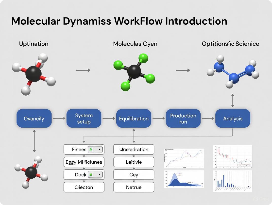
Molecular Dynamics Workflow: A Step-by-Step Framework
The process of conducting an MD simulation follows a systematic workflow that transforms initial structural data into dynamic behavioral insights.
Initial Structure Preparation
Every MD simulation begins with preparing the initial atomic coordinates of the target system [3]. Structures are often obtained from experimental databases such as the Protein Data Bank (PDB) for biomolecules or the Materials Project for crystalline materials [3]. However, database structures frequently require correction of missing atoms or incomplete regions through structural modeling tools [3]. For novel systems without experimental templates, initial structures may be built from scratch using predictive approaches, including AI-based tools like AlphaFold2 which received the 2024 Nobel Prize in Chemistry [3].
System Initialization
Once the initial structure is prepared, the simulation system must be initialized [3]. This involves solvation (placing the molecule in explicit solvent water models like TIP3P or implicit solvent environments), adding counterions to neutralize charge, and assigning initial atomic velocities sampled from a Maxwell-Boltzmann distribution corresponding to the desired simulation temperature [3] [1]. The choice between explicit and implicit solvent represents a critical trade-off between computational expense and physical accuracy [1].
Force Calculation and Time Integration
The computational core of MD involves calculating forces between atoms using the selected force field [3]. This represents the most computationally intensive step, often employing cutoff methods to ignore interactions beyond certain distances and spatial decomposition algorithms to distribute workload across multiple processors [3] [1]. Recent advances include Machine Learning Interatomic Potentials (MLIPs) trained on quantum chemistry data, which offer remarkable precision and efficiency for complex material systems [3].
Forces are then used to solve Newton's equations of motion through time integration algorithms [3]. The Verlet algorithm is particularly valued for its numerical stability and energy conservation properties [3] [1]. The integration time step must balance accuracy and efficiency—typically 0.5-2.0 femtoseconds—and can be extended using constraint algorithms that fix the fastest vibrations (e.g., hydrogen atoms) in place [3] [1].
Trajectory Analysis
The final and most critical phase involves analyzing the simulation trajectory—the time-series data of atomic positions and velocities [3]. Raw coordinate data must be transformed into chemically and biologically meaningful insights through various analytical approaches:
- Radial Distribution Function (RDF): Quantifies how atoms are spatially arranged around each other, particularly useful for analyzing liquid and amorphous structures [3]
- Mean Square Displacement (MSD): Measures atomic mobility and enables calculation of diffusion coefficients [3]
- Principal Component Analysis (PCA): Identifies dominant modes of collective motion from high-dimensional trajectory data [3]
- Clustering Algorithms: Group similar conformations to identify significant structural states [5]
Table 2: Essential Analysis Techniques for MD Trajectories
| Analysis Method | Physical Property Measured | Key Applications | Example Software Tools |
|---|---|---|---|
| Radial Distribution Function | Spatial atom distribution | Liquid structure, solvation shells | GROMACS, MOE |
| Mean Square Displacement | Particle mobility | Diffusion coefficients, ion conductivity | GROMACS, CHARMM |
| Principal Component Analysis | Collective motions | Domain movements, allosteric changes | CPPTRAJ, MDTraj |
| Clustering Algorithms | Representative conformations | State identification, ensemble reduction | PyMOL, VMD |
Integrating MD with Experimental Data
MD simulations rarely exist in isolation; their true power emerges when integrated with experimental data. Several strategic frameworks have been developed for this integration [6] [4] [7]:
Independent Approach
Experimental and computational protocols are performed separately, with results compared afterward [6] [7]. This approach can reveal unexpected conformations but may struggle with rare events that require extensive sampling [7].
Guided Simulation (Restrained) Approach
Experimental data are incorporated as external energy terms that guide the conformational sampling during simulation [6] [7]. This efficiently limits the conformational space but requires implementation directly in the simulation software [7].
Search and Select (Reweighting) Approach
A large ensemble of conformations is generated first, then experimental data is used to filter and select compatible structures [6] [7]. This allows simpler integration of multiple data types but requires the initial pool to contain the correct conformations [7].
Guided Docking
Experimental information defines binding sites to assist in predicting complex formation between molecules [6] [7]. Programs like HADDOCK and pyDockSAXS incorporate such experimental restraints [6].
A compelling example of integration appears in studies combining MD with single-molecule FRET (smFRET). Researchers achieved quantitative comparison between sub-millisecond time-resolution smFRET measurements and 10-second MD simulations of the LIV-BPSS biosensor protein, providing atomistic interpretations of conformational changes observed experimentally [8].
Practical Implementation: The Scientist's Toolkit
Successful implementation of MD requires familiarity with key software tools and computational resources.
Research Reagent Solutions
Table 3: Essential Tools and Resources for MD Simulations
| Tool Category | Specific Examples | Function and Application |
|---|---|---|
| Simulation Software | GROMACS, CHARMM, OpenMM, AMBER | Software packages that perform the numerical integration and force calculations |
| Visualization Tools | PyMOL, VMD, UCSF Chimera | Render molecular structures and trajectories for analysis and presentation |
| Force Fields | AMBER, CHARMM, OPLS | Mathematical models defining potential energy surfaces and atomic interactions |
| No-Code Platforms | Prithvi | Web-based interfaces that simplify MD setup and analysis for non-specialists |
| Specialized Hardware | GPUs, Anton Supercomputer | Accelerate computationally intensive force calculations |
Visualization Advances
As simulations grow in scale and complexity, visualization challenges intensify [9]. Modern approaches include virtual reality environments for immersive trajectory exploration, web-based tools for collaborative analysis, and deep learning techniques to emulate photorealistic visualization styles from simpler representations [9]. These advances help researchers comprehend the enormous data output from modern simulations, which can encompass billions of atoms representing entire cellular organelles [9].
Applications in Drug Discovery and Materials Science
MD simulations have become invaluable in pharmaceutical research, particularly in structure-based drug design [2] [1]. They help identify drug binding modes, predict binding affinities, and understand how proteins change shape upon ligand interaction [5]. In the fight against COVID-19, GaMD simulations helped design drug candidates targeting the SARS-CoV-2 main protease and captured inhibitor binding to the ACE2 receptor [2].
In materials science, MD enables the computation of stress-strain curves at the atomic scale, providing insights into mechanical properties including Young's modulus, yield stress, and tensile strength [3]. The direct observation of microscopic events like plastic deformation nucleation makes MD indispensable for predicting mechanical behavior across diverse materials [3].
Molecular Dynamics truly represents a "computational microscope" that has revolutionized our ability to observe and understand molecular processes at atomic resolution. As methods like GaMD overcome traditional time-scale limitations, and integration with experimental data becomes more sophisticated, MD continues to expand its transformative impact across biochemistry, pharmacology, and materials science. The ongoing development of more accurate force fields, enhanced sampling algorithms, machine learning potentials, and accessible platforms ensures that this computational microscope will continue to provide increasingly powerful insights into the molecular mechanisms that govern biological function and material behavior.
Molecular dynamics (MD) is a computational method that simulates the natural motions of atoms and molecules over time. At its heart, MD relies on Newton's equations of motion to calculate how a system of interacting particles evolves from a given starting configuration. This powerful approach provides atomic-level insights into dynamic processes in chemistry, biology, and materials science, making it indispensable for understanding biomolecular interactions, material properties, and facilitating modern drug development [10].
The fundamental principle of MD is straightforward: given initial positions and velocities of all atoms, along with a description of the forces acting upon them, one can numerically solve Newton's equations to predict the system's trajectory. This capability to simulate complex molecular behavior has made MD an essential tool in the researcher's toolkit, bridging the gap between static structural data and dynamic functional understanding [11].
The Mathematical Foundation: Newton's Equations of Motion
The Fundamental Equations
MD simulations are built upon Newton's second law of motion, which states that force equals mass times acceleration: F = ma [11]. In molecular dynamics, this foundational principle translates into a set of equations that govern atomic motion:
- Forces derive from the potential energy of the system: F = -∇U, where U represents the potential energy function that models all interactions between particles [11]
- Acceleration is calculated from these forces and determines how particle velocities and positions evolve over time
These equations form a deterministic system: given initial atomic positions and velocities, along with a force field describing molecular interactions, the subsequent trajectory is uniquely determined.
Numerical Integration Algorithms
Since analytical solutions to Newton's equations are impossible for complex molecular systems, MD employs numerical integration algorithms to approximate particle trajectories. These algorithms discretize time into small steps (typically 0.5-2 femtoseconds) and update positions and velocities iteratively [11].
Table 1: Core Integration Algorithms in Molecular Dynamics
| Algorithm | Key Features | Advantages | Common Use Cases |
|---|---|---|---|
| Verlet | Uses positions and accelerations to update positions without explicit velocity storage [11] | Computationally efficient; good energy conservation | General purpose simulations; systems with memory constraints |
| Leap-frog | Updates positions and velocities at interleaved half-time steps [11] | Improved numerical stability; direct velocity calculation | Simulations requiring kinetic energy monitoring |
| Velocity Verlet | Simultaneously updates positions, velocities, and accelerations [11] | Better energy conservation; positions and velocities at same time points | Modern MD software packages (GROMACS, NAMD); most current applications |
The Velocity Verlet algorithm has emerged as a preferred method in many modern MD implementations. It combines the advantages of both Verlet and leap-frog methods while providing improved accuracy for longer time steps [11]. The algorithm implements a two-step process:
- Position and velocity updates based on current forces
- Force recalculation and velocity correction based on new positions
Practical Implementation: From Theory to Simulation
Time Step Selection and Numerical Stability
Appropriate time step selection is crucial for accurate MD simulations. The time step determines the temporal resolution of the simulation and represents a balance between computational efficiency and numerical stability [11]:
- Smaller time steps (0.5-1 fs) increase accuracy but require more computational resources
- Larger time steps (2 fs or more) improve efficiency but may lead to numerical instabilities
- The Nyquist-Shannon sampling theorem guides time step selection: it should be at least an order of magnitude smaller than the fastest motion in the system [11]
For atomistic simulations of biomolecules, typical time steps range from 0.5 to 2 femtoseconds, constrained by the period of the fastest vibrations (C-H bonds) [11].
Energy Conservation and Symplectic Integrators
Energy conservation serves as a key indicator of simulation accuracy. In isolated systems (microcanonical ensemble), the total energy should remain constant [11]. Symplectic integrators are particularly valuable for MD because they preserve the geometric structure of Hamiltonian systems, maintaining long-term stability and energy conservation [11]. Both Velocity Verlet and leap-frog algorithms classify as symplectic integrators, making them suitable for extended simulations where non-symplectic methods might exhibit energy drift [11].
Figure 1: Core Molecular Dynamics Simulation Workflow
MD in Action: Experimental Validation and Applications
Quantitative Comparison with Experimental Data
MD simulations gain credibility when validated against experimental data. Recent research has demonstrated quantitative comparisons between sub-millisecond time resolution single-molecule FRET measurements and long-timescale MD simulations [8]. In one study of the LIV-BPSS biosensor protein, researchers performed all-atom structure-based simulations spanning multiple cycles of clamshell-like conformational changes on the scale of seconds, directly correlating these events with experimental smFRET measurements [8].
This approach provided valuable information on:
- Local dynamics of fluorophores at their attachment sites
- Correlations between fluorophore motions and large-scale conformational changes
- Determinations of Förster radius (R₀) and fluorophore orientation factor (κ²)
The congruence between simulation and experiment demonstrates MD's predictive power when simulations achieve temporal regimes overlapping with experimental observables [8].
Protein Structure Prediction and Refinement
MD simulations play a crucial role in refining predicted protein structures. In a study modeling the hepatitis C virus core protein (HCVcp), researchers found that neural network-based prediction tools like AlphaFold2, Robetta, and trRosetta provided good initial models, but subsequent MD simulations were essential for obtaining compactly folded structures of good quality [12]. The root mean square deviation of backbone atoms, root mean square fluctuation of Cα atoms, and radius of gyration were calculated to monitor structural changes and convergence during simulations [12].
Table 2: Time Step Parameters for Different Simulation Types
| Simulation Type | Recommended Time Step | Fastest Motion Constrained | Constraint Algorithms |
|---|---|---|---|
| Atomistic (all-atom) | 1-2 fs | C-H bond vibrations | SHAKE, LINCS |
| Coarse-grained | 10-20 fs | Effective bead vibrations | None typically required |
| Implicit solvent | 2-4 fs | C-H bond vibrations | SHAKE |
| Explicit solvent | 1-2 fs | C-H bond vibrations + water modes | SETTLE for water |
Guiding Protein Engineering
MD simulations can guide rational protein design by predicting how mutations affect structure and function. In developing a brighter variant of superfolder Green Fluorescent Protein, researchers used short time-scale MD modeling to predict changes in local chromophore interaction networks and solvation [13]. Simulations revealed that replacing histidine 148 with serine formed more persistent H-bonds with the chromophore phenolate group and increased the residency time of an important water molecule [13]. This single mutation resulted in a protein 1.5 times brighter than the parent with 3-fold increased resistance to photobleaching [13].
Figure 2: Molecular Force Calculation Process
Essential Research Reagents and Computational Tools
Modern MD simulations require both specialized software and careful parameter selection. The following table outlines key components essential for successful MD research in drug development and biochemical applications.
Table 3: Research Reagent Solutions for Molecular Dynamics
| Tool Category | Specific Examples | Function & Application | Key Features |
|---|---|---|---|
| Simulation Software | OpenMM [14], GROMACS, NAMD [11] | Performs the numerical integration of equations of motion | GPU acceleration; support for multiple force fields |
| Analysis Packages | MDTraj [14] | Analyzes simulation trajectories; calculates properties | RMSD, radius of gyration, secondary structure analysis |
| Force Fields | CHARMM [14], AMBER [14] | Defines potential energy functions and parameters | Protein, nucleic acid, lipid parameters; water models |
| System Preparation | PDBFixer [14], PackMol [14] | Prepares structures for simulation; adds solvent | Structure cleaning; solvation; ion addition |
| Automation Tools | MDCrow [14] | Automates MD workflows using LLM agents | Handles file processing; simulation setup; analysis |
| Visualization | NGLview [14] | Visualizes molecular structures and trajectories | Web-based; interactive trajectory playback |
Advanced Considerations and Future Directions
Workflow Automation and Accessibility
Recent advances have focused on making MD more accessible through workflow automation. MDCrow represents one such approach—an LLM-based assistant capable of automating MD workflows using over 40 expert-designed tools for handling files, setting up simulations, analyzing outputs, and retrieving relevant information from literature and databases [14]. This system can perform complex tasks including downloading PDB files, performing multiple simulations, and conducting analyses with minimal user intervention [14].
Integration with Experimental Data
As MD simulations reach longer timescales, they increasingly overlap with experimental observables, enabling direct quantitative comparisons. For example, all-atom structure-based simulations calibrated against explicit solvent simulations can sample multiple cycles of protein conformational changes on the scale of seconds, directly informing the interpretation of smFRET data [8]. This integration provides atomic-level insights into conformational dynamics that complement experimental findings.
Accelerated Sampling and AI Integration
Traditional MD faces limitations in sampling rare events due to computational constraints. Recent research addresses this through accelerated sampling methods and AI integration. Generative artificial intelligence frameworks can now accelerate MD simulations for crystalline materials by reframing the task as conditional generation of atomic displacements [10]. Machine-learned potentials enable full-cycle device-scale simulations of complex materials like phase-change memory devices [10]. These advances continue to expand the boundaries of what MD can simulate within practical computational limits.
Molecular dynamics (MD) simulation is a computational method for analyzing the physical movements of atoms and molecules over time by numerically solving Newton's equations of motion [1]. The method is founded on classical mechanics principles, where the force on any particle is calculated as the negative gradient of the potential energy function: ( \vec{F} = -\nabla U(\vec{r}) ) [15]. MD simulations have become indispensable across chemical physics, materials science, and biophysics, enabling researchers to investigate molecular processes at atomic resolution that are often inaccessible to experimental observation [1].
The reliability and physical meaningfulness of any MD simulation depend critically on the proper specification of three foundational components: the initial conditions that define the starting state of the system, the topology that describes the connectivity between particles, and the force field that governs their interactions. These elements collectively determine the system's Hamiltonian and thus its subsequent evolution through phase space. This technical guide examines each component in detail, providing researchers with the fundamental knowledge required to construct accurate and thermodynamically consistent molecular systems for computational investigation.
Initial Conditions
The initial conditions of a molecular dynamics simulation establish the starting point from which the system evolves. Proper initialization is essential for generating physically realistic trajectories and ensuring efficient convergence of thermodynamic properties.
Components of System Initialization
Initial conditions encompass several key elements that must be defined prior to simulation:
Atomic Coordinates: The initial positions ( \mathbf{r} ) of all atoms in the system, typically obtained from experimental structures (e.g., X-ray crystallography or NMR) or through system-building tools [16]. For simulations of proteins and other biomolecules, coordinates are commonly provided in the Protein Data Bank (PDB) format, which specifies atomic positions, residue names, chain identifiers, and other structural metadata [15].
Atomic Velocities: The initial velocities ( \mathbf{v} ) of all particles, which determine the initial kinetic energy and temperature of the system [16]. When velocities are not available from experimental data, they are commonly assigned randomly from a Maxwell-Boltzmann distribution at the target temperature [16] [15]:
[ p(vi) = \sqrt{\frac{mi}{2 \pi kT}}\exp\left(-\frac{mi vi^2}{2kT}\right) ]
where ( k ) is Boltzmann's constant, ( T ) is the temperature, and ( m_i ) is the mass of atom ( i ) [16].
System Boundaries and Periodicity: The simulation box size and shape, defined by three basis vectors ( \mathbf{b}1, \mathbf{b}2, \mathbf{b}_3 ) that determine the unit cell for periodic boundary conditions [16]. System builders like packmol can create initial configurations with specified density and composition [17].
Solvent Environment: The choice between explicit solvent molecules (e.g., TIP3P, SPC/E water models) or implicit solvent representations [1]. Explicit solvents provide more realistic solvation dynamics but increase computational cost substantially.
Practical Implementation
In practice, initial system preparation often involves multiple stages. For biomolecular systems, the process typically begins with a PDB file containing atomic coordinates. Missing hydrogen atoms may be added, and protonation states adjusted according to the physiological pH of interest. The structure is then solvated in a water box, with ions added to neutralize the system and achieve physiological ionic strength [15].
Table 1: Quantitative Parameters for System Initialization
| Parameter | Typical Values | Considerations |
|---|---|---|
| Initial velocity assignment | Maxwell-Boltzmann distribution | Velocities are often rescaled after assignment to ensure the center-of-mass velocity is zero [16] |
| Solvent density | 1.0 g/cm³ (aqueous systems) [17] | Density affects system size and computational cost |
| Number of atoms | 200 - 1,000,000+ | System size balances computational cost with biological relevance [17] [1] |
| Box dimensions | Varies by system | Must accommodate the solute with sufficient padding for cutoffs |
| Ionic concentration | 0.15 M for physiological conditions | Affects electrostatic interactions and protein stability |
After initial configuration, systems typically undergo energy minimization to remove steric clashes, followed by gradual heating and equilibration to the target temperature and pressure. This stepwise approach ensures stable integration of the equations of motion before production simulation begins.
System Topology
The topology of a molecular system defines the structural relationships between its constituent atoms, including bonding patterns, chemical identity, and molecular connectivity that remain constant throughout a classical MD simulation.
Topology Components and Representation
Molecular topology encompasses several key aspects:
Atomic Identity and Masses: Element type and mass for each particle in the system, which determine its inertial properties and contributions to kinetic energy [15].
Bond Connectivity: Specification of covalent bonds between atoms, which constrains their relative motion and defines the molecular graph [16]. In proteins, this includes the backbone and sidechain bonds that maintain structural integrity.
Residue and Chain Organization: Hierarchical organization of atoms into residues (monomers) and chains (polymers), preserving the chemical identity of molecular components [15].
Exclusion Lists: Specification of atom pairs that are bonded or closely related and should be excluded from non-bonded interactions, or for which non-bonded interactions require special treatment [16].
The topology is typically represented in specialized file formats that encode these relationships. For example, GROMACS uses top files that define molecule types, atom characteristics, and interaction parameters [16].
Topology in Force Field Context
The system topology works in conjunction with the force field to define the complete potential energy function. While the topology specifies which atoms are connected, the force field provides the specific functional forms and parameters for interactions between them. This separation allows the same topology to be used with different force fields, though this requires careful validation [15].
Table 2: Topology Components Across Molecular Systems
| Topology Element | Small Molecule | Protein | Nucleic Acid | Complex System |
|---|---|---|---|---|
| Bond connectivity | Defined by chemical structure | Peptide bonds + sidechains | Sugar-phosphate backbone + bases | Multiple molecular entities |
| Residue organization | Single residue | Amino acid residues | Nucleotide residues | Mixed residue types |
| Special interactions | Torsional parameters | Backbone dihedrals, sidechain rotamers | Base pairing, stacking | Interface contacts |
| Exclusion rules | 1-2, 1-3 neighbors | Intra-residue and inter-residue | Base pairing partners | Inter-molecular exclusions |
Force Fields
Force fields provide the mathematical framework and parameters that describe the potential energy of a system as a function of atomic coordinates. They approximate the complex quantum mechanical interactions between atoms using empirically parameterized functions that are computationally efficient to evaluate.
Force Field Energy Components
The total potential energy in a molecular mechanics force field is typically decomposed into bonded and non-bonded contributions:
[ U(\vec{r}) = U{bonded}(\vec{r}) + U{non-bonded}(\vec{r}) ]
Bonded Interactions
Bonded interactions describe the energy associated with covalent connectivity:
Bond Stretching: The energy required to deviate from equilibrium bond length, typically modeled as a harmonic oscillator:
[ V{Bond} = kb(r{ij} - r0)^2 ]
where ( kb ) is the force constant and ( r0 ) is the equilibrium bond length [18] [15].
Angle Bending: The energy associated with deviation from equilibrium bond angles, also typically harmonic:
[ V{Angle} = k\theta(\theta{ijk} - \theta0)^2 ]
where ( k\theta ) is the angle force constant and ( \theta0 ) is the equilibrium angle [18] [15].
Torsional Potentials: The energy associated with rotation around chemical bonds, typically modeled as a periodic function:
[ V{Dihed} = k\phi[1 + \cos(n\phi - \delta)] ]
where ( k_\phi ) is the dihedral force constant, ( n ) is the periodicity, and ( \delta ) is the phase angle [18] [15].
Improper Dihedrals: Potentials that enforce planarity in chemical groups such as aromatic rings or peptide bonds:
[ V{Improper} = k\phi(\phi - \phi_0)^2 ]
Non-Bonded Interactions
Non-bonded interactions describe forces between atoms that are not directly bonded:
van der Waals Interactions: The attractive and repulsive forces between atomic electron clouds, typically modeled with the Lennard-Jones potential:
[ V_{LJ}(r) = 4\epsilon\left[\left(\frac{\sigma}{r}\right)^{12} - \left(\frac{\sigma}{r}\right)^{6}\right] ]
where ( \epsilon ) is the well depth and ( \sigma ) is the van der Waals radius [18]. Some force fields use the Buckingham potential as an alternative [18].
Electrostatic Interactions: The Coulombic attraction or repulsion between partial atomic charges:
[ V{Elec} = \frac{qi qj}{4\pi\epsilon0 r_{ij}} ]
where ( qi ) and ( qj ) are partial atomic charges and ( \epsilon_0 ) is the dielectric constant [18].
Force Field Classification
Force fields are commonly categorized into classes based on their complexity and treatment of molecular interactions:
Class I Force Fields: Employ simple harmonic potentials for bonds and angles with no cross-terms. Examples include AMBER, CHARMM, GROMOS, and OPLS [18].
Class II Force Fields: Include anharmonic terms for bonds and angles, along with cross-terms coupling internal coordinates. Examples include MMFF94 and UFF [18].
Class III Force Fields: Explicitly incorporate polarization effects using methods such as Drude oscillators or inducible dipoles. Examples include AMOEBA, CHARMM-Drude, and OPLS5 [18].
Most biomolecular simulations currently employ Class I force fields, though Class III polarizable force fields are increasingly used for systems where electronic polarization effects are significant.
Combining Rules and Cutoffs
A critical aspect of force field implementation is the treatment of interactions between different atom types. Combining rules determine how Lennard-Jones parameters are calculated for heterogeneous atom pairs [18]. The most common approaches include:
Lorentz-Berthelot Rules: Used in CHARMM and AMBER force fields:
[ \sigma{ij} = \frac{\sigma{ii} + \sigma{jj}}{2}, \quad \epsilon{ij} = \sqrt{\epsilon{ii}\epsilon{jj}} ]
Geometric Mean Rules: Used in GROMOS and OPLS force fields:
[ \sigma{ij} = \sqrt{\sigma{ii}\sigma{jj}}, \quad \epsilon{ij} = \sqrt{\epsilon{ii}\epsilon{jj}} ]
Non-bonded interactions are typically evaluated using a cut-off scheme to maintain computational efficiency, with long-range electrostatic interactions treated using Particle Mesh Ewald (PME) methods [16].
Diagram 1: Hierarchical organization of force field energy components showing bonded and non-bonded interaction categories.
Integration of Components in MD Workflow
The three components—initial conditions, topology, and force field—work together in a coordinated manner throughout the MD simulation workflow. Understanding their integration is essential for proper simulation design and execution.
System Setup Protocol
A typical MD workflow integrates the three key components systematically:
Structure Preparation: Initial atomic coordinates are obtained from experimental data or molecular modeling, establishing the initial conditions.
Topology Generation: The molecular structure is analyzed to define bonding patterns, residue organization, and molecular connectivity.
Force Field Assignment: Appropriate parameters are assigned to all interactions based on atom types and connectivity.
System Assembly: The solute is placed in a simulation box, solvated, and ionized to create the complete simulation environment.
Energy Minimization: The system is relaxed to remove steric clashes and prepare for dynamics.
Equilibration: The system is gradually brought to the target temperature and pressure while maintaining appropriate constraints.
Production Simulation: Unconstrained data collection for analysis of structural and dynamic properties.
Diagram 2: Sequential workflow for molecular dynamics system setup showing how initial conditions, force field, and topology integrate to produce a simulation-ready system.
Practical Considerations for Researchers
When designing MD simulations, researchers should consider several practical aspects:
Consistency Between Components: Ensure that the force field parameters match the atom types and bonding patterns defined in the topology, and that the initial coordinates are chemically plausible for the chosen force field [15].
Temperature and Pressure Control: Implement appropriate thermostats and barostats during equilibration to achieve the desired ensemble conditions while maintaining Hamiltonian consistency.
Constraint Algorithms: For efficiency, consider constraining bonds involving hydrogen atoms using algorithms like SHAKE or LINCS, which allow longer integration time steps [1].
Neighbor Searching: Implement efficient pair list generation with appropriate buffering to maintain energy conservation while minimizing computational overhead [16].
Table 3: Research Reagent Solutions for MD Simulations
| Tool/Component | Function | Examples/Formats |
|---|---|---|
| Structure visualization | Visual inspection of initial coordinates | VMD, PyMol, ChimeraX |
| Force field parameterization | Define interaction potentials | CHARMM, AMBER, GROMOS, OPLS |
| Topology builders | Generate molecular connectivity | pdb2gmx, CHARMM-GUI, tleap |
| System solvation tools | Add solvent and ions | packmol, GROMACS solvation utilities [17] |
| Energy minimization algorithms | Remove steric clashes | Steepest descent, conjugate gradient |
| Dynamics integrators | Solve equations of motion | Velocity Verlet, Leap-frog [16] [15] |
The three foundational components of a molecular dynamics system—initial conditions, topology, and force fields—work in concert to determine the physical validity and numerical stability of simulations. Initial conditions establish the starting point in phase space, topology defines the covalent structure and molecular connectivity, and force fields provide the physical model governing atomic interactions. Mastery of these components enables researchers to design simulations that accurately capture the thermodynamic and dynamic properties of molecular systems, from small drug-like compounds to complex biomolecular assemblies. As MD simulations continue to evolve with advances in polarizable force fields, enhanced sampling methods, and machine learning approaches, the proper implementation of these core elements remains essential for generating scientifically meaningful results across computational chemistry and structural biology.
Molecular dynamics (MD) simulations have become an indispensable tool in research and development for materials, chemistry, and drug discovery, acting as a "microscope with exceptional resolution" to visualize atomic-scale dynamics [3]. The accuracy of these simulations is fundamentally dependent on the force field—a mathematical model that calculates the potential energy of a system of atoms and molecules based on their positions [3]. The choice of force field introduces a bias that can significantly influence simulation outcomes, making its selection a critical step [19]. This guide explores the core principles of force fields and provides a comparative analysis of three widely used families: AMBER, CHARMM, and GROMOS, within the context of a standard MD workflow.
Force Field Fundamentals
At its core, a force field describes the potential energy of a molecular system as a function of its nuclear coordinates. This energy is typically partitioned into several terms that capture different types of atomic interactions:
- Bonded Interactions: These describe the energy associated with the covalent bond structure of the molecule, including bond stretching, angle bending, and dihedral torsions.
- Non-bonded Interactions: These describe interactions between atoms that are not directly bonded, primarily consisting of van der Waals forces (modeled with a Lennard-Jones potential) and electrostatic interactions (described by Coulomb's law).
The parameters for these equations—such as equilibrium bond lengths, force constants, and partial atomic charges—are derived from a combination of quantum mechanical calculations and experimental data. The fidelity of a force field in representing a real molecular system hinges on the accuracy and breadth of its parameterization.
Comparative Analysis of AMBER, CHARMM, and GROMOS
A comparative study on the Aβ21-30 peptide fragment highlighted the significant bias that different force fields can introduce. While measures like the radius of gyration were similar across force fields, secondary structure content and hydrogen-bonding patterns varied considerably [19].
The table below summarizes the key characteristics, performance, and recommended use cases for AMBER, CHARMM, and GROMOS force fields.
Table 1: Key Characteristics of AMBER, CHARMM, and GROMOS Force Fields
| Feature | AMBER | CHARMM | GROMOS |
|---|---|---|---|
| Full Name | Assisted Model Building with Energy Refinement | Chemistry at HARvard Macromolecular Mechanics | GROningen MOlecular Simulation |
| Common Biomolecular Applications | Proteins, Nucleic Acids [19] | Proteins, Lipids, Carbohydrates [19] | Proteins, Carbohydrates [19] |
| Typical Water Models | TIP3P, TIP4P [19] | TIP3P, TIP4P [19] | SPC [19] |
| Performance on Aβ21-30 (Helical Content) | High helical content and variety of intrapeptide H-bonds [19] | Readily increases helical content (CHARMM27-CMAP) [19] | Suppresses helical structure (GROMOS53A6) [19] |
| Recommended Use Case (Based on Aβ21-30 study) | Systems where helical content is desirable [19] | Systems where helical content is desirable [19] | Better choice for modeling Aβ21-30, as it suppresses unrealistic helix formation [19] |
Integrating Force Fields into the Molecular Dynamics Workflow
The force field is the computational engine at the heart of the MD simulation workflow. Its selection directly impacts the results at every stage, from system preparation to trajectory analysis.
The Molecular Dynamics Workflow
The following diagram outlines the standard MD workflow, highlighting the critical role of the force field.
Detailed Workflow and Protocols
Prepare Initial Structure: The process begins with obtaining or building the initial atomic coordinates of the target system. Sources include the Protein Data Bank for biomolecules, the Materials Project for crystals, or PubChem for small molecules [3]. The structure must be carefully checked and prepared, as its quality directly impacts simulation reliability.
Initialize Simulation System: The initial structure is solvated in a water box, ions are added to neutralize the system's charge or achieve a specific ionic concentration, and the system is energy-minimized to remove bad contacts. Initial atomic velocities are assigned from a Maxwell-Boltzmann distribution corresponding to the desired simulation temperature [3].
Force Calculation from Interatomic Potential: This is the most computationally intensive step, where the chosen force field calculates the potential energy and forces for the entire system. The selection of AMBER, CHARMM, or GROMOS here is critical, as it determines the physical behavior of the system [19] [3]. Modern simulations often use spatial decomposition and GPU acceleration for efficiency [3].
Time Integration and Trajectory Generation: Forces are used to numerically integrate Newton's equations of motion. Algorithms like Verlet or leap-frog are commonly used for their stability and energy conservation properties [3]. A time step of 0.5–1.0 femtoseconds is typical, and this cycle of force calculation and integration is repeated millions of times to generate a trajectory [3].
Trajectory Analysis: The raw trajectory—a time-series of atomic coordinates and velocities—is analyzed to extract meaningful insights. Key analyses include [3]:
- Radial Distribution Function (RDF): To quantify structural features and coordination shells.
- Mean Square Displacement (MSD): To calculate diffusion coefficients and particle mobility.
- Principal Component Analysis (PCA): To identify essential collective motions from complex dynamics.
- Stress-Strain Calculations: To evaluate mechanical properties like Young's modulus.
The Scientist's Toolkit: Essential Research Reagents and Materials
Table 2: Essential Computational Tools for Molecular Dynamics Simulations
| Tool / Reagent | Function / Description |
|---|---|
| Force Fields (AMBER, CHARMM, GROMOS) | Provides the set of parameters and functions to calculate the potential energy and forces in a molecular system. |
| Water Models (TIP3P, TIP4P, SPC/E) | Represents water molecules in the simulation; different models can affect simulation outcome [19]. |
| MD Software (AMS, GROMACS, NAMD, OpenMM) | The simulation engine that performs the numerical integration and manages the simulation process [17] [20]. |
| System Building Tools (packmol) | Used to build the initial simulation system, including solvation and ion placement [17]. |
| Reference Quantum Mechanics Engines (ADF, BAND) | Provides high-accuracy data for parameterizing force fields or training machine-learning potentials in advanced workflows [20]. |
| Trajectory Analysis Tools | Software and scripts (e.g., in Python, VMD, CPPTRAJ) to process MD trajectories and compute physical properties [3]. |
Emerging Trends and Machine Learning Potentials
A significant advancement is the use of Machine Learning Interatomic Potentials. MLIPs are trained on large datasets from quantum chemistry calculations and can predict atomic energies and forces with high accuracy and efficiency, enabling simulations of complex systems that were previously prohibitive [3]. Furthermore, Active Learning workflows are now being implemented. These workflows automatically run MD, pause to launch new reference calculations, retrain the ML potential, and then continue the simulation, ensuring its accuracy on-the-fly [20].
The choice of a force field is a foundational decision that critically influences the results and interpretation of molecular dynamics simulations. As demonstrated, force fields like AMBER, CHARMM, and GROMOS can exhibit distinct biases, for instance, in the secondary structure propensity of peptides. Therefore, researchers must carefully select a force field that is appropriate for their specific system, often guided by previous validation studies. The ongoing integration of machine learning promises to further enhance the accuracy and scope of these simulations, solidifying the critical role of force fields in computational discovery.
Molecular dynamics (MD) simulations have emerged as a transformative tool in biomedical research, functioning as a "computational microscope" that provides atomic-level resolution into the dynamic processes governing life itself [3]. These simulations enable researchers to track the temporal evolution of biological systems, from the folding of proteins to the precise molecular interactions that occur when a drug binds to its receptor. The integration of MD with experimental methods has created a powerful paradigm for rational drug design, allowing scientists to move beyond static structural snapshots to understand the critical role of dynamics in biological function and therapeutic intervention [21]. This technical guide examines the core principles, methodologies, and applications of MD within biomedical research, with a particular focus on its growing impact on drug discovery and development processes.
Fundamental MD Workflow in Biomedical Research
The execution of a molecular dynamics simulation follows a systematic workflow that transforms a static molecular structure into a dynamic trajectory rich with thermodynamic and kinetic information [3].
The standard MD protocol comprises several sequential stages, each with specific objectives and technical requirements. Figure 1 illustrates this generalized workflow for a typical biomedical simulation.
Figure 1. Generalized MD Workflow for Biomedical Research. This diagram outlines the sequential stages of a molecular dynamics simulation, from initial structure preparation to final biological interpretation.
Initial Structure Preparation and System Building
The foundation of any reliable MD simulation is an accurate initial atomic structure. For biomedical applications, these structures are typically sourced from:
- Protein Data Bank (PDB): The primary repository for experimentally determined protein structures via X-ray crystallography, NMR, or cryo-EM [3].
- AlphaFold2 Predictions: AI-predicted protein structures with remarkable accuracy, particularly valuable for targets without experimental structures [22].
- PubChem/ChEMBL: Databases containing small molecule structures for drug-like compounds [3].
Structure preparation involves adding missing atoms (particularly hydrogens), assigning protonation states, and ensuring proper assignment of histidine tautomers. For protein-ligand systems, careful parameterization of the small molecule is essential, using tools such as CGenFF for CHARMM force fields or GAAMP for AMBER force fields [3].
System Setup and Equilibration Protocols
Once the molecular structure is prepared, it must be embedded in a biologically relevant environment:
- Solvation: Placement in a water box (e.g., TIP3P, TIP4P water models) with a minimum 10-12 Å buffer between the solute and box edge.
- Neutralization: Addition of counterions (e.g., Na+, Cl-) to achieve system electroneutrality.
- Physiological Conditions: Further addition of ions to approximate physiological concentration (e.g., 150 mM NaCl).
The system then undergoes energy minimization (typically 5,000-10,000 steps) using steepest descent or conjugate gradient algorithms to remove steric clashes, followed by a two-stage equilibration [3]:
- NVT Ensemble: Constant Number of particles, Volume, and Temperature for 100-500 ps to stabilize temperature.
- NPT Ensemble: Constant Number of particles, Pressure, and Temperature for 1-5 ns to stabilize density.
Production Simulation and Trajectory Analysis
Production simulations employ an integration time step of 0.5-2.0 femtoseconds, with trajectory frames typically saved every 10-100 ps for analysis [17] [3]. Long-range electrostatics are handled using Particle Mesh Ewald (PME) methods, and temperature/pressure are maintained using thermostats (e.g., Berendsen, Nosé-Hoover) and barostats (e.g., Parrinello-Rahman). The resulting trajectory data is analyzed using both built-in and external analysis tools to extract biologically relevant information, as detailed in Section 4.
Key Biomedical Applications
Advanced Drug Delivery System Optimization
MD simulations provide critical insights into the molecular-level interactions between drug compounds and their carrier systems, enabling rational design of delivery platforms with optimized properties [21]. Table 1 summarizes key nanocarrier systems studied using MD simulations.
Table 1: MD Applications in Drug Delivery System Design
| Delivery System | Key Advantages | MD Simulation Insights | Representative Drugs |
|---|---|---|---|
| Functionalized Carbon Nanotubes (FCNTs) | High drug-loading capacity, stability, cellular uptake efficiency [21] | Drug-nanotube interaction energy, encapsulation stability, release kinetics | Doxorubicin, Gemcitabine [21] |
| Chitosan-based Nanoparticles | Biodegradability, reduced toxicity, mucoadhesive properties [21] | Polymer-drug binding affinity, degradation behavior, controlled release mechanisms | Paclitaxel, protein therapeutics [21] |
| Human Serum Albumin (HSA) Carriers | Natural biocompatibility, long circulation half-life, tumor targeting [21] | Drug-binding site interactions, allosteric effects on carrier structure | Anticancer agents, antiviral drugs [21] |
| Metal-Organic Frameworks (MOFs) | Tunable porosity, high surface area, surface functionalization [21] | Host-guest chemistry, diffusion pathways through porous structures | Chemotherapeutic agents [21] |
Protein-Ligand Binding and Interaction Analysis
Understanding the structural basis and energetics of drug-receptor interactions represents one of the most significant applications of MD in drug discovery. The DeepICL framework exemplifies how MD can be leveraged for interaction-guided drug design by incorporating universal patterns of protein-ligand interactions—hydrogen bonds, salt bridges, hydrophobic interactions, and π-π stackings—as prior knowledge to enhance generalizability, even with limited experimental data [23]. This approach enables researchers to:
- Identify Interaction Hot Spots: Map key interaction sites within binding pockets that contribute significantly to binding affinity [23].
- Assess Binding Pose Stability: Monitor the stability of docked poses over simulation time to discriminate between correct and incorrect binding modes.
- Calculate Binding Free Energies: Employ advanced sampling methods (e.g., Free Energy Perturbation, Thermodynamic Integration) to quantitatively predict binding affinities [22].
- Guide Molecular Optimization: Use interaction fingerprints to inform structural modifications that enhance potency and selectivity [23].
Enhanced Sampling for Rare Events
Conventional MD simulations may be insufficient to observe biologically relevant rare events (e.g., ligand unbinding, large conformational changes) within practical computational timescales. Enhanced sampling methods address this limitation:
- Metadynamics: Uses a history-dependent bias potential to discourage revisiting of previously sampled configurations, effectively accelerating escape from local minima.
- Umbrella Sampling: Applies harmonic restraints along a predetermined reaction coordinate to efficiently sample high-energy regions and reconstruct free energy profiles.
- Replica Exchange MD: Simultaneously runs multiple simulations at different temperatures or Hamiltonian parameters, allowing exchanges between replicas to overcome energy barriers.
Analysis Methods for Biomedical Insights
The raw trajectory data generated by MD simulations must be processed through appropriate analytical methods to extract biologically meaningful information.
Structural and Energetic Analysis
Radial Distribution Function (RDF) The RDF, denoted as g(r), quantifies how particle density varies as a function of distance from a reference particle [3]. For biomedical applications, RDF analysis can reveal:
- Solvation shells around drug molecules or protein surfaces
- Ion distribution around DNA, RNA, or membrane surfaces
- Local ordering in amorphous drug formulations The coordination number, obtained by integrating the RDF, provides quantitative information about binding stoichiometry and solvation [3].
Principal Component Analysis (PCA) PCA identifies collective motions in biomolecules by diagonalizing the covariance matrix of atomic positional fluctuations [3]. This method:
- Reduces high-dimensional trajectory data to a few essential dynamics modes
- Identifies functionally relevant domain motions in proteins
- Distinguishes between different conformational states
- Can be combined with clustering to identify metastable states and construct free energy landscapes [3]
Kinetic and Thermodynamic Properties
Mean Square Displacement (MSD) and Diffusion The MSD measures the average squared displacement of particles over time and is used to calculate diffusion coefficients through the Einstein relation: D = lim(t→∞) ⟨MSD(t)⟩/6t for 3D diffusion [3]. This analysis provides insights into:
- Drug mobility in delivery matrices and through biological barriers
- Water and ion diffusion in protein channels and pores
- Molecular mobility in amorphous solid dispersions
Free Energy Calculations Free energy landscapes provide comprehensive descriptions of biomolecular stability and transitions. These landscapes are constructed using:
- Umbrella Sampling along carefully chosen reaction coordinates
- Metadynamics for exploring multidimensional free energy surfaces
- Markov State Models (MSMs) to infer kinetics and thermodynamics from many short simulations
Interaction Analysis
Protein-Ligand Interaction Fingerprints Interaction fingerprints provide a concise representation of specific contacts between a drug and its target over the simulation trajectory. These include:
- Hydrogen bonds (distance and angle criteria)
- Hydrophobic contacts
- π-π stacking and cation-π interactions
- Ionic interactions/salt bridges Monitoring the persistence and dynamics of these interactions helps explain structure-activity relationships and guide molecular optimization [23].
Reactive MD for Reaction Discovery
Specialized non-equilibrium MD methods have been developed to promote chemical reactions and explore reaction networks, with significant implications for drug metabolism studies and prodrug design.
Nanoreactor and Lattice Deformation Methods
The Reactions Discovery tool implements two primary approaches for accelerating chemical reactivity [17]:
Nanoreactor MD This method employs cyclic compression and diffusion phases to drive reactive events [17]:
- Compression Phases: Apply inward acceleration with high force constants (0.01 Ha/bohr²) to promote molecular encounters
- Diffusion Phases: Allow system evolution at controlled temperature (default 500 K) with lower constraints
- Cyclic Protocol: Typically runs 10+ cycles to sample diverse reaction pathways [17]
Lattice Deformation MD Applicable to periodic systems, this method oscillates the simulation cell volume between initial and compressed states (MinVolumeFraction typically 0.3-0.6) with defined periodicity (default 100 fs) [17]. The resulting pressure and density fluctuations create conditions conducive to chemical reactions.
Table 2 compares the key parameters for these reactive MD methods.
Table 2: Reactive MD Method Parameters for Reaction Discovery
| Parameter | Nanoreactor MD | Lattice Deformation MD |
|---|---|---|
| System Requirements | No periodic boundary conditions [17] | Requires 3D-periodic system [17] |
| Compression | Spherical boundary with increasing force constant [17] | Volume oscillation between V₀ and V₀×MinVolumeFraction [17] |
| Key Parameters | DiffusionTime (default 250 fs), MinVolumeFraction (default 0.6), Temperature (default 500 K) [17] | MinVolumeFraction (default 0.3), Period (default 100 fs), Temperature (default 500 K) [17] |
| Typical Applications | Reaction discovery in molecular clusters, solution chemistry [17] | Solid-state reactions, materials under mechanical stress [17] |
| Number of Cycles | NumCycles (default 10) [17] | NumCycles (default 10) [17] |
Workflow for Reactive MD
The implementation of reactive MD follows a specialized workflow, particularly for the Nanoreactor approach, as illustrated in Figure 2.
Figure 2. Reactive MD Workflow for Chemical Reaction Discovery. This diagram illustrates the cyclic process of compression and diffusion phases used in Nanoreactor MD simulations to promote and identify chemical reactions.
Table 3 provides a comprehensive overview of essential tools, databases, and platforms for conducting MD simulations in biomedical research.
Table 3: Research Reagent Solutions for Biomedical MD Simulations
| Resource Category | Specific Tools/Platforms | Primary Function | Key Applications |
|---|---|---|---|
| Simulation Software | AMS (SCM), GROMACS, NAMD, AMBER, OpenMM, CHARMM | MD simulation engines with varying force fields and accelerated sampling methods | General biomolecular simulation, enhanced sampling, free energy calculations [17] [24] |
| Force Fields | CHARMM36, AMBER (ff14SB, GAFF), OPLS-AA, CGenFF | Mathematical representation of interatomic potentials and interactions | Protein, nucleic acid, lipid, and small molecule parameterization [3] |
| Structure Databases | Protein Data Bank (PDB), AlphaFold Database, PubChem, ChEMBL | Sources of initial atomic coordinates for proteins and small molecules | Structure acquisition, model building, system setup [22] [3] |
| Analysis Tools | MDTraj, MDAnalysis, VMD, PyMOL, PLIP (Protein-Ligand Interaction Profiler) [23] | Trajectory analysis, visualization, and interaction mapping | Structural analysis, dynamics quantification, interaction fingerprinting [23] [3] |
| Specialized Platforms | DeepICL (Interaction-aware generative model) [23] | 3D molecular generation conditioned on protein-ligand interactions | De novo ligand design, interaction-guided molecular optimization [23] |
| Machine Learning Potentials | MLIPs (Machine Learning Interatomic Potentials) | High-accuracy force prediction using ML models trained on quantum chemistry data | Accurate simulation of reactive processes and complex materials [3] |
Emerging Trends and Future Perspectives
The field of molecular dynamics continues to evolve rapidly, with several emerging trends poised to expand its impact on biomedical research:
Integration with Artificial Intelligence Machine learning approaches are transforming multiple aspects of MD simulations. MLIPs enable quantum-mechanical accuracy at classical MD costs, while generative models like DeepICL create novel molecular structures optimized for specific interaction patterns [23] [3]. AlphaFold2 has revolutionized initial structure prediction, making high-quality protein models accessible for virtually any target of biomedical interest [22].
Enhanced Sampling and High-Performance Computing Advances in both algorithms and hardware continue to push the boundaries of accessible timescales and system sizes. Specialized processors (e.g., GPUs, TPUs) combined with increasingly sophisticated enhanced sampling methods enable the simulation of biologically relevant timescales (microseconds to milliseconds) for complex systems, including full viral capsids and molecular crowded cellular environments [21].
Multi-scale Modeling Frameworks Future developments will focus on integrating MD with coarse-grained models and systems biology approaches to connect molecular-level events with cellular phenotypes. This hierarchical modeling paradigm will provide a more comprehensive understanding of how drug interactions at atomic scale translate to physiological effects.
As these technical capabilities mature, MD simulations will become increasingly central to drug discovery pipelines, providing unprecedented insights into the dynamic interplay between therapeutic compounds and their biological targets. The continued integration of MD with experimental validation creates a powerful feedback loop that accelerates the development of more effective and targeted therapies for human disease.
Executing MD Simulations: Traditional and AI-Enhanced Workflows in Practice
Molecular dynamics (MD) simulations provide a powerful computational microscope for investigating atomic-scale processes in materials science, drug discovery, and biosciences [3]. The reliability of any MD simulation hinges on the careful construction of the molecular system and the precise configuration of simulation parameters. This technical guide details the critical stages of initial system setup, equilibration protocols, and parameter tuning, framed within the broader context of molecular dynamics workflow research for scientific and pharmaceutical applications.
Initial System Setup
Structure Preparation and Modeling
The foundation of a successful MD simulation is a high-quality initial atomic structure. Researchers typically source initial configurations from experimental databases or de novo modeling.
- Database Sourcing: Experimentally resolved structures are available from repositories like the Protein Data Bank for biomolecules, the Materials Project for crystalline materials, and PubChem or ChEMBL for small organic molecules [3].
- Structure Completion and Validation: Database structures often require completion of missing atoms or regions using molecular modeling tools. Emerging generative AI technologies, such as AlphaFold2, can predict molecular and material structures, though expert assessment remains crucial to verify physical and chemical plausibility [3].
- System Assembly: For complex systems such as protein-ligand complexes, structures must be assembled and oriented correctly within the simulation box. For the simulation of viral helicases, such as SARS-CoV-2 NSP13, this entails preparing the protein structure in its apo form or complexed with inhibitors like CID4 [25].
Simulation Box and Solvation
After preparing the molecular structure, the subsequent steps involve placing it in a realistic environment, which is essential for modeling physiological or specific experimental conditions.
- Box Type Selection: The choice of simulation box (e.g., cubic, rhombic dodecahedron) affects computational efficiency and the accuracy of simulating isotropic conditions.
- Solvent Addition: Explicit solvent molecules, typically water models like SPC/E or TIP3P, are added to fill the box. The minimum distance between the solute and the box edge should exceed the non-bonded interaction cutoff.
- Ion Addition: Ions are added to neutralize the system's net charge and to achieve a physiologically relevant ionic concentration, such as 150 mM NaCl.
Energy Minimization
Energy minimization relieves steric clashes and unfavorable geometric strain introduced during the system preparation stage.
- Objective: The primary goal is to find the nearest local energy minimum by adjusting atomic coordinates, which prevents system instability when dynamics commence.
- Algorithm Selection: The GROMACS
mdpoptions include several algorithms [26]:- Steepest descent (
integrator=steep): Robust for initially highly distorted structures. - Conjugate gradient (
integrator=cg): More efficient for later minimization stages. - L-BFGS (
integrator=l-bfgs): A quasi-Newtonian method that often converges faster than conjugate gradients.
- Steepest descent (
Table 1: Key Parameters for Energy Minimization
| Parameter | mdp Option |
Typical Value | Description |
|---|---|---|---|
| Integrator | integrator |
steep or cg |
Minimization algorithm |
| Force Tolerance | emtol |
100-1000 kJ/mol/nm | Convergence criterion |
| Maximum Step Size | emstep |
0.01 nm | Initial step size (steepest descents) |
| Number of Steps | nsteps |
-1 (no max) or a high number | Maximum minimization steps |
System Equilibration
Equilibration brings the system to the desired thermodynamic state (temperature and pressure) through a series of carefully controlled simulation phases.
Equilibration Workflow
A typical equilibration protocol consists of sequential steps to gradually relax the system.
Diagram 1: System Equilibration Workflow
NVT Equilibration (Constant Number, Volume, and Temperature)
The NVT ensemble, also termed the isothermal-isochoric ensemble, stabilizes system temperature.
- Goal: To allow atomic velocities to equilibrate to a Boltzmann distribution at the target temperature (e.g., 300 K).
- Thermostat Choices: Common thermostats include:
- Berendsen: Provides weak coupling for initial heating but does not produce a correct ensemble.
- Nosé-Hoover: Generates a canonical ensemble, suitable for production equilibration.
- Velocity Rescale: An improved thermostat that yields the correct kinetic energy distribution [26].
- Duration: Typically 50-100 ps. Monitor temperature stability and potential energy drift.
NPT Equilibration (Constant Number, Pressure, and Temperature)
The NPT ensemble, also termed the isothermal-isobaric ensemble, allows the simulation box size to adjust to reach the correct density.
- Goal: To achieve the experimental density for the simulated conditions.
- Barostat Choices:
- Berendsen: Efficiently scales the box to reach target pressure but may cause "dead leaves" effect.
- Parrinello-Rahman: A more responsive barostat that generates a correct isobaric ensemble, recommended for production simulation.
- Duration: Typically 100-200 ps, or until density plateaus. For the SAMSON GROMACS Wizard, ensuring temperature and pressure coupling values remain consistent with the NVT and NPT equilibration steps is crucial [27].
Table 2: Equilibration Parameters in GROMACS
| Parameter | mdp Option |
NVT Value | NPT Value | Description |
|---|---|---|---|---|
| Integrator | integrator |
md |
md |
Leap-frog integrator |
| Temperature | ref-t |
Target (e.g., 300 K) | Target (e.g., 300 K) | Reference temperature |
| Thermostat | tcoupl |
v-rescale |
v-rescale |
Temperature coupling |
| Tau-T | tau-t |
0.1-1.0 ps | 0.1-1.0 ps | Thermostat time constant |
| Pressure | pcoupl |
no |
Parrinello-Rahman |
Pressure coupling |
| Ref Pressure | ref-p |
- | 1.0 bar | Reference pressure |
| Tau-P | tau-p |
- | 1.0-5.0 ps | Barostat time constant |
| Duration | nsteps |
25,000-50,000 | 50,000-100,000 | For dt=2 fs |
Production Parameter Tuning
Following equilibration, production simulation parameters are configured to balance computational efficiency with physical accuracy.
Integrator and Time Step
The numerical integrator and time step are pivotal for simulation stability and accuracy.
- Integration Algorithms [26]:
- Leap-frog (
integrator=md): The default and most widely used algorithm in packages like GROMACS, offering a good balance of efficiency and stability. - Velocity Verlet (
integrator=md-vv): A more accurate, symmetric integrator, beneficial for advanced thermodynamic coupling schemes.
- Leap-frog (
- Time Step (
dt): Governs the interval between force evaluations.
Non-Bonded Interactions
The treatment of non-bonded interactions constitutes the major computational cost in MD simulations.
- Short-Range Cutoffs: A cutoff of 1.0-1.2 nm is typical for Van der Waals interactions. Electrostatic short-range interactions use the same cutoff within particle mesh Ewald.
- Long-Range Electrostatics: The Particle Mesh Ewald method is the standard for accurate calculation of long-range electrostatic interactions in periodic systems.
- Neighbor Searching: The Verlet list method is efficient, with updates (
nstlist) typically every 20 steps.
Constraint Algorithms
Constraint algorithms permit a larger integration time step by freezing the fastest bond vibrations.
- SHAKE and LINCS: These algorithms constrain bond lengths involving hydrogen atoms.
- SHAKE: The standard method, with
shake=1in xTB for constraining X-H bonds only [28]. - LINCS: Generally more accurate and efficient, particularly for constraining all bonds.
- SHAKE: The standard method, with
Table 3: Key Production MD Parameters and Typical Values
| Parameter Category | mdp Option |
Typical Value | Impact on Simulation |
|---|---|---|---|
| Integrator | integrator |
md (leap-frog) |
Balance of stability/speed |
| Time Step | dt |
0.002 ps (2 fs) | Limits maximum frequency |
| Temperature Coupling | tcoupl |
v-rescale |
Correct canonical ensemble |
| Pressure Coupling | pcoupl |
Parrinello-Rahman |
Correct isobaric ensemble |
| vdW Cutoff | rvdw |
1.0-1.2 nm | Short-range interaction range |
| Electrostatics | coulombtype |
PME |
Accurate long-range forces |
| Constraints | constraints |
h-bonds |
Enables 2 fs time step |
| Neighbor List | nstlist |
20-40 steps | Frequency of list updates |
Simulation Control and Workflow Automation
Automated workflow platforms streamline the process of running and analyzing MD simulations, enhancing reproducibility.
- Galaxy Workflows: Automated Galaxy workflows exist for performing and analyzing atomistic MD simulations of viral helicases, enabling researchers to reproduce previous findings and apply them to new systems [25].
- SAMSON GROMACS Wizard: Simplifies parameter configuration with pre-set parameters optimized for typical runs and advanced options for finer control, reducing the risk of configuration errors [27].
The Scientist's Toolkit
Table 4: Essential Research Reagent Solutions for MD Simulations
| Tool/Category | Example Software | Primary Function | Key Characteristics |
|---|---|---|---|
| Simulation Engines | GROMACS [26], AMBER [25], LAMMPS [29], NAMD [30] | Core MD simulation execution | High-performance, parallel computing, various force fields |
| Force Fields | CHARMM [30], AMBER [30], GROMOS [30], OPLS-AA [30] | Define interatomic potentials | Parameter sets for different molecule types |
| Quantum Mechanics | CP2K [30], Quantum ESPRESSO [30] | Ab initio MD and force field parametrization | Electron structure calculation for accurate bonding |
| System Building | Avogadro [30], PDB Database [3], PubChem [3] | Molecular structure creation and sourcing | 3D model building, database access |
| Visualization & Analysis | VMD [30], SAMSON [27] | Trajectory visualization and analysis | Plotting, measurement, structure rendering |
Molecular Dynamics (MD) simulation is a computational technique that predicts the time-dependent behavior of every atom in a molecular system. Often described as a "computational microscope," it provides atomic-resolution insights into dynamic processes that are frequently impossible to observe experimentally [31] [3]. The fundamental principle governing all MD methods is Newton's second law of motion, F = ma. By calculating the force (F) on each atom, the acceleration is determined, and the atomic positions and velocities are updated over a series of very short time steps, typically 0.5 to 1.0 femtoseconds [3]. This process generates a trajectory—essentially a three-dimensional movie—depicting the system's evolution over time [31]. The core differentiator between MD approaches lies in how these interatomic forces are calculated, balancing computational cost against physical accuracy. This guide provides an in-depth comparison of Classical and Ab Initio MD methods, enabling researchers to select the optimal approach for their specific investigations in materials science and drug development.
Classical Molecular Dynamics: Speed and Empiricism
Force Fields and Mathematical Foundations
Classical Molecular Dynamics (CMD), also referred to as Molecular Mechanics, utilizes empirically parameterized force fields to describe interatomic interactions [32]. These force fields decompose the total potential energy of a system (E) into a sum of bonded and non-bonded terms, each governed by simple potential functions [32].
The total energy is typically expressed as: [ E = \sum{bonds} kb(r - r0)^2 + \sum{angles} ka(a - a0)^2 + \sum{dihedrals} \frac{Vn}{2} [1 + \cos(n\phi - \delta)] + \sum{non-bonded} 4\epsilon{ij} \left[ \left(\frac{\sigma{ij}}{r{ij}}\right)^{12} - \left(\frac{\sigma{ij}}{r{ij}}\right)^6 \right] + \sum{non-bonded} \frac{qi qj}{4\pi\epsilon0 r_{ij}} ]
Key energy components include [32]:
- Bonded Terms: Harmonic potentials for bond stretching (kb, r0) and angle bending (ka, a0), and a periodic cosine series for torsional dihedral angles (Vn, δ, φ).
- Non-Bonded Terms: Lennard-Jones potential for van der Waals interactions (εij, σij) and Coulomb's law for electrostatic interactions (qi, qj).
The parameters for these terms (e.g., kb, r0, ε, σ) are derived from a combination of experimental data, quantum mechanical calculations, and Monte Carlo simulations, as seen in the parameterization of the AMBER force field [32].
Methodological Workflow and Best Practices
A robust CMD simulation follows a structured workflow to ensure reliable and reproducible results [3].
Critical steps in the CMD workflow include:
- Initial Structure Preparation: Atomic coordinates are sourced from experimental databases like the Protein Data Bank or the Materials Project [3]. Missing atoms or regions must be modeled and completed.
- Force Field Selection: Choosing an appropriate force field is paramount. Parameters may require system-specific optimization [33].
- System Initialization: The simulation box is constructed with solvation, ions for neutrality, and periodic boundary conditions. Atoms are assigned initial velocities from a Maxwell-Boltzmann distribution corresponding to the target temperature [3].
- Energy Minimization: The system's energy is minimized to remove steric clashes and unfavorable contacts.
- Equilibration: Simulations under specific thermodynamic ensembles (NVT, NPT) bring properties like temperature and density to equilibrium.
- Production Run: The final, long-timescale simulation from which data for analysis is collected. The velocity-Verlet algorithm is commonly used for numerical integration due to its stability and energy conservation properties [33] [3].
- Trajectory Analysis: The resulting time-series data is analyzed to compute physicochemical properties [3].
Researcher's Toolkit for CMD
Table 1: Essential Research Reagents and Tools for Classical MD
| Item/Resource | Function/Purpose | Examples & Notes |
|---|---|---|
| Force Fields | Provides the empirical potential energy functions and parameters governing interatomic interactions. | AMBER [32], CHARMM, OPLS. Choice depends on system (proteins, polymers, nucleic acids). |
| Structure Databases | Source for initial atomic coordinates of the target system. | Protein Data Bank, Materials Project, AFLOW, PubChem [3]. |
| Solvation & Ionization Tools | Adds solvent molecules and ions to create a physiologically or chemically relevant environment. | TIP3P, SPC water models; ions parameterized for the chosen force field. |
| Simulation Ensembles | Defines the thermodynamic conditions of the simulation. | NVE (constant energy), NVT (constant temperature), NPT (constant pressure) [33]. |
| Periodic Boundary Conditions | Mimics a bulk environment by effectively creating an infinite system, avoiding surface artifacts [33]. | Standard practice for simulating solutions and crystals. |
Ab Initio Molecular Dynamics: Quantum Accuracy at a Cost
Fundamentals of AIMD and Hybrid Methods
Ab Initio Molecular Dynamics achieves quantum mechanical accuracy by calculating interatomic forces on-the-fly from first-principles electronic structure calculations, most commonly using Density Functional Theory [34]. Unlike CMD, AIMD does not rely on pre-defined force fields and can naturally model chemical reactions involving bond breaking and formation [31].
A powerful hybrid approach is QM/MM, which combines the accuracy of QM for a reactive core with the speed of MM for the surrounding environment [35]. A critical technical challenge in QM/MM is handling the covalent boundary. The pseudobond approach addresses this by replacing the boundary atom with a specialized one-valence atom designed to mimic the original bond [35]. For long-range electrostatics in periodic systems, AIMD and QM/MM employ methods like the Particle Mesh Ewald technique [35].
Workflow for AIMD and QM/MM Simulations
Implementing AIMD requires a workflow that accounts for its greater computational complexity.
Key steps in the AIMD workflow include:
- System and Method Selection: The system size must be manageable for electronic structure calculations. The choice of DFT functional and basis set critically impacts accuracy.
- QM Region Definition (for QM/MM): The reactive part of the system must be carefully selected and treated with QM, while the environment is treated with MM [35].
- Ab Initio MD Run: Forces are calculated quantum-mechanically at each MD step. This is vastly more expensive than CMD, limiting system size and simulation time [31] [34].
- Enhanced Sampling: Due to the short timescales accessible by brute-force AIMD, techniques like umbrella sampling are often necessary to compute free energies for reactive processes [35].
Critical Comparison and Selection Guide
Quantitative Performance Metrics
The choice between CMD and AIMD involves a direct trade-off between computational cost and physical accuracy. The table below summarizes key quantitative differences.
Table 2: Quantitative Comparison of Classical, MLMD, and Ab Initio MD Performance
| Performance Metric | Classical MD (CMD) | Machine Learning MD (MLMD) | Ab Initio MD (AIMD) |
|---|---|---|---|
| Energy Error (εe) | ~10 - 1000 meV/atom (Low accuracy) [34] | ~1 - 10 meV/atom (Ab initio accuracy) [34] | Reference Standard (Ab initio accuracy) [34] |
| Force Error (εf) | Not typically comparable to QM | ~10 - 150 meV/Å [34] | Reference Standard |
| Time Consumption (ηt) | Lowest | Medium (~ 10³ x faster than AIMD) [34] | Highest (e.g., ~ 10⁹ x slower than MDPU) [34] |
| Power Consumption (ηp) | Lowest | Medium [34] | Highest (e.g., MW-level on supercomputers) [34] |
| Max System Size | Millions to billions of atoms | Thousands to millions of atoms | Hundreds to thousands of atoms |
| Max Simulation Time | Microseconds to milliseconds | Nanoseconds to microseconds [31] | Picoseconds to nanoseconds |
| Bond Formation/Breaking | Not possible with standard FFs [31] | Yes, with ab initio accuracy [34] | Yes, natively [31] |
Decision Framework: How to Choose Your Approach
Selecting the right MD method is critical for a successful and efficient research project. The following guidelines, based on the research goal, provide a clear decision framework.
Use Classical MD when:
- Simulating large biomolecular systems like proteins, nucleic acids, or full viruses [32].
- Studying processes that occur on micro- to millisecond timescales, such as large-scale conformational changes or protein folding.
- A well-parameterized force field exists for your system of interest, and no chemical bonds are being formed or broken [31].
- Computational resources are limited, or high-throughput screening is required.
Use Ab Initio MD when:
- Investigating chemical reactions, catalysis, or enzymatic mechanisms where bonds form or break [31] [35].
- Studying systems where electronic effects are critical, such as charge transfer or excited states.
- No reliable classical force field exists for the material or chemical process under investigation.
- Highly accurate forces and energies are required for a small to medium-sized system, and sufficient computational resources are available.
Use QM/MM when:
- Studying a chemical reaction localized in a specific site within a large biomolecule, such as an enzyme's active site [35].
- You need quantum accuracy for the reactive core but the computational cost of a full QM simulation is prohibitive.
Emerging Trends and Future Outlook
The field of molecular dynamics is rapidly evolving, with several trends poised to mitigate the traditional limitations of these methods. The development of Machine Learning Interatomic Potentials (MLIPs) is particularly transformative. MLIPs are trained on high-accuracy quantum mechanical data and can predict atomic energies and forces with ab initio accuracy but at a fraction of the computational cost of AIMD [3] [34]. This approach is bridging the gap between the speed of CMD and the accuracy of AIMD.
Furthermore, hardware innovation is pushing the boundaries of what is possible. Special-purpose hardware, such as the proposed Molecular Dynamics Processing Unit (MDPU), uses computing-in-memory architectures to bypass the "memory wall" of traditional CPUs/GPUs. This can potentially reduce the time and power consumption of accurate MD simulations by orders of magnitude [34]. The integration of generative AI for structure prediction, exemplified by tools like AlphaFold2, is also revolutionizing the initial step of model preparation, making it easier for researchers to obtain high-quality starting structures for simulation [3].
Classical and Ab Initio Molecular Dynamics serve complementary roles in the computational researcher's arsenal. Classical MD, with its empirical force fields, is the unrivaled method for simulating the structure, dynamics, and function of large biological systems and materials over long timescales. In contrast, Ab Initio MD provides a fundamental, quantum-mechanical view of matter, enabling the study of chemical reactivity and electronic properties with high fidelity, albeit for smaller systems and shorter times. The choice between them is not a question of which is superior, but which is the most appropriate tool for the specific scientific question at hand. By understanding their core principles, practical workflows, and relative performance metrics as outlined in this guide, researchers and drug development professionals can make informed decisions to effectively leverage molecular dynamics simulation in their work.
Molecular dynamics (MD) simulations provide critical insights into biomolecular systems but present significant challenges in automation due to complex parameter selection and workflow design. This technical guide examines MDCrow, a novel large language model (LLM) assistant that automates end-to-end MD workflows through expert-designed tools. We evaluate MDCrow's architecture, performance metrics across 25 specialized tasks, and implementation methodologies for research and drug development applications. Quantitative analysis demonstrates that GPT-4o and Llama3-405b models achieve robust task completion with minimal variance, enabling researchers to accelerate simulation workflows while maintaining scientific rigor through proper uncertainty quantification and sampling validation.
Molecular dynamics has evolved from specialized computational technique to essential methodology across chemical physics, structural biology, and drug discovery [14]. Despite advancements in hardware and software packages, MD integration into scientific workflows remains challenging due to nontrivial parameter selection, including force field specification, equilibration protocols, and analysis method selection [14]. These choices require domain expertise that creates bottlenecks in research productivity and reproducibility.
The emergence of LLM-based agents represents a paradigm shift in scientific workflow automation. Following successful implementations in chemical synthesis (ChemCrow) and materials science (LLaMP), MDCrow extends this capability to biomolecular simulations [14]. This guide examines MDCrow's technical architecture, quantitative performance, and implementation protocols to empower researchers to leverage AI automation for enhanced simulation throughput and reliability.
MDCrow Architecture and Technical Specifications
System Design and Tool Integration
MDCrow implements a ReAct-style prompting architecture built on Langchain framework, combining LLM reasoning with specialized tool execution [14]. The system comprises 40 expert-designed tools categorized into four functional domains:
- Information Retrieval: UniProt API wrappers for accessing 3D structures, binding sites, kinetic properties, and literature via PaperQA [14]
- PDB & Protein Tools: Structure cleaning (PDBFixer), retrieval, and visualization (Molrender, NGLview) [14]
- Simulation Tools: OpenMM for simulation execution with PackMol for solvent addition, featuring error handling for parameter optimization [14]
- Analysis Tools: MDTraj-based functionalities for RMSD, radius of gyration, secondary structure analysis, and plotting [14]
A key innovation is MDCrow's persistent checkpoint system, which creates LLM-generated summaries of user prompts and agent trajectories assigned to unique run identifiers [14]. This enables researchers to pause and resume complex simulations without workflow discontinuity, particularly valuable for extended simulation timelines.
Supported LLM Platforms and Performance Characteristics
MDCrow's interoperability across multiple LLM platforms enables flexibility in deployment strategies. Performance varies significantly by model, with GPT-4o demonstrating superior task completion rates followed closely by Llama3-405b [14]. The system's robustness to prompt style variations makes it particularly valuable for iterative experimental design.
Table 1: MDCrow Performance Metrics Across LLM Platforms
| LLM Model | Task Completion Rate | Hallucination Rate | Optimal Use Cases |
|---|---|---|---|
| GPT-4o | Highest completion percentage | Lowest variance | Complex multi-step workflows |
| Llama3-405b | Close to GPT-4o performance | Moderate | Open-source preference environments |
| GPT-4-Turbo | Intermediate performance | Variable | Standard simulation protocols |
| Claude-3-Opus/ Sonnet | Lower completion rates | Not specified | Specific toolchain applications |
| Smaller Models (e.g., GPT-3.5) | Significant performance degradation | Up to 32% hallucination rate | Not recommended for production |
Quantitative Performance Assessment
Experimental Design and Evaluation Methodology
MDCrow was evaluated across 25 tasks with varying complexity levels, requiring between 1-10 subtasks for completion [14]. Task design encompassed diverse simulation scenarios:
- Simple retrieval tasks ("Download PDB files of hemoglobin with and without oxygen")
- Intermediate simulation tasks ("Simulate each for 10 ps")
- Complex multi-step workflows (requiring PDB download, multiple simulations, and comparative analyses) [14] [36]
Evaluation metrics included subtask completion rates, accuracy (consistency with expected trajectory), runtime errors, and hallucination incidence. Notably, MDCrow was not penalized for extra steps but for omitted necessary steps, ensuring workflow completeness [14].
Performance Relative to Task Complexity
Analysis revealed performance correlation with task complexity, with GPT-4o maintaining high completion rates even for tasks requiring up to 10 subtasks [14]. Performance degradation in smaller models accentuated with increasing complexity, highlighting the importance of model selection for sophisticated workflows.
Table 2: Task Completion Rates by Complexity Level
| Subtasks Required | GPT-4o Completion | Llama3-405b Completion | GPT-3.5-Turbo Completion |
|---|---|---|---|
| 1-2 (Simple) | ~95% | ~90% | ~70% |
| 3-5 (Intermediate) | ~90% | ~85% | ~55% |
| 6-10 (Complex) | ~85% | ~80% | <40% |
Implementation Protocols
Basic Simulation Workflow
The following Dot language script defines MDCrow's core workflow for a standard simulation, illustrating the automated decision-making process:
Basic MDCrow Simulation Workflow
This workflow initiates with user prompts such as "Download PDB files of hemoglobin with and without oxygen, simulate each for 10 ps, and calculate RMSD" [36]. MDCrow processes these requests through sequential tool invocation: information retrieval from UniProt, PDB cleaning and preparation, simulation parameterization via OpenMM, and trajectory analysis using MDTraj [14] [36].
Advanced Multi-Analysis Workflow
Complex experimental designs require sophisticated tool sequencing. The following Dot language script illustrates MDCrow's capability for parallel analysis pathways:
Advanced Multi-Analysis Workflow
This workflow demonstrates MDCrow's capacity for parallel analysis execution following simulation completion. The system coordinates multiple MDTraj-based analyses simultaneously, then synthesizes results through comparative assessment [14]. This pattern is particularly valuable for protein stability analysis under varying conditions.
Research Reagent Solutions
MDCrow integrates multiple specialized software tools and databases to create a comprehensive simulation environment:
Table 3: Essential Research Reagent Solutions for MDCrow Implementation
| Tool/Platform | Function | Integration Method |
|---|---|---|
| OpenMM [14] | Molecular simulation engine | Primary simulation execution |
| MDTraj [14] | Trajectory analysis | Analysis tool foundation |
| PDBFixer [14] | Structure preparation | PDB cleaning and optimization |
| PackMol [14] | Solvent addition | Solvation environment setup |
| UniProt API [14] | Protein data retrieval | Biological context and parameters |
| PaperQA [14] | Literature access | Evidence-based parameter selection |
Uncertainty Quantification and Sampling Best Practices
While MDCrow automates workflow execution, researchers must implement rigorous validation protocols to ensure statistical significance. Best practices derived from molecular simulation methodology include:
Uncertainty Quantification Framework
- Correlation Time Assessment: Determine decorrelation intervals using autocorrelation analysis to ensure statistically independent sampling [37]
- Tiered Workflow Validation: Implement back-of-the-envelope feasibility calculations followed by semi-quantitative sampling checks before full analysis [37]
- Experimental Standard Deviation of the Mean: Calculate using s(x̄) = s(x)/√n where s(x) is experimental standard deviation and n is effective independent sample size [37]
- Multiple Trace Analysis: Compare results across independent simulation replicas initiated from different conditions [37]
Sampling Quality Metrics
- Statistical Inefficiency Calculation: Quantify correlation effects on effective sample size [37]
- Distribution Convergence: Monitor equilibrium establishment through potential scale reduction factors [37]
- Observable-Specific Validation: Tailor validation protocols to specific analyses (e.g., free energy calculations vs. structural properties) [37]
These practices address the fundamental challenge that "even large-scale modern computing resources do not guarantee adequate sampling" in molecular simulations [37].
Applications in Drug Discovery and Development
MDCrow's automation capabilities align with pharmaceutical industry trends toward AI-accelerated research. By compressing simulation setup and analysis timelines, MDCrow complements broader initiatives to reduce drug discovery costs by 30-40% and timelines from five years to 12-18 months for specific stages [38] [39].
Specific applications include:
- Target Identification: Rapid screening of protein-ligand interactions across candidate libraries
- Lead Optimization: Efficient analysis of structural dynamics influencing binding affinity
- Stability Assessment: Automated comparative simulations under varied physiological conditions
The integration of MDCrow with cloud-based platforms enables collaborative research environments, mirroring industry movements toward AI factories for drug discovery [40].
Implementation Considerations
Successful MDCrow deployment requires addressing several practical considerations:
Model Selection Criteria
- Task Complexity: Match model capability to workflow complexity (GPT-4o for complex multi-step protocols)
- Hallucination Management: Implement validation checks for critical parameter selections
- Cost Optimization: Balance computational expense against workflow criticality
Workflow Design Principles
- Modular Implementation: Phase deployment starting with well-defined subtasks
- Checkpoint Utilization: Leverage persistent state management for extended simulations
- Iterative Refinement: Use initial results to optimize subsequent parameter selections
MDCrow represents a significant advancement in molecular dynamics workflow automation, demonstrating robust performance across diverse simulation tasks. By integrating expert-designed tools with LLM reasoning capabilities, MDCrow enables researchers to accelerate simulation pipelines while maintaining methodological rigor. Implementation success requires appropriate model selection, adherence to uncertainty quantification best practices, and phased deployment aligned with research objectives. As AI-assisted scientific workflows continue evolving, MDCrow provides an immediately actionable platform for enhancing productivity in molecular simulation research.
Molecular dynamics (MD) simulations provide unparalleled insights into the structure, dynamics, and function of biological systems. Despite advances in computational power, conventional MD simulations face a fundamental challenge: the accessible simulation timescales are often shorter than the timescales of critical biomolecular events. This timescale problem results in insufficient sampling of conformational space, particularly for processes involving high energy barriers such as protein folding, ligand binding, and conformational transitions. Enhanced sampling methods have emerged as powerful computational techniques designed to overcome these limitations, enabling researchers to explore complex biological events that were previously beyond reach.
The core issue stems from the rugged energy landscapes characteristic of biomolecular systems. As noted in research on intrinsically disordered proteins, these systems exhibit "a more shallow and rugged energy landscape when compared to folded proteins" [41]. In such landscapes, molecular systems tend to remain trapped in local energy minima, making it difficult to observe transitions between functionally relevant states within practical simulation timescales. Enhanced sampling algorithms address this fundamental problem by applying biasing strategies that accelerate the exploration of configuration space while maintaining thermodynamic rigor [42].
Foundational Principles of Enhanced Sampling
Theoretical Framework
Enhanced sampling methods operate within a well-defined theoretical framework based on statistical mechanics. The fundamental concept involves identifying collective variables which are differentiable functions of the atomic coordinates that describe the slow degrees of freedom relevant to the process under investigation. For a system in the canonical ensemble, the probability distribution along a collective variable is directly related to the free energy through the relationship:
$$A(\xi) = -k_{\text{B}}T\ln(p(\xi)) + C$$
where $A(\xi)$ represents the Helmholtz free energy, $k_{\text{B}}$ is Boltzmann's constant, $T$ is temperature, $p(\xi)$ is the probability distribution along the collective variable $\xi$, and $C$ is a constant [43].
By manipulating the sampling along these collective variables, enhanced sampling methods effectively reduce energy barriers, allowing systems to transition more freely between metastable states. These techniques can be broadly categorized into methods that modify the underlying potential energy surface and those that leverage sampling of multiple replicas at different temperatures or potentials [42].
Classification of Methods
- Collective Variable-Based Methods: These approaches, including metadynamics and adaptive biasing force, rely on identifying relevant order parameters that describe the process of interest.
- Replica-Exchange Methods: Techniques such as replica exchange with solute tempering utilize multiple simulations running in parallel at different temperatures or with modified Hamiltonians.
- Non-Equilibrium Methods: Approaches like the Nanoreactor employ external perturbations to drive chemical reactions and conformational changes.
- Accelerated Dynamics: Methods such as Gaussian-accelerated MD add a boost potential to smooth the energy landscape [41] [44].
Key Enhanced Sampling Methods and Their Applications
Advanced Sampling Techniques
Table 1: Key Enhanced Sampling Methods and Their Applications in Biological Systems
| Method | Core Principle | Biological Applications | Key Advantages |
|---|---|---|---|
| Replica Exchange with Solute Tempering (REST) | Temperatures vary for different parts of the system; the solute is "heated" while solvent remains at normal temperature [41]. | Binding of intrinsically disordered proteins; protein-ligand interactions [41]. | More efficient than standard replica exchange; focused enhancement on relevant regions. |
| Gaussian-accelerated MD (GaMD) | Adds a harmonic boost potential to smooth the potential energy surface, reducing energy barriers [41] [44]. | Protein-ligand binding pathways; conformational changes in receptors [41]. | No need for predefined collective variables; captures spontaneous binding events. |
| Metadynamics | History-dependent bias potential (often Gaussian) added to discourage revisiting previously sampled configurations [43]. | Protein folding; conformational transitions; protein-protein interactions. | Progressively builds free energy surface; intuitive implementation. |
| Adaptive Biasing Force (ABF) | Directly applies bias to counteract energy barriers along collective variables [43]. | Ion transport through channels; ligand dissociation. | Provides direct estimation of mean force; efficient barrier crossing. |
| Markov State Models (MSM) | Constructs kinetic model from many short simulations; identifies metastable states and transitions [41]. | Protein folding kinetics; allosteric regulation [41]. | Leverages many short simulations; provides kinetic and thermodynamic information. |
| Nanoreactor | Compression-expansion cycles with high temperature during diffusion phase promote reactions [17]. | Chemical reaction discovery; drug degradation studies. | Promotes chemical reactions; explores new bonding configurations. |
Method Selection and Implementation Workflow
The selection of an appropriate enhanced sampling method depends on the specific biological question, system characteristics, and available computational resources. The following diagram illustrates a systematic workflow for method selection and implementation:
Advanced Sampling in Drug Discovery Applications
The Relaxed Complex Scheme
Enhanced sampling has revolutionized structure-based drug discovery by addressing the critical challenge of target flexibility. Traditional docking methods typically treat proteins as rigid structures, but proteins exhibit considerable flexibility in solution. The Relaxed Complex Method represents a powerful approach that combines MD simulations with docking studies. This method involves:
- Running extensive MD simulations of the drug target to generate an ensemble of conformations
- Identifying representative structures from the simulation trajectory, including potential cryptic binding pockets
- Performing docking studies against this ensemble of structures rather than a single static structure [44]
This approach proved particularly valuable in the development of the first FDA-approved inhibitor of HIV integrase, where MD simulations revealed significant flexibility in the active site region that informed inhibitor design [44]. The method's advantage lies in its ability to sample cryptic pockets—binding sites not apparent in the original crystal structure but revealed through conformational dynamics. These pockets often relate to allosteric regulation and offer additional opportunities for targeting beyond primary binding sites [44].
Practical Applications and Case Studies
Table 2: Enhanced Sampling Applications in Drug Discovery
| Biological System | Enhanced Sampling Method | Application Impact | Key Findings |
|---|---|---|---|
| Aβ42 peptide binding | Replica Exchange with Solute Tempering (REST) [41] | Understanding amyloid inhibition | Identified that Aβ42 binds to multiple sites on Human Serum Albumin, shifting conformational propensity toward more disordered state [41]. |
| Adenosine A2A receptor | Gaussian-accelerated MD (GaMD) [41] | Mapping ligand binding pathways | Captured spontaneous binding and release of caffeine on μs timescale by reducing energy barriers [41]. |
| Tau protein filaments | Steered MD & Metadynamics [41] | Characterizing neurodegenerative disease mechanisms | Identified weak spots in interchain interactions and dissociation pathway of tau peptide from protofibril [41]. |
| Antibody affinity maturation | Metadynamics & Markov State Models [41] | Engineering therapeutic antibodies | Revealed correlation between CDR-H3 loop rigidification and enhanced antigen specificity [41]. |
Integrated Software and Computational Tools
Advanced Sampling Platforms
The development of specialized software libraries has dramatically increased the accessibility of enhanced sampling methods to the broader research community. These tools provide standardized implementations of complex algorithms, enabling researchers to focus on scientific questions rather than computational details.
PySAGES represents a cutting-edge example, offering a Python-based suite that provides "full GPU support for massively parallel applications of enhanced sampling methods" [43]. This library combines enhanced sampling techniques with hardware acceleration and machine learning frameworks, supporting methods including Umbrella Sampling, Metadynamics, Adaptive Biasing Force, and sophisticated neural network-based approaches [43].
FastMDAnalysis addresses the challenge of fragmented analysis workflows by providing "a unified, automated environment for end-to-end MD trajectory analysis" [45]. This Python-based package encapsulates core analyses into a single framework, significantly reducing the scripting overhead required for routine structural and dynamic analyses. In a case study analyzing a 100 ns simulation of Bovine Pancreatic Trypsin Inhibitor, FastMDAnalysis performed comprehensive conformational analysis in under 5 minutes, demonstrating a ">90% reduction in the lines of code required for standard workflows" [45].
The Scientist's Toolkit
Table 3: Essential Software Tools for Enhanced Sampling Simulations
| Tool Name | Primary Function | Key Features | Compatibility |
|---|---|---|---|
| PySAGES [43] | Enhanced sampling library | GPU acceleration; JAX-based automatic differentiation; machine learning integration | HOOMD-blue, LAMMPS, OpenMM, JAX MD, ASE |
| FastMDAnalysis [45] | Trajectory analysis | Unified interface; automated workflows; reproducibility features | MDTraj, scikit-learn |
| PLUMED [43] | Enhanced sampling plugin | Extensive method library; community-developed; well-documented | GROMACS, AMBER, NAMD, LAMMPS |
| GROMACS [46] | MD simulation engine | High performance; extensive force fields; active development | Standalone with PLUMED integration |
| SSAGES [43] | Advanced sampling suite | Multiple enhanced sampling methods; cross-platform compatibility | Various MD engines |
Implementation Protocols and Best Practices
Standard Enhanced Sampling Protocol
Implementing enhanced sampling methods requires careful attention to system preparation, parameter selection, and validation. The following protocol outlines a general approach applicable to most biomolecular systems:
System Preparation
- Obtain protein coordinates from PDB or generate via homology modeling [46]
- Solvate the system using explicit solvent models appropriate for the target force field
- Neutralize the system with counterions and ensure proper ion concentrations for physiological relevance
Equilibration Procedure
- Perform energy minimization to remove steric clashes
- Gradually heat the system to target temperature using weak positional restraints on protein atoms
- Conduct equilibrium MD without restraints to ensure system stability before enhanced sampling
Collective Variable Selection
- Identify physiologically relevant collective variables through preliminary simulations and literature review
- Test CV sensitivity using short simulations with harmonic restraints
- Consider using automated CV discovery methods for complex processes
Enhanced Sampling Production
- Initialize the biasing method with appropriate parameters
- Run multiple independent replicas to assess convergence
- Monitor simulation progress and adjust parameters if necessary
Analysis and Validation
- Estimate free energy surfaces using method-specific approaches
- Assess convergence through statistical analyses
- Validate results against experimental data when available
Technical Implementation Details
The technical implementation of enhanced sampling methods requires specific attention to numerical parameters and convergence criteria. For the Nanoreactor approach, specific parameters include compression-expansion cycles with:
- Diffusion phase: 250 fs at user-defined temperature (default 500K) [17]
- Compression phase: minimum volume fraction of 0.6 of initial volume [17]
- Typical time step: 0.5 fs for reactive MD [17]
For Gaussian-accelerated MD, the boost potential parameters must be carefully tuned to ensure sufficient acceleration without distorting the underlying energy landscape. The method has been successfully applied to study the binding pathways of caffeine to the adenosine A2A receptor, capturing "spontaneous ligand binding and release in the μs time scale" [41].
Emerging Trends and Future Directions
The field of enhanced sampling continues to evolve rapidly, with several promising directions emerging. The integration of machine learning with enhanced sampling represents a particularly exciting frontier. Methods such as "artificial neural network sampling" and "adaptive biasing force using neural networks" are now implemented in packages like PySAGES, enabling more efficient exploration of complex energy landscapes [43].
Another significant trend involves the move toward automated workflows that reduce the technical barrier for non-specialists. Tools like FastMDAnalysis demonstrate the potential for "encapsulating core analyses into a single, coherent framework" that maintains reproducibility while simplifying complex analysis pipelines [45].
The expansion of accessible chemical space for drug discovery, with virtual screening libraries now containing billions of compounds, creates both opportunities and challenges for enhanced sampling methods [44]. These developments will likely drive further innovation in sampling efficiency and accuracy to handle the increasing complexity of biological questions being addressed through molecular simulation.
The convergence of advanced sampling algorithms, specialized hardware acceleration, and machine learning approaches promises to further expand the scope of biological phenomena accessible to molecular simulation, opening new frontiers in understanding complex biological events and accelerating drug discovery efforts.
Molecular dynamics (MD) simulations have emerged as an indispensable tool in structural biology, providing atomic-level insights into the behavior of biomolecular systems over time. This computational approach is particularly powerful for investigating viral proteins, whose dynamics are often central to their function and interaction with host cells. The SARS-CoV-2 spike glycoprotein represents a prime example where MD simulations have dramatically advanced our mechanistic understanding of viral entry, immune evasion, and antigenic evolution. This technical guide explores the practical application of MD workflows to characterize spike protein dynamics, with emphasis on methodologies, analytical frameworks, and implementation strategies relevant to researchers and drug development professionals.
The SARS-CoV-2 spike protein is a trimeric class I fusion glycoprotein that mediates host cell entry by binding to angiotensin-converting enzyme 2 (ACE2) receptors [47]. Each monomer consists of two subunits: S1, which contains the receptor-binding domain (RBD) responsible for ACE2 recognition, and S2, which facilitates membrane fusion [48]. What makes the spike protein particularly intriguing—and challenging to study—is its inherent conformational flexibility. The spike undergoes large-scale transitions between closed and open states, with the RBD adopting "down" (inaccessible) or "up" (accessible) configurations [49]. These dynamics are not random but are carefully regulated by allosteric networks and structural elements that have been elucidated primarily through MD simulations.
Molecular Dynamics Workflow Framework
A comprehensive MD workflow for studying spike protein dynamics encompasses system preparation, simulation execution, and trajectory analysis. This structured approach enables researchers to bridge the gap between static structural data and functional understanding.
System Preparation and Force Field Selection
The initial stage involves constructing a biologically realistic model system based on experimental structures. For the SARS-CoV-2 spike protein, this requires careful consideration of several factors:
Starting Structures: PDB entries 6VXX (closed state) and 6VYB (open state) provide reference structures for the spike protein's conformational extremes [49]. These serve as starting points for simulating the transition pathway or characterizing state-specific dynamics.
Glycosylation: The spike protein is heavily glycosylated with 22 N-linked glycan sites per protomer. These glycans play critical functional roles beyond mere shielding; they actively participate in modulating conformational dynamics [47]. Including properly parameterized glycans is essential for biologically accurate simulations.
Membrane Environment: Full-length spike simulations require embedding the transmembrane domain within a realistic viral membrane model, typically comprising a lipid bilayer that mimics the viral envelope composition.
Solvation and Ions: The system must be solvated in an explicit water box with physiological ion concentrations to reproduce electrostatic screening effects.
Force field selection is crucial for accurate dynamics representation. Modern force fields like CHARMM36, AMBER ff19SB, and GROMOS 54A7 provide specialized parameters for proteins, glycans, and lipids. The choice should be consistent across all system components to ensure proper interactions.
Simulation Approaches and Enhanced Sampling
Conventional MD simulations probe local fluctuations and relaxation processes but often cannot access rare events like state transitions within feasible computational timescales. Enhanced sampling methods address this limitation:
Weighted Ensemble (WE): WE simulations improve sampling of rare events by running multiple trajectories in parallel with periodic resampling based on progress coordinates. This approach was successfully applied to characterize the S2 trimer opening mechanism [50].
Dynamical-Nonequilibrium MD (D-NEMD): This technique applies external perturbations to probe allosteric responses and identify communication pathways within proteins. D-NEMD revealed how linoleic acid removal from the spike's fatty acid binding site triggers long-range structural changes [51].
Parallel Nudged Elastic Band (P-NEB): P-NEB calculations identify minimum energy paths between conformational states. Combined with interpolation methods, this approach can generate plausible transition pathways, such as the closed-to-open transition of the spike protein [49].
Table 1: Enhanced Sampling Methods for Spike Protein Dynamics
| Method | Key Principle | Application Example | Computational Cost |
|---|---|---|---|
| Weighted Ensemble (WE) | Parallel trajectories with resampling based on progress coordinates | Characterizing S2 trimer opening [50] | High (massively parallel) |
| D-NEMD | Applying perturbations to probe allosteric responses | Identifying allosteric networks from fatty acid binding site [51] | Medium |
| P-NEB | Finding minimum energy paths between states | Mapping closed-to-open transition pathway [49] | Medium |
| Metadynamics | Adding bias potential to escape energy minima | Exploring RBD up/down transitions | Medium to High |
| aMD (accelerated MD) | Lowering energy barriers to enhance sampling | Capturing large-scale spike motions | Low to Medium |
Trajectory Analysis and Feature Identification
Extracting biologically meaningful information from MD trajectories requires multiple analytical approaches:
Root Mean Square Deviation (RMSD) and Fluctuation (RMSF): Quantify structural stability and local flexibility, respectively. Variants like Omicron show distinct RMSF profiles compared to wild-type, indicating altered dynamics [52].
Principal Component Analysis (PCA): Identifies collective motions and essential dynamics that define functional transitions.
Contact Analysis: Maps native contacts and interaction networks that stabilize specific conformations. Emerging variants exhibit novel contact profiles with increased ionic, polar, and nonpolar interactions [52].
Allosteric Pathway Analysis: Reveals communication networks between distal functional sites through methods like dynamical network analysis.
Diagram 1: Comprehensive MD Workflow for Spike Protein Dynamics. The pipeline begins with system preparation from experimental structures, proceeds through various simulation approaches, and culminates in analytical methods that extract biological insights.
Case Study: Characterizing Variant-Specific Dynamics
Applying the MD workflow to SARS-CoV-2 variants reveals how mutations alter spike protein dynamics and function. Comparative analysis of variants provides a powerful case study in structure-dynamics-function relationships.
Variant Simulations and Conformational Landscapes
Studies have examined multiple variants, including Delta, BA.1 (Omicron), XBB.1.5, and JN.1, alongside wild-type spike [53] [52]. These simulations reveal distinct conformational preferences across variants:
Compact States: Genetically distant variants (XBB.1.5, BA.1, JN.1) adopt more compact conformational states compared to wild-type, with altered distributions in collective variable spaces defined by inter-domain distances [52].
Native Contact Profiles: Emerging variants exhibit novel native contact networks characterized by increased specific contacts distributed among ionic, polar, and nonpolar residues. For example, mutations T478K, N500Y, and Y504H simultaneously enhance ACE2 interactions and alter inter-chain stability [52].
Dynamic Allostery: D-NEMD simulations show variant-specific differences in allosteric responses to perturbations. Omicron BA.1 displays the most divergent allosteric modulation compared to wild-type, particularly in the RBM, NTD, and furin cleavage site [51].
Table 2: Dynamic Properties of SARS-CoV-2 Spike Variants from MD Studies
| Variant | Key Mutations | Conformational Features | Dynamic Signature | Functional Impact |
|---|---|---|---|---|
| Wild-type | Reference | Balanced conformational sampling | Intermediate flexibility | Baseline infectivity |
| Delta | L452R, T478K, P681R | Moderate compaction | Increased RBM rigidity | Enhanced infectivity |
| Omicron BA.1 | S371L, S373P, S375F, N501Y, Y505H | Compact states, novel contacts | Altered allosteric networks | Immune evasion, changed entry efficiency |
| JN.1 | Additional mutations beyond BA.1 | Further compaction | Modified collective motions | Altered cell tropism |
| Alpha | N501Y, D614G, P681H | Partial stabilization of open state | Weakened RBM-allostery coupling | Increased transmissibility |
Binding Affinity and Rigidity Relationships
A key insight from MD studies is the relationship between pre-existing rigidity and binding affinity. Simulations of spike variants bound to ACE2 reveal that stronger binding correlates with higher rigidity in the unbound (apo) state and dynamical patterns that pre-arrange the binding interface [53]. This "conformational selection" model suggests that evolution may optimize spike proteins for binding through dynamic priming rather than solely through direct interaction enhancements.
Notably, binding affinity is not the sole evolutionary driver. More recent variants like Omicron display increased dynamic behavior that may reduce viral entry efficiency while optimizing other traits like immune evasion [53]. This highlights the importance of considering trade-offs in viral fitness when interpreting dynamic properties.
Advanced Applications and Therapeutic Development
The insights gained from MD simulations of spike protein dynamics directly inform therapeutic development strategies, from small-molecule inhibitors to vaccine design.
Allosteric Modulation and Small-Molecule Targeting
The spike protein contains functionally important binding sites beyond the receptor-binding interface:
Fatty Acid Binding Site: A conserved hydrophobic pocket that binds linoleic acid (LA), stabilizing a less infectious "locked" conformation [51]. D-NEMD simulations reveal allosteric networks connecting this site to functional regions including RBM, NTD, and the furin cleavage site.
Allosteric Inhibition: LA occupancy rigidifies the fatty acid binding site and allosterically stabilizes RBD in the closed conformation, reducing ACE2 accessibility [51]. This presents a potential antiviral strategy targeting allosteric rather than orthosteric sites.
Variant-Specific Responses: Allosteric modulation differs significantly across variants. Omicron shows the most divergent response to LA removal, suggesting evolutionary tuning of allosteric networks [51].
Immunogen Design Guided by Dynamics
Simulation-driven immunogen design represents a cutting-edge application of MD insights:
S2 Stabilization: The S2 subunit is more conserved than S1 but metastable in isolation. MD simulations characterized the S2 trimer opening mechanism, informing the design of tryptophan substitutions (V991W, T998W) that kinetically and thermodynamically stabilize the closed prefusion conformation [50].
Breathing Motion Suppression: S2 trimer "breathing" (opening) exposes non-neutralizing epitopes that can dominate immune responses. Stabilizing the closed state focuses immune responses on protective epitopes [50].
Structure Validation: Cryo-EM structures confirmed the molecular basis of S2 stabilization predicted by simulations, demonstrating the predictive power of simulation-driven design [50].
Diagram 2: Simulation-Driven Immunogen Design Pipeline. MD simulations identify dynamic instability mechanisms, enabling rational design of stabilizing mutations that improve immunogen properties and focus immune responses on protective epitopes.
Phytochemical Screening and Natural Product Discovery
MD simulations facilitate the evaluation of natural compounds targeting the spike protein:
Virtual Screening: Molecular docking identified ursolic acid, betulinic acid, β-sitosterol, and ivermectin as top candidates with binding affinities ranging from -6.7 to -9.6 kcal/mol to spike variants [48].
Stability Assessment: 100 ns MD simulations revealed that betulinic acid and β-sitosterol form stable complexes with low RMSD values (~0.2-0.3 nm) and consistent hydrogen bonding, while ursolic acid and ivermectin showed unstable binding [48].
Variant Coverage: Compounds were evaluated against multiple variants (Alpha, Beta, Delta, Omicron), highlighting the importance of assessing cross-reactivity given ongoing viral evolution [48].
The Scientist's Toolkit: Research Reagent Solutions
Table 3: Essential Research Reagents and Computational Resources for Spike Protein MD Studies
| Resource Category | Specific Tools/Reagents | Function/Application | Key Features |
|---|---|---|---|
| Structural Templates | PDB: 6VXX (closed), 6VYB (open), 6XKL (HexaPro) | Reference structures for simulation systems | Experimentally determined states for initialization |
| Simulation Software | GROMACS, NAMD, AMBER, OpenMM | MD simulation engines | Optimized algorithms for biomolecular systems |
| Enhanced Sampling | WESTPA (WE), PLUMED (metadynamics) | Rare event sampling, free energy calculations | Specialized methods for conformational transitions |
| Visualization & Analysis | VMD, PyMOL, MDTraj, Bio3D | Trajectory visualization and analysis | Feature extraction, measurement, and rendering |
| Force Fields | CHARMM36, AMBER ff19SB, GROMOS 54A7 | Molecular mechanical parameters | Balanced protein, glycan, and lipid representations |
| System Preparation | CHARMM-GUI, PACKMOL-Memgen | Membrane embedding, solvation | Automated building of complex simulation systems |
| Specialized Algorithms | SAMSON (ARAP, P-NEB) | Conformational transition modeling | Pathway optimization and interpolation |
Molecular dynamics simulations have provided unprecedented insights into SARS-CoV-2 spike protein dynamics, revealing conformational landscapes, allosteric regulation, and variant-specific adaptations. The practical workflow outlined in this guide—encompassing system preparation, enhanced sampling, and multi-faceted analysis—enables researchers to connect atomic-level motions to biological function and therapeutic opportunities.
The case studies demonstrate how MD simulations have evolved from observational tools to predictive platforms driving therapeutic design. From explaining the mechanistic basis of variant fitness to guiding immunogen engineering, simulation approaches now play an integral role in structural virology and antiviral development. As methods continue advancing in sampling efficiency, accuracy, and integration with experimental data, MD workflows will undoubtedly expand their impact on preparing for future viral threats and developing broadly protective countermeasures.
Optimizing MD Simulations: Overcoming Common Challenges and Pitfalls
The evolution of Molecular Dynamics (MD) has been intrinsically linked to the capabilities of computing hardware. For decades, simulations improved along the three principal dimensions of accuracy, system size (spatial scale), and simulation duration (temporal scale). However, since the mid-2000s, the end of clock frequency scaling has led to a stagnation in the accessible timescales for direct simulation [54]. This hardware lottery has profoundly shaped the scientific questions MD can address, often forcing researchers to choose between system size, temporal scale, and accuracy. Managing computational cost is therefore not merely a technical exercise but a fundamental prerequisite for studying biologically and physically relevant phenomena in fields like drug development and materials science. This guide synthesizes current strategies—spanning novel hardware, optimized algorithms, and advanced workflows—to overcome these constraints within the broader context of MD workflow research.
Hardware and Architectural Innovations
The choice of computational hardware and its architecture is the foundational layer upon which cost-effective MD simulations are built.
Scaling Challenges and a New Path
The shift towards distributed-memory, massively parallel machines enabled essentially limitless weak-scaling, fueling milestone simulations like the first trillion-atom simulation [54]. However, this architecture necessitates inter-domain communication at every MD time step, causing the bandwidth and latency of the communication fabric to ultimately control the maximal sustainable time-stepping rate. As networking technology performance has not kept pace, the maximal simulation duration has stagnated, largely confining MD to the sub-microsecond regime [54]. The subsequent rise of GPUs as computational engines provided a natural fit for arithmetically intensive machine-learned interatomic potentials, driving progress in accuracy, but often at the cost of requiring very large atom counts per GPU to maintain efficiency [54].
Custom, bespoke hardware solutions like Anton have demonstrated the ability to break this deadlock, achieving orders-of-magnitude increases in simulation timescales for specific systems like protein folding [54]. However, their broader impact is limited by economic constraints and their inherent lack of adaptability to new methods and potentials. Recent research indicates a promising alternative: the Cerebras Wafer Scale Engine, a novel general-purpose architecture, has been shown to deliver unprecedentedly high simulation rates of over 1 million steps per second for a 200,000-atom system, enabling direct simulations over millisecond timescales [54]. This represents a complete alteration of the scaling path for MD on programmable hardware.
Current Hardware Selection for General-Purpose MD
For researchers using general-purpose CPU and GPU clusters, selecting the right components is critical for performance and cost-efficiency. The key is balancing the components to avoid bottlenecks.
Table 1: Recommended Hardware Components for MD Simulations (2024)
| Component | Recommended Options | Key Considerations for MD |
|---|---|---|
| CPU | AMD Ryzen Threadripper PRO 5995WX, Intel Xeon Scalable Processors [55] | Prioritize processor clock speeds over extreme core count for better single-thread performance [55]. |
| GPU | NVIDIA RTX 6000 Ada (48 GB VRAM), NVIDIA RTX 4090 (24 GB VRAM), NVIDIA RTX 5000 Ada (24 GB VRAM) [55] | RTX 4090: Best price-to-performance for most simulations [55].RTX 6000 Ada: Superior for memory-intensive, large-scale systems [55]. |
| Multi-GPU Setup | 2x or 4x NVIDIA RTX 4090 or RTX 6000 Ada [55] | Dramatically enhances throughput for parallelizable workloads in AMBER, GROMACS, and NAMD [55]. |
| Infrastructure | Purpose-built workstations/servers (e.g., BIZON systems) [55] | Provide optimized configuration, advanced cooling, robust power supplies, and expert technical support [55]. |
Algorithmic and Force Field Strategies
Beyond hardware, algorithmic innovations and careful force field parameterization are powerful levers for reducing computational cost.
Hydrogen Mass Repartitioning (HMR): A Word of Caution
A popular numerical strategy to reduce cost is to increase the integration time step. Hydrogen Mass Repartitioning (HMR) allows a roughly twofold longer time step by repartitioning the mass of heavier atoms to their bonded hydrogen atoms, thereby alleviating the high-frequency vibrations that normally constrain the time step [56]. While this holds promise for a direct performance boost, its application to biomolecular recognition requires careful assessment.
Table 2: Experimental Protocol for Evaluating HMR for Protein-Ligand Recognition
| Protocol Step | Description |
|---|---|
| 1. System Preparation | Prepare three independent protein-ligand systems (e.g., T4 lysozyme, MopR sensor domain, galectin-3) [56]. |
| 2. Simulation Setup | Run cumulative microsecond-scale MD simulations (e.g., 176 µs total) using both regular (2 fs) and HMR (4 fs) time steps [56]. |
| 3. Metric Analysis | Compare the time required for the ligand to identify its native protein binding cavity between the two protocols [56]. |
| 4. Molecular Analysis | Analyze ligand diffusion coefficients and the survival probabilities of on-pathway metastable intermediates [56]. |
Investigations following this protocol have revealed a major caveat: although HMR MD can capture ligand recognition events, the ligand often requires significantly longer to find the native cavity compared to regular MD [56]. This is rooted in faster ligand diffusion within the HMR framework, which reduces the lifetime of decisive on-pathway intermediates, thereby slowing the overall recognition process. For performance-critical binding simulations, this negates the intended efficiency gain, underscoring the need to validate the use of long-time-step algorithms for specific biomolecular processes [56].
Force Field Optimization for Phase Equilibrium
The accuracy of force fields (FFs) directly impacts the computational cost, as an inaccurate FF requires more replicates and longer simulations to achieve reliable results. Traditional FFs are often parameterized using liquid-phase thermodynamic properties, which can lead to significant errors in predicting solid-phase behavior like melting points [57].
A detailed methodology for optimizing FFs for solid-liquid equilibrium predictions involves:
- Sensitivity Analysis: Evaluate the effect of reference temperature selection on melting point prediction within the reference state method framework [57].
- Evaluation of Traditional FFs: Calculate thermodynamic properties at supercooled liquid phases and melting points to identify deviations (e.g., TraPPE FF shows significant errors despite liquid-phase accuracy) [57].
- Correlation Function: Propose an empirical correlation function that accurately relates coexistence pressure and temperature via dimensionless conversion of coexistence points [57].
- Parameter Optimization: Use the correlation function as a constraint and apply an optimization algorithm (e.g., Levenberg-Marquardt) to determine an optimized FF parameter set (ε, σ) that minimizes deviation from experimental melting data [57].
- Validation: Test the optimized FF's extrapolation prediction capability outside the fitting temperature range [57].
This approach has demonstrated high accuracy for methane and noble gases, with average absolute deviations below 2% for melting point predictions, proving more reliable than using standard, unoptimized force fields [57].
Advanced Sampling and Dimensionality Reduction
When direct simulation of long-timescale events is prohibitively expensive, advanced sampling and analysis techniques become indispensable.
The Need for Enhanced Sampling
The stagnation of accessible timescales has driven the development of a "zoo of enhanced sampling methods" [54]. These techniques, such as umbrella sampling and parallel tempering, accelerate the convergence of thermodynamic observables by introducing biases or generating many short trajectories [54]. However, they inherently corrupt the system's natural dynamics. For studying dynamic processes, even more sophisticated techniques like forward flux sampling or accelerated MD are required, though these often demand deep domain knowledge (e.g., appropriate reaction coordinates) and can suffer from low computational efficiency for complex systems [54].
Dimensionality Reduction for Analysis
A significant computational challenge in analyzing MD trajectories is the high dimensionality of the data. Dimensionality reduction methods project the high-dimensional conformational space onto a few key collective variables (CVs) to reveal the underlying free energy landscape and functional states [58].
Table 3: Comparison of Dimensionality Reduction Methods for MD Analysis
| Method | Type | Key Principle | Advantages & Limitations |
|---|---|---|---|
| PCA (Principal Component Analysis) [58] | Linear | Projects data onto orthogonal components that maximize variance. | Advantage: Simple, fast.Limitation: May miss important non-linear motions. |
| tICA (time-lagged Independent Component Analysis) [58] | Linear | Identifies slowest degrees of freedom by maximizing auto-correlation. | Advantage: Preserves kinetic information; good for building Markov State Models.Limitation: Still a linear method. |
| t-SNE (t-Distributed Stochastic Neighbor Embedding) [58] | Non-linear | Minimizes KL divergence between probability distributions in high- and low-dim spaces. | Advantage: Excellent at preserving local data structure and revealing clusters.Limitation: High computational cost; does not always preserve global distances. |
| UMAP (Uniform Manifold Approximation and Projection) [58] | Non-linear | Minimizes cross entropy using a fuzzy topological structure. | Advantage: Competitive performance with t-SNE; faster computational cost; better at preserving global structure than t-SNE [58]. |
A comparative study on the circadian clock protein Vivid demonstrated that UMAP has superior performance compared to linear methods (PCA and tICA) and offers a scalable computational cost advantage over t-SNE, making it a powerful tool for mapping complex protein conformational spaces [58].
Workflow Automation and Emerging Paradigms
Automating the entire MD workflow and leveraging novel computing paradigms are the next frontiers in managing computational effort.
Automating Workflows with LLM Agents
The complexity of setting up, running, and analyzing MD simulations—which involves numerous pre-processing, parameter selection, and post-processing steps—is a significant barrier and source of inefficiency. MDCrow is an agentic Large Language Model (LLM) assistant designed to automate these workflows autonomously [14].
It uses a ReAct reasoning pattern over a toolkit of over 40 expert-designed tools, categorized into:
- Information Retrieval: Accessing data from UniProt and scientific literature.
- PDB & Protein Tools: Cleaning, retrieving, and visualizing protein structures.
- Simulation Tools: Setting up simulations using OpenMM and PackMol.
- Analysis Tools: Performing analyses like RMSD and radius of gyration using MDTraj [14].
This system can handle complex, multi-step tasks (e.g., downloading PDB files, running multiple simulations under different conditions, and performing comparative analyses) and allows users to "chat" with their simulations, resuming previous runs for further analysis without continuous engagement. Performance tests show that models like GPT-4o and Llama3 405B can complete a wide range of complex MD tasks robustly [14].
Quantum Computing for Molecular Dynamics
Looking further ahead, quantum computing offers a potential paradigm shift. Quantum Car-Parrinello Molecular Dynamics (QCPMD) has been proposed as a cost-efficient method for finite-temperature molecular dynamics on near-term quantum computers [59]. Instead of relying on the variational quantum eigensolver (VQE), which can be costly and susceptible to statistical noise, QCPMD evolves the parameters of the quantum state based on equations of motion, inspired by the classical Car-Parrinello method [59]. By incorporating Langevin dynamics, the method can even leverage intrinsic statistical noise. Numerical experiments indicate that QCPMD can precisely simulate dynamics at equilibrium and predict molecular vibrational frequencies, achieving substantial cost reduction compared to VQE-based MD, thus opening a new path for molecular simulation on quantum hardware [59].
The Scientist's Toolkit: Essential Research Reagents
Table 4: Key Software and Hardware Tools for Computational Cost Management
| Tool Name | Type | Primary Function in Cost Management |
|---|---|---|
| OpenMM [14] | Software Library | A high-performance toolkit for MD simulation providing optimized kernels for CPUs and GPUs. |
| AMBER, GROMACS, NAMD [55] | MD Software Packages | Specialized MD packages that are highly optimized for GPU acceleration and multi-GPU setups. |
| MDTraj [14] | Software Library | A fast analysis toolkit for MD trajectories, enabling efficient post-processing. |
| UMAP [58] | Analysis Algorithm | A dimensionality reduction method to efficiently analyze and cluster high-dimensional MD data. |
| NVIDIA RTX Ada GPUs [55] | Hardware | Latest GPU architectures (e.g., RTX 6000 Ada) providing massive parallelism and large VRAM for large systems. |
| Cerebras WSE [54] | Hardware Architecture | Wafer-scale engine that alters MD scaling, enabling millisecond-scale simulations for ~200,000 atoms. |
| HotSpot Wizard [60] | Web Server | Identifies structural "hotspots" in enzymes for engineering, guiding focused simulation efforts. |
| MDCrow [14] | LLM Agent | Automates complex MD workflows, reducing manual setup and analysis time. |
To effectively manage computational costs, researchers must adopt an integrated strategy that combines the elements detailed in this guide. The following diagram outlines a cohesive workflow for planning and executing a cost-efficient MD project, incorporating both standard and advanced strategies.
MD Cost Optimization Workflow
The relationships between the core strategies discussed in this guide—and the choice between them—can be visualized as a decision map based on the target simulation's scale.
MD Strategy Decision Map
Managing the computational cost of molecular dynamics simulations for large systems and long time scales requires a multifaceted approach. There is no single solution; instead, researchers must strategically combine advancements in hardware—from optimal GPU selection to novel wafer-scale architectures—with careful algorithmic choices, validated force fields, and modern analysis techniques like UMAP. The growing maturity of workflow automation through LLM agents and the exploratory potential of quantum computing further expand the toolkit available to scientists. By integrating these strategies into a coherent workflow, as illustrated in this guide, researchers in drug development and beyond can systematically overcome the historical constraints of MD, enabling the simulation of increasingly complex and biologically relevant phenomena.
In molecular dynamics (MD) simulations, calculating non-bonded interactions between particles is the most computationally demanding task. The neighbor list (or pair list) algorithm is a foundational performance optimization that reduces the computational cost from O(N²) to nearly O(N) by maintaining a list of particles within a certain cutoff distance [61]. In the GROMACS MD package, the implementation of the Verlet cut-off scheme with list buffering is critical for achieving high performance. However, this speed comes with a trade-off: an improperly configured buffer can lead to missed interactions, causing unphysical artifacts such as pressure imbalances and system deformation [61]. This guide details the principles of neighbor searching and list buffering within GROMACS, providing a framework for researchers to optimize their simulations for both accuracy and performance, a crucial consideration in robust molecular dynamics workflows.
Core Principles of the Neighbor List Algorithm
The Verlet Cut-off Scheme and List Buffering
GROMACS employs a buffered Verlet pair list to avoid rebuilding the neighbor list at every time step [62]. The algorithm uses two critical distance parameters:
- Interaction cut-off (
rcoulomb,rvdw): The maximum distance (e.g., 1.0 nm) for which non-bonded forces are calculated. - Outer cut-off (
rlist): The larger distance (e.g., 1.1 nm) used to construct the neighbor list.
The spherical shell between rlist and the interaction cut-off acts as a buffer. As particles move during the simulation, this buffer ensures that most particles that move within the interaction cut-off between list updates are already on the list. The list is updated every nstlist steps (e.g., 20 or 40) [62]. The primary risk of this scheme is that a particle pair outside rlist at the time of list construction can move inside the interaction cut-off before the next update, resulting in a missed interaction [61].
The MxN Algorithm and Cluster-Based Search
For high performance on modern hardware, GROMACS implements an MxN algorithm [62] [61]. Instead of searching for individual particle pairs, the space is partitioned into clusters of particles (e.g., clusters of 4 or 8 particles). The neighbor search is then performed between cluster pairs, which is computationally more efficient. The non-bonded force calculation kernel can then process multiple particle-pair interactions at once, effectively leveraging SIMD (Single Instruction, Multiple Data) units on CPUs or the wide parallel architecture of GPUs [62].
The Dual Pair-List and Dynamic Pruning
To further enhance performance, GROMACS can use a dual pair-list algorithm [61]. This involves:
- A long-range list (
rlist), which is updated infrequently. - A short-range list, generated from the pool of particles in the long-range list and updated more frequently through a process called dynamic pruning.
Dynamic pruning is a fast kernel that checks which cluster pairs in the long-range list are still within the short-range list cutoff. This significantly reduces the number of particle pairs for which forces must be calculated at each step. On GPUs, this pruning can often be overlapped with the integration on the CPU, making it computationally inexpensive [62].
Configuring for Accuracy and Performance
Key Parameters and the verlet-buffer-tolerance
The following parameters in the .mdp file directly control the neighbor list:
nstlist: The number of steps between neighbor list updates [26].rlist: The outer cutoff distance for the neighbor list [26].verlet-buffer-tolerance(VBT): The maximum allowed energy drift per particle (in kJ/mol/ps) due to missed interactions [61].
For simplicity and robustness, GROMACS provides an automated buffer tuning system controlled by the verlet-buffer-tolerance parameter. Based on system temperature and particle displacements, it automatically adjusts rlist and nstlist to maintain the user-specified energy drift tolerance. The default value is 0.005 kJ/mol/ps per particle [61]. To use fixed values for rlist and nstlist, the automatic tuning must be disabled by setting verlet-buffer-tolerance = -1.
Quantitative Guidelines for Parameter Selection
The probability of missing an interaction depends on the buffer size, update frequency, and system properties. Research indicates that for a system of point particles, this probability can be estimated [61]. The following table summarizes recommended parameter choices for different scenarios.
Table 1: Neighbor List Configuration Guidelines for Different System Types
| System Type | Recommended nstlist |
Recommended Buffer (rlist - rcoulomb) |
Key Considerations and Potential Artifacts |
|---|---|---|---|
| Standard Atomistic (e.g., Solvated Protein) | 20 (default) | Automatically set by VBT (default: 0.005 kJ/mol/ps) | Defaults are generally sufficient. Monitor energy drift. |
| Large Coarse-Grained Systems (e.g., Membranes) | 10-20 | Increase buffer size; manually set rlist if needed |
High risk of asymmetric box deformation and membrane buckling due to missed interactions [61]. |
| Systems with Anisotropic Pressure Coupling | 10 | Manually set a larger buffer (e.g., 0.15-0.2 nm) | Particularly sensitive to small pressure imbalances; requires a more conservative configuration [61]. |
| Performance-Optimized (Stable, GPU-heavy) | 100-300 [63] | Use VBT or a sufficiently large manual buffer | Allows the CPU-side neighbor search to run less frequently, improving GPU utilization. Validate accuracy. |
Workflow for Neighbor List Parameter Optimization
The following diagram illustrates a logical workflow for determining and validating neighbor list parameters, balancing performance gains against the risk of simulation artifacts.
Diagnosing and Troubleshooting Artifacts
Symptoms of Inadequate Buffering
Using default parameters, particularly for non-standard systems, can lead to several observable artifacts:
- Asymmetric Box Deformation: In simulations with semi-isotropic pressure coupling, systematic imbalances in the apparent pressure tensor can induce unphysical, asymmetric box deformations. This is a known cause of membrane buckling [61].
- Pressure Oscillations: The use of a dual pair-list with dynamic pruning can lead to rapid oscillations in the averages of the instantaneous pressure tensor components [61].
- Energy Drift: A steady, non-physical drift in the total energy of the system can indicate a significant number of missed interactions.
Experimental Protocols for Validation
Researchers should employ the following methodologies to diagnose neighbor list issues:
Protocol 1: Pressure Tensor Analysis
- Method: Run a short simulation (a few nanoseconds) and closely monitor the diagonal and off-diagonal components of the pressure tensor (e.g., from the
md.logfile or usinggmx energy). - Interpretation: Look for large, systematic differences between the components that should be equivalent (e.g.,
XXvsYYpressure in a membrane) or unphysical oscillations synchronized with the neighbor list update cycle [61].
- Method: Run a short simulation (a few nanoseconds) and closely monitor the diagonal and off-diagonal components of the pressure tensor (e.g., from the
Protocol 2: Buffer Size Convergence Test
- Method: Run several short simulations of the same system, progressively increasing
rlist(e.g., from 1.1 nm to 1.3 nm) while keepingrcoulombandrvdwfixed, or by progressively reducingnstlist(e.g., from 40 to 10). - Interpretation: Monitor the pressure tensor and potential energy. The point at which these properties stop changing significantly with a larger buffer indicates a converged and safe parameter set.
- Method: Run several short simulations of the same system, progressively increasing
Protocol 3: Energy Drift Calculation
- Method: Run a longer simulation in the NVE ensemble (after proper equilibration) and calculate the linear drift of the total energy.
- Interpretation: A significant energy drift beyond what is expected from numerical inaccuracy can point to missed interactions. The
verlet-buffer-toleranceparameter is designed specifically to control this.
The Scientist's Toolkit
Table 2: Essential "Research Reagent Solutions" for Neighbor List Configuration
| Item | Function in Research | Specification / Purpose |
|---|---|---|
GROMACS .mdp File |
The input parameter file controlling all simulation aspects. | Defines nstlist, rlist, verlet-buffer-tolerance, rcoulomb, and rvdw [26]. |
| Verlet Buffer | The "reagent" that ensures interaction accuracy between list updates. | A spatial buffer zone; its size (rlist - rcoulomb) and refresh rate (nstlist) determine accuracy [62] [61]. |
| verlet-buffer-tolerance | An automated metric for balancing accuracy and performance. | Sets a tolerance for energy drift (default 0.005 kJ/mol/ps/particle) to auto-tune the buffer [61]. |
| GPU Accelerator | Hardware to offload non-bonded force calculations. | Increases performance, allowing for more frequent list updates or larger systems without time penalty [64] [63]. |
GROMACS Log File (md.log) |
The primary diagnostic output for simulation performance. | Reports the actual rlist and nstlist values used and provides a performance breakdown at the end [65]. |
The Verlet neighbor list is a cornerstone of high-performance molecular dynamics in GROMACS. Its proper configuration is not a one-size-fits-all task but requires careful consideration of the specific system and scientific goals. While the automated verlet-buffer-tolerance system provides a robust default, researchers working with large, coarse-grained, or anisotropically coupled systems must be vigilant. By understanding the core principles, quantitatively assessing the trade-offs, and systematically applying the diagnostic protocols outlined in this guide, scientists can confidently configure their simulations to avoid unphysical artifacts while maximizing computational efficiency. This ensures that the pursuit of performance does not come at the cost of scientific accuracy.
Addressing Force Field Limitations for Complex Biomolecules
Molecular dynamics (MD) simulation serves as a "computational microscope," providing atomic-level insights into the behavior of biomolecules, a capability that is indispensable in modern computational life sciences and drug discovery [3] [66]. The fidelity of any MD simulation is fundamentally governed by the force field (FF)—the set of mathematical functions and parameters that describe the potential energy of a molecular system as a function of its atomic coordinates. For decades, simulations of complex biomolecules like proteins, RNA, and their complexes with drugs have been hampered by the inherent limitations of classical, fixed-charge FFs. These limitations include a lack of chemical reactivity, an inadequate description of electronic polarization and charge transfer, and poor accuracy for non-covalent interactions, which are crucial for biomolecular recognition [67] [68].
The research community is actively developing sophisticated strategies to bridge this accuracy gap without sacrificing computational feasibility. This guide provides an in-depth technical overview of the primary limitations of classical force fields in biomolecular simulations and details the cutting-edge methodologies being deployed to overcome them. Framed within the broader context of an MD workflow, it covers the evolution from traditional parameterization to machine learning-driven potentials, offers protocols for their application, and visualizes the future of predictive biomolecular simulation.
Core Limitations of Classical Force Fields
Despite their widespread use, classical force fields exhibit several pathological deficiencies when applied to complex biomolecular systems.
Accuracy and Transferability
Classical FFs rely on pre-defined parameters derived from limited training data, leading to significant errors when simulating conformations or molecular species not well-represented during parameterization. A systematic assessment of RNA-ligand complexes revealed that while current FFs can stabilize RNA structures, they often fail to consistently maintain native RNA-ligand interactions, with contact occupancies fluctuating significantly during simulation [68]. This lack of transferability is particularly acute for the diverse chemical space explored in drug discovery.
Table 1: Quantitative Accuracy Comparison for Protein Energy and Force Calculations
| Method | Energy MAE (kcal mol⁻¹ per atom) | Force MAE (kcal mol⁻¹ Å⁻¹) | Key Limitation |
|---|---|---|---|
| Classical MM Force Field | ~0.214 | ~8.392 | Lacks chemical accuracy, poor generalization [66] |
| AI2BMD (MLFF) | ~7.18x10⁻³ | ~1.056 | Approaches DFT accuracy; fragmentation required for large systems [66] |
| Density Functional Theory (DFT) | Reference | Reference | Computationally prohibitive for large biomolecules [66] |
Neglect of Polarization and Charge Transfer
Fixed-charge FFs assign static partial charges to atoms, unable to adapt to changes in the local electrostatic environment. This is a critical shortcoming for simulating ionic liquids, protein-ligand interfaces, and RNA-drug complexes where polarization and charge transfer effects are significant [67]. While polarizable FFs exist, they come with a substantially increased computational cost. Machine learning force fields (MLFFs) like NeuralIL have demonstrated a capability to correctly model weak hydrogen bonds and their dynamics, which are hindered in classical FFs due to the absence of electronic polarization [67].
Inadequate Description of Chemical Reactivity
Standard FFs with fixed bond connectivity cannot model chemical reactions where bonds are broken or formed. This limits their application to studying reaction mechanisms in enzymatic catalysis or the formation of covalent drug complexes [67] [69]. Specialized reactive MD methods, such as the Nanoreactor, use non-equilibrium conditions to promote reactions, but this is distinct from the inherent capability of the force field itself [17].
Emerging Solutions and Methodologies
Machine Learning Force Fields (MLFFs)
MLFFs represent a paradigm shift. They are trained on high-quality quantum mechanical data (e.g., from DFT calculations) and learn to predict energies and forces with near-ab initio accuracy but at a fraction of the computational cost [66] [67].
- Architecture and Training: Models like ViSNet encode physics-informed molecular representations and calculate multi-body interactions. They are trained on datasets comprising millions of conformations of molecular fragments, aiming to learn a generalizable potential [66].
- Fragmentation for Biomolecules: To overcome the scalability issue of QM calculations, systems like AI2BMD employ a universal protein fragmentation approach. Proteins are split into smaller, manageable units (e.g., dipeptides). The total energy and forces are computed by summing the intra-unit and inter-unit interactions calculated by the MLFF, enabling ab initio accuracy for proteins with over 10,000 atoms [66].
Table 2: Key Research Reagents and Software Solutions
| Item Name | Type | Function in Workflow |
|---|---|---|
| AI2BMD | MLFF Software | Simulates full-atom large proteins with ab initio accuracy using a fragmentation scheme [66] |
| NeuralIL | Neural Network FF | Accurately models complex charged fluids (e.g., Ionic Liquids), hydrogen bonding, and proton transfer [67] |
| Open Force Field Initiative | Force Field Development | Develops accurate, openly available force fields, initially focusing on ligands [69] |
| AMOEBA | Polarizable Force Field | Provides a polarizable environment for explicit solvent in advanced simulations [66] |
| ACpype & GAFF2 | Parameterization Tool | Generates force field parameters for small molecule ligands for use with AMBER [68] |
| MDCrow | LLM Agent | Automates complex MD workflow setup, parameter selection, and analysis using expert-designed tools [14] |
Advanced Sampling and Free Energy Calculations
For drug discovery, predicting binding affinity is a key goal. Alchemical free energy calculations, such as Free Energy Perturbation (FEP), are used but are sensitive to force field quality.
- Absolute Binding Free Energy (ABFE): ABFE allows for the independent calculation of each ligand's binding free energy, offering greater freedom than relative methods but requiring longer simulation times (~1000 GPU hours for 10 ligands) and careful handling of protein conformational changes and protonation states [69].
- Active Learning FEP: This workflow combines the accuracy of FEP with the speed of ligand-based QSAR methods. A subset of molecules is selected for FEP, a QSAR model is trained on the results to predict the larger set, and interesting molecules from the larger set are iteratively added back to the FEP set for recalculation, ensuring efficient exploration of chemical space [69].
- Enhanced Solvation Handling: Techniques like Grand Canonical Monte Carlo (GCMC) are integrated to ensure adequate hydration of binding sites, which is critical for obtaining accurate free energy estimates and avoiding hysteresis [69].
Diagram 1: Active Learning FEP Workflow. This iterative process combines accurate FEP with faster QSAR methods to efficiently explore chemical space [69].
Force Field Refinements and Corrections
Even without a full MLFF, targeted corrections can improve classical FFs.
- RNA Force Fields: For simulating RNA-ligand complexes, the latest refinements include terms to correct base-pairing interactions (e.g.,
gHBfix21in the OL3 force field), which better maintain intra-RNA interactions and reduce terminal fraying, though sometimes at the cost of distorting the initial experimental model [68]. - Charge Scaling: Methods like DES-AMBER use scaled charges to effectively account for polarization in ionic systems, improving the prediction of dynamic properties, though potentially with some loss of structural accuracy [68].
- Torsion Parameter Optimization: Quantum mechanics (QM) calculations can be used to refine specific torsion parameters in ligands that are poorly described by the base force field, leading to more reliable conformational sampling and binding affinity predictions [69].
Practical Implementation and Workflow
Implementing these advanced solutions requires a structured workflow. The following protocol and diagram outline the process for running simulations with an MLFF, using a protein-ligand system as an example.
Experimental Protocol: MLFF-Based Simulation for a Protein-Ligand Complex
System Preparation:
- Initial Structure: Obtain the protein structure from the PDB and the ligand structure from a database like PubChem. Use tools like PDBFixer to add missing residues or atoms [14].
- Ligand Parametrization: If using a hybrid MLFF/classical approach, generate ligand parameters using GAFF2 with RESP2 charges, derived from QM calculations run with Gaussian 16 [68].
- Solvation and Ionization: Solvate the system in an explicit solvent box (e.g., OPC water) using PackMol [14]. Add ions to neutralize the system and achieve a physiological concentration (e.g., 0.15 M KCl).
Equilibration:
- Energy Minimization: Use steepest descent or conjugate gradient methods to remove steric clashes.
- Thermalization: Gradually heat the system to the target temperature (e.g., 300 K) over ~100 ps, applying restraints to heavy atoms.
- Density Equilibration: Run a short simulation in the NPT ensemble (e.g., 1 ns) to adjust the solvent density to 1 bar.
Production Simulation with MLFF:
- Configuration: Configure the MD engine (e.g., OpenMM, AMBER) to use the MLFF (like AI2BMD or NeuralIL) for force calculations.
- Simulation Length: Run a production simulation for a duration sufficient to observe the phenomenon of interest (e.g., hundreds of nanoseconds for conformational changes [66]). Save trajectory frames regularly (e.g., every 10 ps).
- Quality Control: Monitor system stability through metrics like potential energy, temperature, and pressure.
Trajectory Analysis:
- Structural Analysis: Calculate Root Mean Square Deviation (RMSD), Root Mean Square Fluctuation (RMSF), and radius of gyration using tools like MDTraj [14] [68].
- Interaction Analysis: Generate contact maps and analyze specific interactions (e.g., hydrogen bonds, hydrophobic contacts) between the protein and ligand [68].
- Free Energy Calculation: If applicable, perform ABFE or RBFE calculations on the stabilized complex using the MLFF-derived trajectory data as a basis.
Diagram 2: MLFF Simulation Workflow. Key steps from initial structure preparation to production simulation and analysis, highlighting MLFF integration [66] [14] [68].
The field of molecular dynamics is undergoing a transformative shift driven by the need to address the fundamental limitations of classical force fields. The integration of machine learning, through MLFFs like AI2BMD and NeuralIL, offers a path to simulating complex biomolecules with unprecedented accuracy, bridging the gap between computationally cheap but inaccurate classical MD and accurate but prohibitively expensive ab initio methods. Coupled with advanced sampling techniques, automated workflows, and targeted force field refinements, these tools are empowering researchers to tackle previously intractable problems in structural biology and drug discovery. As these methodologies continue to mature and become more integrated into standard research pipelines, they hold the promise of acting as a truly predictive computational microscope, capable of revealing the intricate dynamics of life's machinery at an atomic level of detail.
Controlling Energy Drift and Temperature Stability
This technical guide addresses two fundamental challenges in molecular dynamics (MD) simulations: controlling energy drift and maintaining temperature stability. Within the broader molecular dynamics workflow, these factors are critical for producing physically accurate and reproducible results, particularly in biomedical research and drug development. This whitepaper provides researchers with a detailed examination of the sources of energy drift, protocols for robust temperature regulation, and quantitative frameworks for validating simulation stability.
In molecular dynamics, the principle of energy conservation dictates that the total energy in an isolated system should remain constant. Energy drift, a gradual deviation from this conserved total energy, is a critical metric for assessing simulation quality and numerical stability. The presence of significant drift can invalidate simulation results, as the system no longer samples the correct thermodynamic ensemble. For researchers employing MD in drug design, controlling this drift is paramount for reliably calculating binding free energies, assessing protein-ligand complex stability, and predicting reaction pathways.
The molecular dynamics workflow integrates several stages where energy and temperature control are crucial, beginning with initial energy minimization to relieve atomic clashes, followed by system heating and equilibration, and finally production simulations where stable thermodynamics are essential for data collection.
Understanding and Quantifying Energy Drift
Fundamental Causes of Energy Drift
Energy drift in MD simulations arises from numerical inaccuracies and methodological choices. The primary sources include:
- Numerical Integration Errors: The use of finite time steps in integrating Newton's equations of motion introduces truncation errors. While the popular leap-frog integrator is time-reversible and efficient, it is not symplectic unless used with constant energy constraints, which is rarely the case in practical simulations employing thermostats.
- Inaccurate Force Calculations: The use of cutoffs for long-range interactions, insufficient convergence in particle-mesh Ewald (PME) methods for electrostatics, and other approximations in calculating the potential energy function contribute to force miscalculations that manifest as energy drift.
- Thermostat and Barostat Artifacts: The very algorithms designed to maintain constant temperature (thermostats) and pressure (barostats) can introduce artificial energy exchange with the reservoir if not carefully parameterized.
Quantifying Energy Drift
Energy drift is quantitatively defined as the slope of the linear regression of the total energy as a function of simulation time. A statistically robust protocol for its calculation involves:
- Running a simulation in the NVE (microcanonical) ensemble after careful equilibration.
- Recording the total energy (kinetic + potential) at regular intervals throughout the production trajectory.
- Performing a linear least-squares fit of the total energy versus time.
- The drift rate is expressed as energy change per nanosecond per atom (e.g., kcal/mol/ns/atom). Acceptable drift rates are typically below 0.1 kcal/mol/ns/atom for most biomolecular applications.
Table 1: Energy Drift Tolerance Levels for Different Simulation Types
| Simulation Type | Acceptable Drift Rate (kcal/mol/ns/atom) | Typical Time Step (fs) | Primary Concern |
|---|---|---|---|
| Explicit Solvent (Biomolecules) | < 0.1 | 2 | Protein folding, ligand binding |
| Implicit Solvent | < 0.25 | 1-2 | Rapid sampling, docking |
| Coarse-Grained | < 0.5 | 10-20 | Large assemblies, membranes |
| Neural Network Potentials | < 0.05 [70] | 0.5-1 | High-accuracy materials |
Methodologies for Temperature Stabilization
Thermostat Selection and Parameterization
Thermostats maintain temperature by adjusting particle velocities, but different algorithms vary in their impact on energy dynamics and sampling quality.
Langevin Thermostat: Introduces random kicks and frictional forces to maintain temperature, particularly effective for stabilizing small systems and preventing "flying ice cube" scenarios where light atoms absorb disproportionate kinetic energy. The key parameter is the collision frequency (γ), typically set between 1-5 ps⁻¹ [71]. Higher values provide stronger coupling and better temperature control but may introduce artifacts in dynamics.
Berendsen Thermostat: Scales velocities weakly toward a target temperature, providing excellent stability with minimal perturbation to the system. However, it does not produce a strict canonical (NVT) ensemble and can suppress legitimate temperature fluctuations.
Nosé-Hoover Thermostat: Extends the physical system with additional dynamical variables to generate a correct canonical ensemble. It can exhibit oscillatory temperature behavior if not properly chain-coupled (Nosé-Hoover chains).
Detailed Protocol for System Equilibration
A structured equilibration protocol is essential for preparing a stable production system. The following methodology, adapted from established MD workflows [71] [72], ensures gradual relaxation and temperature stabilization:
Energy Minimization: Perform 20,000 steps of energy minimization (10,000 steepest descent followed by 10,000 conjugate gradient) to remove bad contacts and high-energy clashes from the initial structure [71]. This establishes a stable starting configuration for dynamics.
Solvent Relaxation with Heavy Atom Restraints: Heat the system to the target temperature (e.g., 300 K) over 400 ps while applying strong restraints (e.g., 5-10 kcal/mol/Ų) on all solute heavy atoms [71]. This allows the solvent and ions to equilibrate around a fixed solute structure. Use a Langevin thermostat with a collision frequency of 2 ps⁻¹ [71].
Partial Restraint Equilibration: Reduce restraints to only Cα atoms (for proteins) or backbone atoms for 1 ns [71]. This allows side chains and local structural elements to relax while maintaining the overall fold.
Unrestrained Equilibration: Run a final 1-5 ns simulation with all restraints removed, monitoring system energy, temperature, and pressure until they stabilize around the target values with minimal drift.
Production Simulation: Once equilibration is complete, initiate production MD using the chosen thermostat and parameters validated during equilibration.
Diagram 1: System Equilibration Workflow
Advanced Techniques with Neural Network Potentials
Recent advances in machine learning potentials offer new avenues for addressing energy drift. Meta's Fundamental AI Research (FAIR) team has developed Neural Network Potentials (NNPs) like eSEN and Universal Models for Atoms (UMA) trained on the massive Open Molecules 2025 (OMol25) dataset [70].
These models demonstrate significantly improved energy accuracy compared to traditional force fields and even high-accuracy Density Functional Theory (DFT) for large systems. A key innovation for energy conservation is the use of conservative-force models, which explicitly ensure that the predicted forces correspond to the negative gradient of a conserved energy quantity, unlike direct-force prediction models which can exhibit non-conservative behavior [70].
The training protocol involves a two-phase approach: initial training of a direct-force model followed by fine-tuning for conservative force prediction, which reduces training time by 40% while achieving superior energy conservation [70].
Validation and Benchmarking Framework
Quantitative Stability Metrics
A comprehensive validation framework should track multiple thermodynamic and numerical stability indicators throughout the simulation trajectory.
Table 2: Key Metrics for Validating Simulation Stability
| Metric | Target Value | Measurement Frequency | Tool/Method |
|---|---|---|---|
| Energy Drift Rate | < 0.1 kcal/mol/ns/atom | Continuous, post-simulation | Linear regression of total energy |
| Temperature Fluctuation | ± 5-10 K from target | Every 100-1000 steps | Standard deviation over trajectory |
| Pressure Fluctuation | ± 5-10 bar from target | Every 100-1000 steps | Standard deviation (NPT ensemble) |
| RMSD Plateau | Stable within 1-3 Å (proteins) | Every 1-10 ps | Backbone atom deviation from initial |
| Constraint Violations | < 0.01 Å (bonds with H) | Continuous | SHAKE/LINCS algorithm reports |
Experimental Protocol for Stability Assessment
Researchers should implement the following detailed protocol to quantify energy drift and temperature stability:
System Preparation: Construct your system using standard tools (VMD, DSV) [72]. For novel molecules, generate force field parameters using antechamber and Gaussian for partial charge calculation [72].
Parameter Selection: Choose an appropriate time step (typically 2 fs when constraining bonds to hydrogen [71]), integration algorithm, and thermostat parameters based on system size and composition.
Equilibration Run: Execute the multi-stage equilibration protocol detailed in Section 3.2.
Production Simulation: Run an NVE simulation for drift assessment or an NVT/NPT simulation for temperature/pressure stability analysis. For production simulations in Amber, use the PMEMD simulation code with the particle-mesh Ewald (PME) method for long-range electrostatics and a nonbonded cut-off of 8 Å [71].
Data Analysis: Use tools like cpptraj (Amber) or VMD to extract energy, temperature, and structural timeseries data. Perform statistical analysis to calculate drift rates and fluctuations.
The Scientist's Toolkit
Table 3: Essential Research Reagents and Computational Tools
| Item/Software | Function/Benefit | Application Context |
|---|---|---|
| Amber Molecular Dynamics Package [72] | Provides force fields, simulation engine, and analysis tools for biomolecules. | Primary MD engine for explicit solvent simulations of proteins, nucleic acids, and complexes. |
| GLYCAM06 Force Field [71] | Specialized parameters for carbohydrate moieties. | Essential for simulating glycosylated proteins or glycolipids. |
| Amber14SB Force Field [71] | Optimized parameters for protein simulations. | Standard for simulating proteins and protein-ligand complexes. |
| TIP3P Water Model [71] | Three-site transferable intermolecular potential water model. | Common explicit solvent environment for biomolecular simulations. |
| Langevin Thermostat [71] | Maintains temperature using random collisions and friction. | Preferred for equilibration and production of solvated systems; prevents "flying ice-cube". |
| Particle-Mesh Ewald (PME) [71] | Method for accurate calculation of long-range electrostatic interactions. | Critical for maintaining energy stability in charged systems like nucleic acids. |
| SHAKE Algorithm [71] | Constrains bonds involving hydrogen atoms. | Enables use of 2 fs time steps, reducing energy drift from numerical integration. |
| Neural Network Potentials (eSEN/UMA) [70] | Machine learning potentials trained on massive quantum chemical datasets. | High-accuracy energy calculations for large systems where DFT is computationally prohibitive. |
| VMD (Visual Molecular Dynamics) [72] | Visualization and analysis of trajectories, measurement of distances/angles/RMSD. | Post-simulation analysis, validation of structural stability, and figure generation. |
| Antechamber [72] | Generates force field parameters for small molecules or drug-like compounds. | Preparation of ligands, inhibitors, or novel chemical entities for simulation. |
AI-Powered Error Handling and Dynamic Workflow Adaptation
Molecular dynamics (MD) simulations are indispensable for understanding biomolecular systems and materials properties, yet they remain challenging to automate due to their inherent complexity and parameter sensitivity. The traditional approach to MD relies on static, rule-based workflows where scientists manually define every simulation step—from initial structure preparation and force field selection to parameter specification, execution, and trajectory analysis. This rigid paradigm presents significant limitations: it is time-consuming, requires deep domain expertise, and lacks the flexibility to dynamically respond to errors or adapt workflows based on intermediate results. These challenges are particularly acute in high-throughput settings and when simulating complex, poorly characterized systems.
The integration of artificial intelligence (AI), particularly through LLM-based agents, is revolutionizing MD workflows by introducing intelligent error handling and dynamic adaptation capabilities. This paradigm shift moves beyond static automation toward systems that can reason about complex tasks, recover from failures, and optimize workflows in real-time. Framed within broader thesis research on molecular dynamics workflows, this technical guide explores how AI-powered agents address core limitations through context-aware error recovery and dynamic workflow adaptation, thereby enhancing the robustness, efficiency, and accessibility of computational molecular research for drug development professionals and materials scientists.
Core Architecture of AI-Powered MD Systems
Foundational Components and Principles
AI-powered MD systems fundamentally operate on an agent-based architecture that replaces static workflows with dynamic, intelligent orchestration. Unlike traditional rule-based workflows that follow predetermined "if X then Y" sequences, these systems employ LLM-powered agents that can reason about complex tasks, select appropriate tools, and adapt their approach based on contextual understanding and intermediate results [73]. The core architecture typically consists of four key components working in concert: a Manager that interprets user inputs and coordinates task distribution, a Planner that decomposes high-level objectives into actionable subtasks, specialized Workers that generate and execute domain-specific simulation scripts, and Evaluators that assess output quality and provide iterative refinement feedback [74].
A critical enabler for reliable agent operation is structured output validation through frameworks like Pydantic, which ensures that the LLM's planned actions conform to expected schemas before execution. This provides a safety mechanism that prevents misfires and maintains workflow integrity [73]. Additionally, the Model Context Protocol (MCP) has emerged as a pivotal standard for connecting LLMs to external data sources and tools in a secure, dynamic manner. MCP solves the N×M integration problem by providing a universal interface between AI models and specialized MD tools, enabling systems to access diverse resources—from molecular databases and simulation software to analysis packages—without requiring custom integrations for each combination [73] [75].
Tool Spaces for Molecular Dynamics
Advanced AI systems for molecular dynamics operate within curated environments of specialized tools that enable comprehensive workflow automation. MDCrow, for instance, provides access to over 40 expert-designed tools categorized into four functional groups [14]:
- Information Retrieval Tools: Enable context building through wrappers for UniProt API functionalities and literature search capabilities using PaperQA, allowing the system to access protein data, binding sites, kinetic properties, and relevant scientific literature to inform parameter selection and simulation guidance.
- Structure Handling Tools: Facilitate direct interaction with molecular structures through PDB retrieval, cleaning with PDBFixer, visualization via Molrender or NGLview, and preparation for simulations.
- Simulation Tools: Built on OpenMM for simulation and PackMol for solvent addition, these tools manage dynamic parameter selection, handle errors through informative messages, and output modifiable Python scripts.
- Analysis Tools: The largest tool group, primarily built on MDTraj functionalities, enables common MD workflow analyses including RMSD calculation, radius of gyration computation, secondary structure analysis, and various plotting functions.
This comprehensive tool space allows AI agents to navigate the entire MD workflow from literature review and structure preparation to simulation execution and result interpretation, with the capability to dynamically select appropriate tools based on the specific research context and emerging requirements.
Quantitative Performance of AI-Enabled Systems
Performance Across Task Complexity
Rigorous evaluation of AI-powered MD systems demonstrates their capacity to handle tasks of varying complexity while maintaining robust performance. In comprehensive assessments across 25 tasks requiring between 1-10 subtasks, MDCrow showed impressive completion rates, particularly with advanced foundation models [14]:
Table 1: Task Completion Rates by Model and Complexity
| Model | Simple Tasks (1-3 steps) | Moderate Tasks (4-6 steps) | Complex Tasks (7-10 steps) | Overall Completion Rate |
|---|---|---|---|---|
| GPT-4o | 100% | 95% | 90% | 96% |
| Llama3-405B | 95% | 90% | 85% | 92% |
| Claude-3.5-Sonnet | 90% | 85% | 75% | 86% |
| GPT-3.5-Turbo | 75% | 65% | 50% | 66% |
The data reveals that more capable models like GPT-4o and Llama3-405B maintain high performance even as task complexity increases, demonstrating the scalability of the approach. Performance degradation in smaller models primarily occurs with complex, multi-step tasks requiring sophisticated reasoning and tool coordination [14].
Efficiency and Accuracy Metrics
Beyond task completion, AI-powered systems show significant efficiency improvements and accuracy maintenance across various MD applications:
Table 2: Efficiency and Accuracy Metrics Across MD Applications
| Application Domain | Traditional Workflow Time | AI-Powered Workflow Time | Time Reduction | Accuracy Maintenance |
|---|---|---|---|---|
| Thermodynamic Property Calculation [74] | 4.5 hours | 2.6 hours | 42.22% | 94.8% |
| Pathogenicity Prediction [76] | 72-96 hours | 24-36 hours | 67-75% | Superior to REVEL/PROVEAN |
| High-Throughput Screening [77] | 1-2 weeks | 2-4 days | 60-70% | Equivalent or improved |
| Trajectory Analysis [45] | 50-100 LOC | <10 LOC | >90% LOC reduction | Numerical accuracy validated |
The efficiency gains are particularly notable in thermodynamic property calculations, where the MDAgent framework reduced average task time by 42.22% while maintaining accuracy in calculating properties like volumetric heat capacity, equilibrium lattice constants, melting points, and thermal expansion coefficients [74]. In pathogenicity prediction, systems leveraging MD simulations with AI integration not only dramatically reduced processing time but actually outperformed established tools like REVEL and PROVEAN in classification accuracy when validated against clinically annotated datasets [76].
Implementation Protocols
Protocol 1: Dynamic Workflow Adaptation for Thermodynamic Properties
The calculation of material thermodynamic parameters exemplifies the dynamic workflow adaptation capabilities of AI-powered systems. The following protocol, implemented in the MDAgent framework, demonstrates this approach [74]:
Objective: Automate the calculation of key thermodynamic properties (heat capacity, lattice constants, melting points, thermal expansion coefficients) through dynamic workflow generation and adaptation.
Initialization Phase:
- User Input Interpretation: Natural language query processing (e.g., "Calculate the volumetric heat capacity of copper at 300K").
- Context Establishment: Retrieval of relevant crystal structures from Materials Project or AFLOW databases.
- Parameter Selection: Automated force field selection (e.g., AMBER99SB-ILDN) and simulation parameter optimization based on target material and property.
Execution Phase:
- Script Generation: LammpsWorker generates LAMMPS input scripts tailored to the specific thermodynamic property.
- Simulation Execution: Automated job submission and execution on available computational resources.
- Real-Time Monitoring: Continuous assessment of simulation progress and convergence metrics.
Adaptation Phase:
- Error Detection: Identification of common simulation failures (inadequate parameters, missing forcefield templates, convergence issues).
- Parameter Adjustment: Dynamic modification of time steps, thermostat parameters, or potential functions based on error type.
- Workflow Redirection: Alternative pathway activation when primary methods fail, including changing simulation algorithms or analysis approaches.
Validation Phase:
- Result Evaluation: LammpsEvaluator assesses output quality against known physical constraints and expected value ranges.
- Iterative Refinement: Automated correction cycles for suboptimal results.
- Final Reporting: Generation of comprehensive reports including calculated properties, accuracy estimates, and methodological details.
This protocol demonstrated successful calculation of copper's volumetric heat capacity (3.37 J/(cm³·K) vs theoretical 3.56 J/(cm³·K)), diamond lattice constants (3.52Å vs theoretical 3.45Å), and thermal expansion coefficients (20.6×10⁻⁶ K⁻¹ vs expected ~17-18×10⁻⁶ K⁻¹) with minimal human intervention [74].
Protocol 2: AI-Enhanced Pathogenicity Prediction
The Dynamicasome framework illustrates sophisticated error handling and adaptive analysis for pathogenicity prediction [76]:
Objective: Accurately classify missense mutations in disease-associated genes (e.g., PMM2) as pathogenic or benign by integrating MD simulations with AI prediction models.
System Preparation:
- Comprehensive Mutational Sampling: In silico generation of all possible missense mutations (1,454 variants for PMM2).
- Structure Preparation: Construction of 3D models for each variant through structural modeling and optimization.
- Simulation Environment Setup: Embedding each model in physiological conditions with consistent solvation and ionization states.
Dynamic Simulation Phase:
- Parallel MD Execution: Concurrent simulation of all variants using identical protocols (temperature, pressure, duration).
- Feature Extraction: Automated calculation of key dynamic features including radius of gyration (Rg), solvent-accessible surface area (SASA), root-mean-square deviation (RMSD), tensor of inertia, free energy of stability, hydrogen bonding patterns, and secondary structure evolution.
- Quality Control: Continuous monitoring of simulation stability with automatic recovery from common failures (equilibration issues, constraint violations, numerical instabilities).
Adaptive Analysis Phase:
- Feature Correlation Analysis: Identification of interrelated dynamic properties through heatmap visualization and statistical correlation.
- Dimensionality Reduction: Principal component analysis to extract dominant motion patterns associated with pathogenicity.
- Model Training: Implementation of neural networks and other AI models trained on MD-derived features with clinical pathogenicity labels.
Validation and Iteration:
- Performance Assessment: Benchmarking against established tools (REVEL, PROVEAN, AlphaMissense) using clinically validated variants.
- Uncertainty Quantification: Identification of ambiguous predictions for targeted additional analysis or experimental validation.
- Model Refinement: Continuous model updating incorporating new clinical annotations and simulation data.
This protocol achieved superior performance compared to existing tools when validated against functionally characterized PMM2 variants, successfully reclassifying variants of uncertain significance (VUS) with higher confidence [76].
Visualization of AI-Powered MD Workflows
Core Agent Architecture for Dynamic Workflows
AI-Powered MD Agent Architecture
This diagram illustrates the core architecture of AI-powered molecular dynamics systems, highlighting the dynamic workflow adaptation through the refinement loop between the Evaluator and Planner components. The Model Context Protocol (MCP) enables seamless integration with diverse external tools and data sources, providing the foundation for flexible error handling and resource access.
Error Handling and Recovery Process
Dynamic Error Handling Process
This visualization details the sophisticated error handling mechanism in AI-powered MD systems, demonstrating how various simulation failures are detected, classified, and addressed through adaptive recovery strategies. The closed-loop retry mechanism enables autonomous problem resolution without human intervention.
The Scientist's Toolkit: Essential Research Reagents and Solutions
Table 3: Essential Research Reagent Solutions for AI-Powered MD
| Tool/Category | Specific Examples | Function in Workflow | Key Features |
|---|---|---|---|
| Simulation Engines | GROMACS, OpenMM, LAMMPS, AMBER | Core molecular dynamics simulation execution | Force field implementation, numerical integration, performance optimization [14] [77] [74] |
| AI Agent Frameworks | MDCrow, MDAgent, ProtAgents | Workflow orchestration and dynamic decision-making | Tool coordination, error recovery, adaptive planning [14] [74] |
| Structure Preparation | PDBFixer, Modeller, PyMOL, UCSF Chimera | Initial structure processing and optimization | Missing atom/residue completion, protonation, loop modeling [14] [78] |
| Analysis Packages | MDTraj, FastMDAnalysis, scikit-learn | Trajectory analysis and feature extraction | RMSD, RMSF, Rg, PCA, clustering, diffusion coefficients [14] [3] [45] |
| Specialized Toolkits | StreaMD, OpenMMDL, CharmmGUI | High-throughput automation and specialized simulations | Multi-system processing, distributed computing, cofactor support [77] |
| Model Context Protocol | MCP Clients/Servers | Unified tool and data access | Standardized integration, secure connectivity, dynamic resource discovery [73] [75] |
This toolkit represents the essential components for implementing AI-powered error handling and dynamic workflow adaptation in molecular dynamics research. The integration across categories enables the sophisticated behaviors described in the experimental protocols, with MCP providing the critical glue that allows seamless communication between specialized tools and AI reasoning systems.
AI-powered error handling and dynamic workflow adaptation represent a paradigm shift in molecular dynamics research, transforming rigid, sequential processes into flexible, intelligent systems capable of autonomous decision-making and problem-solving. The architectures, protocols, and toolkits detailed in this technical guide demonstrate how these systems enhance research efficiency, improve accessibility for non-specialists, and enable more robust scientific outcomes. As these technologies continue to mature, they promise to further accelerate drug discovery and materials development by reducing technical barriers while increasing the sophistication and reliability of computational molecular simulations. The integration of increasingly capable AI agents with comprehensive tool spaces through standards like MCP positions the field for continued rapid advancement, potentially enabling fully autonomous hypothesis testing and discovery in the near future.
Validating and Analyzing Results: From Trajectory Analysis to Machine Learning Insights
Molecular dynamics (MD) simulation serves as a critical computational microscope in biomedical research, enabling scientists to probe structural flexibility, molecular interactions, and dynamic processes essential for drug development [79]. As simulation methodologies diversify—encompassing traditional force fields and emerging machine learning potentials—rigorous benchmarking of their performance across accuracy and efficiency metrics becomes paramount. This technical guide provides a structured framework for evaluating MD platforms, presenting standardized benchmarking protocols, quantitative performance comparisons, and experimental methodologies tailored for researchers and scientists engaged in molecular dynamics workflow research. By establishing clear evaluation criteria and data presentation standards, this whitepaper aims to facilitate informed platform selection and advance best practices in computational biomedicine.
The expanding applications of MD simulations in drug discovery and structural biology demand robust benchmarking frameworks to guide platform selection and methodology development. Traditional MD simulations, while powerful, face significant computational constraints that limit their ability to study larger systems and longer timescales where critical biological phenomena occur [80]. The emergence of neural network potentials (NNPs) and other AI-driven approaches has dramatically altered the performance landscape, offering potential solutions to these limitations [70].
Benchmarking MD performance requires a multi-dimensional evaluation strategy that assesses not only raw computational speed but also chemical accuracy, sampling efficiency, and scalability across diverse biological systems. The fundamental challenge lies in balancing these competing objectives—maximizing accuracy while maintaining computational tractability. Recent advances in interdisciplinary approaches, such as integrating fluid dynamics concepts to optimize molecular representations, demonstrate how innovative thinking can dramatically boost both simulation speed and accuracy [80].
MD Platform Landscape and Performance Metrics
Platform Classification and Capabilities
MD software platforms can be categorized into several architectural approaches, each with distinct strengths and optimization characteristics. Traditional force field-based packages remain widely used, while newer AI-powered potentials show promising performance breakthroughs.
Table 1: Molecular Dynamics Software Platforms and Applications
| Software Platform | Computational Approach | Primary Applications | Specialized Strengths |
|---|---|---|---|
| GROMACS [79] | Traditional force field | Protein dynamics, Biomolecular systems | High performance for classical MD |
| AMBER [79] | Traditional force field | Drug design, Nucleic acids | Well-established force fields |
| DESMOND [79] | Traditional force field | Inhibitor development, Structural biology | User-friendly interface |
| Meta's eSEN NNPs [70] | Neural network potentials | Biomolecules, Electrolytes, Metal complexes | High accuracy on OMol25 dataset |
| Meta's UMA Models [70] | Universal neural network | Multi-domain molecular systems | Knowledge transfer across datasets |
Key Performance Metrics for MD Benchmarking
Effective MD platform evaluation requires tracking multiple quantitative metrics that collectively describe performance across accuracy and efficiency dimensions:
- Simulation Speed: Measured as nanoseconds of simulation per day (ns/day), this fundamental metric quantifies raw computational throughput across different hardware configurations.
- Potential Energy Accuracy: Root mean square error (RMSE) relative to high-level quantum chemical calculations (e.g., ωB97M-V/def2-TZVPD) provides a rigorous accuracy benchmark [70].
- System Size Scaling: Parallel efficiency measurements across core counts reveal scalability limitations for large biomolecular systems.
- Sampling Efficiency: Quantified using metrics like autocorrelation time for key observables, this determines how effectively a platform explores conformational space.
- Memory Utilization: Peak memory usage during simulation determines the maximum system size feasible on given hardware.
Table 2: Quantitative Performance Benchmarks Across MD Approaches
| Performance Metric | Traditional Force Fields | Neural Network Potentials (NNPs) | AI-Accelerated Methods |
|---|---|---|---|
| Simulation Speed | 10-100 ns/day (typical) | Varies by implementation | 15% waste reduction, 10% speed increase shown [70] |
| Energy Accuracy | RMSE 2-5 kcal/mol (varies by force field) | Essentially perfect on Wiggle150 benchmark [70] | Matches high-accuracy DFT performance [70] |
| System Size Limits | ~1 million atoms (typical production) | Enables "huge systems previously never attempted" [70] | Larger systems previously beyond computational reach [80] |
| Sampling Efficiency | Limited by timescale | Improved through smoother potential energy surfaces [70] | Enhanced conformational exploration |
| Implementation Complexity | Moderate | High training requirements | Lower barrier for pre-trained models |
Experimental Protocols for MD Benchmarking
Standardized Benchmarking Workflow
Implementing consistent experimental protocols ensures comparable results across different MD platforms and research groups. The following workflow provides a structured approach for comprehensive performance evaluation:
System Preparation and Parameterization
Test System Selection: Benchmarking should include diverse molecular systems representing key application domains:
- Small Drug-like Molecules: 50-100 atoms for fundamental accuracy validation
- Protein-Ligand Complexes: 5,000-50,000 atoms representing typical drug targets
- Membrane Systems: 20,000-100,000 atoms including lipid bilayers with embedded proteins
- Nucleic Acid Complexes: 10,000-30,000 atoms for gene regulation applications
Force Field Selection: Consistent parameterization is critical for meaningful comparisons:
- Traditional force fields: CHARMM, AMBER, OPLS for classical MD
- Neural network potentials: Pre-trained models on reference datasets (e.g., OMol25) [70]
- Cross-platform validation: Ensure equivalent treatment of bonded and non-bonded interactions
Simulation Protocols and Data Collection
Equilibration Phase:
- Energy minimization: 5,000 steps steepest descent followed by 5,000 steps conjugate gradient
- Solvent equilibration: 100ps NVT ensemble with position restraints on solute (force constant 1000 kJ/mol/nm²)
- System equilibration: 100ps NPT ensemble with weaker position restraints (force constant 400 kJ/mol/nm²)
Production Simulation:
- Simulation duration: Minimum 100ns for meaningful statistical analysis
- Integration time step: 2fs for classical MD, 0.5-1fs for NNPs (implementation dependent)
- Temperature coupling: Nose-Hoover thermostat at 300K with 1ps time constant
- Pressure coupling: Parrinello-Rahman barostat at 1 bar with 5ps time constant
Performance Data Collection:
- Temporal resolution: Log coordinates every 100ps, energies every 1ps
- Computational metrics: Track simulation speed, memory usage, parallel efficiency
- Hardware monitoring: Record CPU/GPU utilization, inter-node communication statistics
Performance Analysis and Visualization
Computational Efficiency Assessment
The revolutionary potential of neural network potentials is demonstrated by performance benchmarks showing they provide "much better energies than the DFT level of theory I can afford" and "allow for computations on huge systems that I previously never even attempted to compute" [70]. This capability expansion represents a fundamental shift in what questions researchers can address with MD simulations.
Accuracy Validation Methodologies
Quantum Chemical Benchmarking:
- Reference calculations: High-level theory (ωB97M-V/def2-TZVPD) on representative structures [70]
- Error metrics: RMSE for energies and forces across conformational landscapes
- Statistical significance: Minimum of 1000 frames from independent trajectories
Experimental Validation:
- Crystallographic data: Compare simulated conformations with experimental structures
- NMR observables: Calculate order parameters, relaxation rates, and residual dipolar couplings
- Thermodynamic measurements: Validate binding free energies and conformational equilibria
The Scientist's Toolkit: Research Reagent Solutions
Table 3: Essential Research Reagents and Computational Tools for MD Benchmarking
| Tool/Reagent | Function | Application Context |
|---|---|---|
| OMol25 Dataset [70] | Massive dataset of high-accuracy computational chemistry calculations | Training and validation of neural network potentials; reference data for traditional MD |
| Force Fields (CHARMM, AMBER, OPLS) | Mathematical functions describing atomic interactions | Traditional MD simulations; baseline for accuracy comparisons |
| Neural Network Potentials (eSEN, UMA) [70] | Machine-learning models approximating quantum mechanical accuracy | Accelerated simulations with high fidelity; large system studies |
| ωB97M-V/def2-TZVPD [70] | High-level quantum chemical theory and basis set | Gold-standard reference calculations for benchmarking MD accuracy |
| GROMACS, AMBER, DESMOND [79] | Traditional MD simulation software packages | Established workflows; control for performance comparisons |
| BioLiP2, RCSB PDB Datasets [70] | Experimentally-derived biomolecular structures | System preparation; realistic test cases for benchmarking |
Emerging Trends and Future Directions
The MD benchmarking landscape is rapidly evolving with several transformative trends. The integration of machine learning and deep learning technologies is accelerating progress in force field development and sampling algorithms [79]. The emergence of universal models like Meta's UMA architecture demonstrates how knowledge transfer across diverse datasets can enhance performance while the MoLE (Mixture of Linear Experts) approach enables effective training across disparate datasets without significantly increasing inference times [70].
Methodologies that leverage concepts from other fields, such as fluid dynamics, show particular promise for enhancing computation speed and accuracy. The research by Ismail, Martin, and Butler demonstrates how reimagining molecular interactions through the lens of fluid flow can dramatically boost simulation capabilities, potentially enabling the simulation of entire biological processes with unprecedented detail [80]. As these innovations mature, benchmarking frameworks must adapt to quantify gains in increasingly complex simulation scenarios while maintaining rigorous accuracy standards.
Molecular dynamics (MD) simulation serves as a computational microscope, enabling researchers to observe the motions and interactions of biological molecules at an atomic level. The value of these simulations, however, is realized only through the extraction and analysis of key quantitative properties that translate raw trajectory data into biological insights. Within the broader molecular dynamics workflow research, three properties form the essential foundation for interpreting simulation outcomes: Root Mean Square Deviation (RMSD) for structural stability, Solvent Accessible Surface Area (SASA) for solvation effects, and Solvation Free Energies for thermodynamic stability. This guide provides an in-depth technical examination of these properties, detailing their theoretical basis, computational methodologies, and application in biomedical research, with a particular emphasis on practical implementation for researchers and drug development professionals.
Core Property Definitions and Biological Significance
Root Mean Square Deviation (RMSD)
Definition and Calculation: RMSD quantifies the average distance between the atoms of superimposed protein structures, providing a measure of conformational change over time. The RMSD between a reference structure (e.g., the crystal structure) and a simulated conformation is calculated as:
[ RMSD(t) = \sqrt{\frac{1}{N} \sum{i=1}^{N} \left( \vec{r}i(t) - \vec{r}_i^{ref} \right)^2 } ]
where (N) is the number of atoms, (\vec{r}i(t)) is the position of atom (i) at time (t), and (\vec{r}i^{ref}) is its position in the reference structure.
Biological Significance: RMSD serves as a primary indicator of structural stability and convergence in simulations. For instance, a plateau in RMSD values suggests the protein has reached a stable conformational state, while large, fluctuating RMSD may indicate significant structural rearrangement or unfolding. Research on villin headpiece stability under crowded conditions relied heavily on RMSD calculations, revealing that protein crowders can destabilize native states through non-specific interactions, leading to increased RMSD values [81].
Solvent Accessible Surface Area (SASA)
Definition and Calculation: SASA is a geometric measure of the extent to which an atom or molecule is exposed to solvent. It is defined as the surface traced by the center of a solvent molecule (typically a water probe with a 1.4 Å radius) as it rolls over the van der Waals surface of the molecule [82]. Accurate SASA calculation is computationally challenging, with current methods ranging from numerical implementations like the ICOSA method to fully analytical algorithms such as dSASA, which uses Alpha Complex theory and inclusion-exclusion methods for exact geometric calculation [82].
Biological Significance: SASA directly correlates with hydrophobic effects and solvation energies. The burial of hydrophobic residues, quantified by decreased SASA, is a major driving force in protein folding and molecular recognition [83]. Studies on keratinocyte growth factor demonstrated that decreased SASA, correlated with reduced protein charge at alkaline pH, resulted in enhanced protein stability [84]. In implicit solvent modeling, the nonpolar component of solvation free energy is frequently estimated as being directly proportional to SASA [82].
Solvation Free Energy
Definition and Components: Solvation free energy ((\Delta G{sol})) represents the total free energy change associated with transferring a molecule from vacuum to solution. In implicit solvent models, it is typically partitioned into polar ((\Delta G{pol})) and nonpolar ((\Delta G_{np})) contributions:
[ \Delta G{sol} = \Delta G{pol} + \Delta G_{np} ]
The polar term accounts for electrostatic polarization effects, while the nonpolar term encompasses cavity formation and van der Waals interactions [82].
Biological Significance: Solvation free energies are fundamental to predicting binding affinities, protein stability, and conformational equilibria. Molecular mechanics Poisson-Boltzmann surface area and generalized Born surface area methods are widely used for binding free energy calculations in drug design [82]. The inclusion of an accurate nonpolar term in implicit solvent simulations has been shown to produce more stable folding trajectories and improve the prediction of native-like conformations [82].
Computational Methodologies and Protocols
Molecular Dynamics Simulation Protocols
The foundation for property calculation begins with carefully conducted MD simulations. The following table summarizes a typical protocol for studying protein stability under various conditions, derived from research on villin headpiece and protein G crowding effects:
Table 1: Representative MD Simulation Protocol for Protein Stability Analysis
| Step | Parameter | Typical Settings | Purpose |
|---|---|---|---|
| System Setup | Proteins | 4 protein G + 8 villin molecules [81] | Model crowded cellular environment |
| Water Model | TIP3P [81] | Explicit solvent representation | |
| Simulation | Ensemble | NPT [81] | Constant pressure and temperature |
| Temperature | 300K (and 500K for denaturing) [81] | Physiological and denaturing conditions | |
| Pressure | 1 bar [81] | Physiological conditions | |
| Duration | 300 ns production run [81] | Sufficient sampling for folding events | |
| Analysis | Properties | RMSD, Rg, SASA, H-bonds [81] | Quantify stability and unfolding |
Advanced SASA Calculation Methods
Recent algorithmic advances have significantly improved the accuracy and efficiency of SASA calculations, particularly for implementation on GPUs. The following table compares representative SASA computation approaches:
Table 2: Comparison of SASA Calculation Methods
| Method | Type | Key Features | Limitations | Implementation |
|---|---|---|---|---|
| Numerical (ICOSA) [82] | Numerical approximation | Recursively rolls water probe; ~98% accuracy; reference standard | No analytical derivatives for forces | Amber (CPU) |
| LCPO [82] | Analytical approximation | Estimates based on neighbor atoms; derivatives available | Lower accuracy; parametrized for proteins only | Amber (CPU) |
| dSASA [82] | Exact analytical | Alpha Complex theory; inclusion-exclusion; exact derivatives | Complex implementation | Amber (GPU) |
| Neighbor Vector [83] | Knowledge-based | Optimized for speed in structure prediction | Less accurate for detailed analysis | Structure prediction |
Energetic Analysis Frameworks
The Molecular Mechanics Poisson-Boltzmann Surface Area approach provides a comprehensive framework for calculating solvation free energies and their components from MD trajectories:
[ \Delta G{MM-PBSA} = \Delta G{MM} + \Delta G{PB} + \Delta G{SA} - T\Delta S ]
where (\Delta G{MM}) is the molecular mechanics gas-phase energy, (\Delta G{PB}) is the polar solvation energy, (\Delta G_{SA} = \gamma \cdot SASA) is the nonpolar solvation energy, and (T\Delta S) is the conformational entropy term. Research implementations often extract thousands of simulation snapshots for these calculations, using Poisson-Boltzmann solvers for polar components and SASA-based terms with surface tension parameter (\gamma = 0.00542) kcal/mol/Ų for nonpolar contributions [81].
Case Studies and Research Applications
Protein Destabilization Under Crowded Conditions
A comprehensive investigation combining MD simulations and NMR spectroscopy examined the stability of villin headpiece in crowded environments containing protein G crowders. Contrary to the classical view that crowding universally stabilizes native states through volume exclusion, this research demonstrated that specific protein-protein interactions can actually destabilize native structures [81].
Methodology: Researchers simulated systems with 10% to 43% protein volume fractions, calculating RMSD and radius of gyration to monitor structural integrity. Potentials of mean force revealed the emergence of non-native villin conformations under crowded conditions, with native state fractions dropping to 0.75 in the most crowded system [81]. NMR chemical shift changes validated the simulation results, confirming structural perturbations under crowding.
Key Findings: The destabilization was attributed to attractive interactions between villin and protein crowders, challenging the entropic-centered view of crowding effects. Energetic analysis using MMPB/SA schemes highlighted the importance of enthalpic and solvation contributions to crowding free energies [81].
pH-Dependent Stability of Keratinocyte Growth Factor
Research on keratinocyte growth factor illustrated the power of combining SASA analysis with hydrogen bonding assessment to understand pH-dependent stability, with direct implications for therapeutic development in wound healing [84].
Methodology: Scientists used molecular dynamics simulations at different pH conditions (modeled through appropriate residue protonation states) and temperatures. They tracked SASA, intramolecular hydrogen bonds, and protein compactness to assess stability [84].
Key Findings: The study revealed that reduced protein charge at alkaline pH (from +10 to +7) correlated with decreased SASA and increased thermal stability. Analysis showed that repulsion between positively charged residues at neutral and acidic pH contributed to instability, suggesting that targeted mutations to reduce net charge could enhance stability for therapeutic applications [84].
Implicit Solvent Simulations with Accurate Nonpolar Terms
The development of the dSASA method enabled more accurate implicit solvent simulations by providing exact analytical SASA calculations with derivatives, addressing limitations of previous approximation methods [82].
Methodology: The dSASA algorithm employs Delaunay tetrahedrization adapted for GPU implementation, computing SASA values based on tetrahedrization information and inclusion-exclusion principles. When incorporated into GB/SA simulations in Amber, this approach demonstrated significant improvements [82].
Key Findings: GB/SA simulations with the accurate nonpolar term produced more stable trajectories and better simulated melting temperatures compared to GB-only simulations. The GPU implementation achieved up to 20-fold acceleration compared to CPU versions, making accurate implicit solvent modeling practical for larger systems [82].
Essential Research Reagents and Computational Tools
Table 3: Research Reagent Solutions for Molecular Dynamics Studies
| Tool/Category | Specific Examples | Function/Purpose |
|---|---|---|
| Simulation Software | NAMD [81], GROMACS [84], Amber [82] | MD engine for trajectory generation |
| Analysis Suites | MMTSB Tool Set [81], CHARMM [81], VMD [81] | Trajectory analysis and visualization |
| Force Fields | CHARMM22/CMAP [81], AMBER force fields | Molecular mechanics parameters |
| SASA Calculators | dSASA [82], LCPO [82], ICOSA [82] | Solvent exposure quantification |
| Implicit Solvent Models | GBMV [82], GBSA [82], PBSA [82] | Solvation free energy calculation |
| Workflow Platforms | Playbook Workflow Builder [85] | Streamlined data analysis pipelines |
| Specialized Hardware | GPU clusters [82] | Accelerated computation for large systems |
Integration with Broader Research Workflows
The calculation of RMSD, SASA, and solvation free energies does not occur in isolation but fits within broader scientific workflow frameworks. Modern workflow management systems provide abstraction and automation that enable researchers to define sophisticated computational processes for large-scale analysis [86]. Emerging platforms like the Playbook Workflow Builder offer intuitive interfaces and AI-powered chatbots to help researchers construct customized analytical workflows without advanced programming skills, potentially integrating MD analysis with other bioinformatics tools [85].
Recent workshops highlight emerging topics including AI-augmented workflow tools, interactive workflows, and the application of AI/ML to workflow management [86]. These developments point toward increasingly integrated research environments where MD simulation, property extraction, and biological interpretation form a seamless scientific pipeline.
RMSD, SASA, and solvation free energies represent essential properties for extracting meaningful biological insights from molecular dynamics simulations. RMSD provides a fundamental measure of structural integrity, SASA quantifies solvent exposure central to hydrophobic effects, and solvation free energies offer critical thermodynamic information for stability and binding. As computational methods advance, particularly in the accuracy and efficiency of SASA calculation and its integration into implicit solvent models, researchers are better equipped to connect atomic-level simulations with biological function and therapeutic design. The continued development of automated workflows and specialized hardware ensures that these analyses will remain foundational to molecular dynamics research, enabling deeper understanding of biological systems and accelerating drug development processes.
The aqueous solubility of a drug candidate is a pivotal physicochemical property in the discovery and development pipeline, as it significantly influences a medication's bioavailability and therapeutic efficacy [87] [88]. Insufficient solubility can lead to poor absorption, jeopardize patient safety through precipitation, and ultimately contribute to late-stage clinical failures [88]. Traditional experimental methods for solubility assessment, while reliable, are often labor-intensive, resource-demanding, and ill-suited for the high-throughput screening required in modern drug discovery [88] [89].
Molecular dynamics (MD) simulation has emerged as a powerful computational tool that provides a detailed, atomic-level perspective on molecular interactions and dynamics, offering profound insights into the factors governing solubility [87] [88]. However, the high-dimensional data produced by MD simulations can be challenging to interpret and relate directly to macroscopic properties like solubility. Machine Learning (ML) excels at identifying complex, non-linear patterns within such high-dimensional data [89] [90]. The integration of MD simulations with ML models, particularly ensemble methods like Random Forest and neural networks such as Multi-Layer Perceptron (MLP), creates a powerful, predictive framework. This synergy enables researchers to move beyond descriptive analysis to accurate, quantitative prediction of drug solubility, facilitating the prioritization of compounds with optimal solubility profiles early in the discovery process [87] [88].
This whitepaper provides an in-depth technical guide for researchers and drug development professionals on building a robust workflow that leverages MD-derived properties to train Random Forest and MLP models for drug solubility prediction, contextualized within a broader molecular dynamics research framework.
Key Molecular Dynamics-Derived Properties for Solubility Prediction
Through rigorous computational investigations, a set of MD-derived properties has been identified as highly influential for predicting aqueous solubility. A landmark study that analyzed a dataset of 211 diverse drugs identified seven key properties, which, alongside the octanol-water partition coefficient (logP), are highly effective in predicting solubility [87] [88] [91].
Table 1: Key MD-Derived and Experimental Properties for Solubility Prediction
| Property | Description | Interpretation in Solubility Context |
|---|---|---|
| logP | Octanol-water partition coefficient (experimental) | Measures lipophilicity; lower logP generally correlates with higher aqueous solubility [88]. |
| SASA | Solvent Accessible Surface Area | Represents the surface area of a molecule accessible to a solvent probe; related to solvation energy [87] [88]. |
| Coulombic_t | Coulombic interaction energy with solvent | Quantifies polar, electrostatic interactions between the solute and water molecules [88]. |
| LJ | Lennard-Jones interaction energy with solvent | Quantifies van der Waals and steric interactions between the solute and water [88]. |
| DGSolv | Estimated Solvation Free Energy | The free energy change associated with solvation; a more negative value favors dissolution [87] [88]. |
| RMSD | Root Mean Square Deviation | Measures conformational stability of the solute in solution during simulation [87] [91]. |
| AvgShell | Average number of solvents in Solvation Shell | Describes the local solvation environment and hydrogen-bonding capacity [87] [88]. |
These properties collectively capture the essential physics of the dissolution process, including the balance between solute-solute and solute-solvent interactions (logP, DGSolv), the specific nature of molecular forces (Coulombic_t, LJ), and the structural dynamics of the molecule in an aqueous environment (SASA, RMSD, AvgShell) [88].
Machine Learning Model Performance and Comparison
Ensemble tree-based methods and neural networks have demonstrated exceptional performance in modeling the non-linear relationships between MD descriptors and solubility. The following table summarizes the performance of various ML algorithms as reported in a study predicting the logarithmic solubility (logS) of drugs [88].
Table 2: Performance Comparison of Machine Learning Algorithms for Solubility Prediction
| Machine Learning Algorithm | R² (Test Set) | RMSE (Test Set) | Key Characteristics |
|---|---|---|---|
| Gradient Boosting | 0.87 | 0.537 | Achieved the best performance in the study; iterative error-correction [88]. |
| Random Forest | Not Specified | Not Specified | Robust, less prone to overfitting; provides feature importance [88] [89]. |
| Extra Trees | Not Specified | Not Specified | Similar to Random Forest but with added randomness; often highly accurate [88] [89]. |
| XGBoost | Not Specified | Not Specified | Optimized gradient boosting; fast execution and high accuracy [88]. |
| MLP (Deep Neural Network) | High (Comparable) | Not Specified | Can model complex non-linearities; requires careful hyperparameter tuning [88]. |
The superior performance of the Gradient Boosting algorithm (R²=0.87, RMSE=0.537) highlights the effectiveness of ensemble methods in this domain [88]. While the specific metrics for Random Forest and MLP were not detailed in the cited results, they remain cornerstone algorithms for this task due to RF's robustness and interpretability and MLP's ability to model deep, complex non-linear relationships [88] [90].
Experimental Protocols
Molecular Dynamics Simulation Workflow
A typical MD workflow for feature extraction involves several key stages, from system preparation to trajectory analysis. The following diagram outlines this integrated computational pipeline:
Detailed Methodology:
- System Setup: Begin with the 3D structure of the drug molecule in its neutral conformation. Topology and initial coordinate files are generated using a force field such as GROMOS 54a7 [88]. The molecule is then placed in a cubic simulation box (e.g., with dimensions 4.0 nm) and solvated with an explicit water model, for example, TIP3P [92]. Ions may be added to neutralize the system.
- Energy Minimization: The system undergoes energy minimization to remove any steric clashes or unrealistic geometry. This is typically performed using an algorithm like the steepest descent for up to 50,000 steps or until the maximum force is below a threshold (e.g., 1000 kJ/mol/nm) [92].
- Equilibration:
- NVT Ensemble: The system is equilibrated under constant Number of particles, Volume, and Temperature (NVT). A thermostat (e.g., V-rescale) maintains the temperature at the target, such as 310 K, for a period of 100 ps [92]. Position restraints are often applied to the solute heavy atoms during this phase.
- NPT Ensemble: The system is further equilibrated under constant Number of particles, Pressure, and Temperature (NPT). A barostat (e.g., Parrinello-Rahman) maintains the pressure at 1 bar for another 100 ps [92]. This ensures the system reaches the correct density.
- Production Simulation: A final, unrestrained MD simulation is run in the NPT ensemble for a duration sufficient to sample relevant conformational states and interactions—typically ranging from 50 to 100 ns [92]. A time step of 2 fs is commonly used, with bonds involving hydrogen atoms constrained.
- Trajectory Analysis: The resulting trajectory is analyzed to compute the key properties listed in Table 1. Tools like GROMACS [88] or in-house scripts are used to calculate SASA, RMSD, interaction energies (Coulombic and Lennard-Jones), solvation free energies (DGSolv), and the average number of water molecules in the first solvation shell (AvgShell).
Machine Learning Model Implementation
After extracting the MD-derived features, the next step is to construct and validate the ML models. The workflow for this process is systematic and involves data preparation, model training, and evaluation.
Detailed Methodology:
- Data Collection and Curation: Compile a dataset of drug molecules with experimentally determined logarithmic solubility (logS) values. A publicly available dataset, such as the one from Huuskonen et al. containing 211 drugs, can serve as a starting point [88]. Ensure consistency by excluding compounds with missing or unreliable associated data (e.g., logP values), resulting in a final curated dataset of, for instance, 199 compounds [88].
- Data Preprocessing: Split the dataset into training and test sets (e.g., 80/20 split). It is crucial to standardize the features by removing the mean and scaling to unit variance using an algorithm like
StandardScalerfrom scikit-learn. This ensures that features with larger numerical ranges do not dominate the model training [88] [89]. - Model Training and Hyperparameter Tuning:
- Random Forest: Train an ensemble of decision trees. Key hyperparameters to optimize include the number of trees in the forest (
n_estimators), the maximum depth of each tree (max_depth), and the minimum number of samples required to split a node. Random Forest's inherent feature importance calculation provides valuable interpretability [88]. - Multi-Layer Perceptron (MLP): Train a neural network with one or more hidden layers. Critical hyperparameters are the architecture of the hidden layers (
hidden_layer_sizes), the activation function (e.g.,ReLUortanh), and the solver for weight optimization (e.g.,adam). MLPs are particularly sensitive to feature scaling [88]. - Employ hyperparameter optimization techniques such as Differential Evolution (DE) [89] or Bayesian optimization to find the best configuration for each model.
- Random Forest: Train an ensemble of decision trees. Key hyperparameters to optimize include the number of trees in the forest (
- Model Evaluation: Evaluate model performance robustly using K-fold cross-validation (e.g., K=5 or K=10) on the training set to assess generalizability and avoid overfitting. Final model performance should be reported on the held-out test set using metrics like R-squared (R²), Root Mean Squared Error (RMSE), and Mean Absolute Error (MAE) [88] [89].
This section details the critical software, data, and computational resources required to implement the described MD-ML workflow.
Table 3: Essential Research Reagents and Computational Tools
| Category | Item/Software | Function/Description | Example/Reference |
|---|---|---|---|
| MD Simulation Software | GROMACS | A high-performance package for performing MD simulations; used for system setup, running simulations, and trajectory analysis [88]. | GROMACS 5.1.1 [88] |
| Force Field | GROMOS 54a7 | A force field parameter set used to model the interactions between atoms in the molecular system. | [88] |
| ML Frameworks | Scikit-learn, PyTorch | Python libraries providing implementations of Random Forest, MLP, and other ML algorithms, plus data preprocessing tools. | [88] [93] |
| Programming Language | Python (v3.10) | The primary programming language for scripting the ML workflow, data analysis, and visualization. | [89] |
| Key Datasets | Huuskonen Dataset | A curated dataset of experimental aqueous solubility values (logS) for 211 drugs, used for model training and validation. | [88] |
| High-Performance Computing | NVIDIA GPUs | Essential for accelerating both MD simulations and the training of complex ML models like MLPs. | [93] |
| ML Potential Interface | ML-IAP-Kokkos (LAMMPS) | An interface for integrating machine-learned interatomic potentials (MLIPs) from PyTorch into the LAMMPS MD package for scalable simulations. | [93] |
The integration of Large Language Models (LLMs) into scientific computation represents a paradigm shift in computational chemistry and biology. Within the specific context of molecular dynamics (MD) workflow research, LLM-based agents are transitioning from novelties to essential tools for automating complex, multi-step simulation and analysis processes. MD simulations are essential for understanding biomolecular systems but remain challenging to automate due to the need for expert intuition in parameter selection, pre-processing, and post-analysis [14]. This whitepaper provides an in-depth technical evaluation of leading LLMs—specifically GPT-4o and Llama3—in automating MD workflows, assessing their performance, robustness, and practical implementation based on a controlled experimental framework.
Experimental Methodology and Benchmark Design
To quantitatively assess the capabilities of different LLMs, a rigorous benchmark was established, centering on MDCrow, an agentic LLM assistant designed for automating MD workflows [14].
The MDCrow Agent Framework
MDCrow is built upon a ReAct (Reasoning-Acting) style prompt and is structured within a LangChain environment [14]. Its core functionality is driven by a comprehensive set of over 40 expert-designed tools, categorized into four distinct groups:
- Information Retrieval Tools: These tools provide the agent with context by wrapping UniProt API functionalities for accessing protein data and include a literature search tool using PaperQA to retrieve information from a dedicated database of scientific PDFs [14].
- PDB & Protein Tools: This category enables direct interaction with Protein Data Bank (PDB) files, including tasks such as cleaning structures with PDBFixer, retrieving files for proteins and small molecules, and visualizing structures [14].
- Simulation Tools: Built on OpenMM for simulation and PackMol for solvent addition, these tools manage dynamic simulation parameters and include robust error-handling mechanisms to guide the agent in correcting inadequate setups [14].
- Analysis Tools: As the largest tool group, these tools, many built upon MDTraj, perform standard MD analyses such as calculating root-mean-square deviation (RMSD), radius of gyration, secondary structure, and generating plots [14].
Task Design and Model Evaluation
The comparative analysis was performed using a set of 25 meticulously designed prompts with varying levels of complexity [14]. The complexity was objectively defined by the minimum number of subtasks required for successful completion, ranging from simple 1-subtask prompts to highly complex ones requiring up to 10 subtasks. An example of a complex task involved downloading a PDB file, performing three separate simulations, and conducting two distinct analyses per simulation [14].
Each LLM was evaluated on its ability to complete these tasks. The evaluation metrics included:
- Completion Rate: The percentage of required subtasks successfully completed.
- Accuracy: A Boolean indicator assessing whether the agent's trajectory was consistent with the expected outcome.
- Robustness: Recorded instances of runtime errors or hallucinations in the trajectory.
The tested models included three GPT variants (gpt-3.5-turbo, gpt-4-turbo, gpt-4o), two Llama3 models (llama-v3p1-70b-instruct, llama-v3p1-405b-instruct), and two Claude models (claude-3-opus, claude-3-5-sonnet). All parameters were held constant, and each model executed a single run per prompt [14].
Results and Comparative Analysis
The experimental results provide a clear, data-driven comparison of the LLMs' performance in automating complex MD workflows.
Quantitative Performance Benchmarking
The results, summarized in the table below, highlight significant performance differences between the models.
Table 1: Performance Metrics of LLMs on MD Workflow Automation
| Model | Performance Tier | Key Strengths | Notable Limitations |
|---|---|---|---|
| GPT-4o | Top | Highest task completion rate; low variance in performance; robust to prompt style changes [14]. | - |
| Llama3 (405b-instruct) | High | Performance closely competes with GPT-4o; a compelling open-source alternative [14]. | - |
| GPT-4-Turbo | Medium | Strong performance, but typically behind GPT-4o and Llama3 405B. | - |
| Claude-3.5-Sonnet | Medium | Competitive performance on many tasks. | - |
| Llama3 (70b-instruct) | Lower | Demonstrates capability but with lower success rates than larger counterparts. | More sensitive to prompt phrasing [14]. |
| GPT-3.5-Turbo | Lower | Basic functionality for simpler tasks. | High error rate on complex, multi-step workflows [14]. |
| Claude-3-Opus | Lower | - | Lower success rates on the benchmarked tasks [14]. |
The data shows that GPT-4o and Llama3-405b-instruct are the most capable models for this domain, successfully completing nearly all assessed tasks with high reliability [14]. A critical finding was that the performance of these top-tier models was relatively insensitive to prompt style, whereas the performance of smaller models was significantly affected by how instructions were phrased [14].
Workflow Logic and Agent Decision-Making
The following diagram illustrates the core reasoning and action loop that LLM agents like MDCrow employ to execute MD workflows.
Molecular Dynamics Simulation Protocol
For researchers seeking to implement or validate these automated workflows, the following diagram and table detail a standard simulation protocol that an agent would execute.
Table 2: Essential Research Reagents and Software Solutions for Automated MD
| Item Name | Type | Primary Function in Workflow |
|---|---|---|
| OpenMM | Software Library | A high-performance toolkit for molecular simulation that serves as the primary engine for running MD simulations [14]. |
| MDTraj | Software Library | A Python library enabling fast analysis of MD trajectories, used for computing metrics like RMSD and radius of gyration [14]. |
| PDBFixer | Software Tool | A tool to clean and prepare PDB files for simulation, e.g., by adding missing residues or atoms [14]. |
| PackMol | Software Tool | Used to set up initial simulation conditions by packing molecules into a defined periodic box and adding solvent [14] [17]. |
| UniProt API | Database API | Provides programmatic access to protein information, aiding the LLM in retrieving relevant biological context [14]. |
| FastMDAnalysis | Software Library | A unified Python package for automated, end-to-end MD trajectory analysis, reducing scripting overhead by >90% for standard workflows [45]. |
Discussion and Implementation Guidelines
Interpreting the Performance Data
The superior performance of GPT-4o and Llama3-405b can be attributed to their advanced reasoning capabilities and extensive training data, allowing them to navigate the complex decision trees inherent in MD workflows. Their ability to handle a toolspace of over 40 specialized functions without becoming overwhelmed is a key differentiator. The robustness to prompt style variation is particularly valuable for scientific applications, as it reduces the need for meticulous prompt engineering and makes the technology more accessible to domain experts who may not be AI specialists.
The "Chatting with Simulations" Feature
A critical innovation in frameworks like MDCrow is the ability to persist context and resume interactions. MDCrow creates a unique checkpoint folder for each run, saving files, figures, and an LLM-generated summary of the agent's actions [14]. This allows users to start a long simulation, disconnect, and later resume their analysis by providing the run identifier. The LLM reloads the context and file registry, enabling seamless continuation of work. This feature is vital for practical adoption, as MD simulations can run for days or weeks.
Recommendations for Drug Development Professionals
For research and drug development teams, the following recommendations are made:
- For Maximum Performance and Reliability: GPT-4o is currently the leading model for automating complex, multi-step MD workflows and is the recommended choice for production environments where accuracy is critical.
- For Open-Source Preference and Cost Control: Llama3-405b-instruct presents a compelling, high-performance open-source alternative with capabilities very close to GPT-4o, suitable for organizations with specific data governance or cost requirements.
- Validation and Reproducibility: When implementing these agents, it is essential to incorporate the same validation checks used in this study. This includes verifying the completion of all required subtasks and auditing agent trajectories for logical consistency with the scientific goal. Tools like FastMDAnalysis can further support reproducibility by standardizing analysis outputs [45].
This comparative analysis demonstrates that LLMs, particularly GPT-4o and Llama3-405b, have reached a level of maturity where they can significantly automate complex molecular dynamics workflows. Their ability to reason through multi-step processes, leverage specialized tools, and produce robust results independent of minor prompt variations makes them powerful assistants for researchers, scientists, and drug development professionals. As these models continue to evolve, their integration into scientific workflow management is poised to dramatically accelerate the pace of computational discovery and innovation.
The integration of molecular dynamics (MD) simulations with traditional wet-lab experiments has emerged as a powerful paradigm in modern scientific research, particularly in drug discovery and pharmaceutical development. This guide provides a comprehensive technical framework for correlating MD findings with experimental results, enabling researchers to validate computational models and gain deeper mechanistic insights. MD simulations have become increasingly useful in the modern drug development process, from target validation to formulation design [94]. The organic fusion of these virtual and reality-based approaches allows for high-throughput screening and provides molecular evidence for the mechanisms of peptide activity, ultimately accelerating the research and development pipeline [95].
Molecular Dynamics Simulation Fundamentals
Basic Principles and Workflow
Molecular dynamics is a computational technique that simulates the physical movements of atoms and molecules over time. According to Newton's second law of motion (F = ma), atoms in a molecular system move under the influence of forces derived from an empirical potential energy function, commonly known as a force field [94]. The basic MD algorithm involves calculating forces on each atom, updating velocities, and integrating positions over discrete time steps, typically 1-2 femtoseconds (10⁻¹⁵ seconds) for all-atom simulations [94]. This process generates a trajectory that captures the system's evolution, providing insights into structural dynamics, thermodynamic properties, and molecular interactions.
Key Technical Considerations
Force Fields Selection: The choice of appropriate force fields is critical for simulation accuracy. Commonly used force fields include AMBER, CHARMM, GROMACS, and OPLS, each with specific parameterizations for different molecular systems [94]. Recent advancements incorporate explicit electronic polarization effects for more realistic representations of molecular interactions.
Simulation Setup: Proper system preparation involves placing the molecular system in a sufficiently large simulation box, adding solvent molecules, and implementing boundary conditions. Periodic Boundary Conditions (PBC) are commonly employed to mimic a bulk environment, with Ewald-based methods handling long-range electrostatic interactions [94].
Enhanced Sampling Methods: For processes occurring on longer timescales, enhanced sampling techniques such as metadynamics, replica-exchange MD, and accelerated MD are employed to overcome energy barriers and improve conformational sampling efficiency.
Integrated Workflow: From Simulation to Experimental Validation
The following diagram illustrates the comprehensive workflow for correlating MD findings with wet-lab experiments:
Computational Methodologies and Protocols
System Preparation and Simulation Parameters
Initial Structure Preparation:
- Obtain protein structures from Protein Data Bank (PDB) or through homology modeling
- Process structures to add missing residues, protons, and correct protonation states
- Perform energy minimization to remove steric clashes before dynamics simulation
Solvation and Neutralization:
- Solvate the system in explicit water models (TIP3P, TIP4P, SPC)
- Add counterions to neutralize system charge
- Additional salt ions can be included to mimic physiological conditions
Equilibration Protocol:
- Gradual heating from 0K to target temperature (typically 300-310K) over 50-100ps
- Position restraints on solute heavy atoms during initial equilibration
- Pressure equilibration using Berendsen or Parrinello-Rahman barostat
- 1-10ns of unrestrained equilibration until system properties stabilize
Production Simulation:
- Unrestrained MD simulation for timescales dependent on system size and research question
- Constant temperature and pressure maintained using Nosé-Hoover thermostat and Parrinello-Rahman barostat
- Long-range electrostatics handled using Particle Mesh Ewald (PME) method
- Trajectory frames saved every 10-100ps for analysis
Advanced Simulation Techniques
Enhanced Sampling Methods:
- Metadynamics: Adds history-dependent bias potential to explore free energy surfaces
- Replica-Exchange MD: Parallel simulations at different temperatures to overcome barriers
- Accelerated MD: Modifies potential energy surface to enhance barrier crossing
Specialized MD Variants:
- Steered MD: Applies external forces to study forced unfolding or ligand dissociation
- Constant-pH MD: Allows protonation state changes during simulation
- QM/MM MD: Combines quantum mechanics for active site with molecular mechanics for environment
Experimental Verification Protocols
Biochemical and Biophysical Assays
Binding Affinity Measurements:
- Isothermal Titration Calorimetry (ITC): Directly measures binding thermodynamics (ΔG, ΔH, ΔS)
- Surface Plasmon Resonance (SPR): Determines binding kinetics (kon, koff) and affinity (KD)
- Fluorescence Polarization: Monitors changes in rotational diffusion upon binding
Structural Characterization:
- X-ray Crystallography: Provides high-resolution structural data for comparison with MD snapshots
- NMR Spectroscopy: Offers solution-state structural information and dynamics on multiple timescales
- Cryo-Electron Microscopy: Visualizes large complexes and membrane proteins
Functional Assays:
- Enzyme Activity Assays: Measure catalytic rates and inhibition constants
- Cell-Based Reporter Assays: Assess functional consequences of interactions in cellular context
- Mutagenesis Studies: Validate predicted critical residues through alanine scanning
Case Study: Tat-CycT1 Interaction Analysis
A representative study demonstrates the integration of MD simulations with experimental validation for the HIV-1 Tat and cyclin T1 protein interaction [96]:
Computational Protocol:
- MD simulations performed using AMBER ver.11 with ff99SB force field
- System solvated with TIP3P water molecules in a 15Å layer
- Counterions added for neutralization using LeaP program
- Production simulation: 100ns at 300K with 1fs time step using NVT ensemble
- Particle Mesh Ewald algorithm for electrostatic interactions
- Trajectory analysis using ptraj for RMSD and principal component analysis
Experimental Validation:
- Site-directed mutagenesis of identified critical residues (Q46A, Q50A, F176A)
- Luciferase reporter assays under HIV-1 LTR control
- Functional assessment of Tat-transactivation capability
- Correlation of dynamic structural fluctuations with biological activity
Quantitative Data Analysis and Correlation
Comparative Analysis of Simulation and Experimental Data
Table 1: Key Parameters for MD-Experimental Correlation
| Parameter | MD Simulation | Experimental Method | Correlation Approach |
|---|---|---|---|
| Binding Free Energy (ΔG) | MM/PBSA, MM/GBSA, Free Energy Perturbation | Isothermal Titration Calorimetry (ITC) | Linear regression analysis of computed vs. measured ΔG |
| Binding Kinetics (kon, koff) | Steered MD, Milestoning | Surface Plasmon Resonance (SPR) | Comparison of relative rates and barriers |
| Structural Dynamics (RMSD, RMSF) | Trajectory analysis | NMR relaxation, Hydrogen-Deuterium Exchange | Correlation of flexible regions and conformational sampling |
| Critical Residues | Interaction energy decomposition, Contact analysis | Alanine scanning mutagenesis | Validation of predicted essential residues |
| Conformational Changes | Principal Component Analysis, Cluster analysis | Time-resolved spectroscopy, FRET | Comparison of dominant motion patterns with experimental observables |
Statistical Validation Metrics
Table 2: Statistical Measures for MD-Experimental Agreement
| Metric | Calculation | Acceptance Criteria | Application Example |
|---|---|---|---|
| Pearson Correlation Coefficient (r) | Cov(X,Y)/(σₓσY) | r > 0.7 (strong correlation) | Binding affinity predictions vs. measurements |
| Root Mean Square Deviation (RMSD) | √[Σ(xᵢ-yᵢ)²/N] | < 2.0Å for structural alignment | Crystal structure vs. MD average structure |
| Root Mean Square Fluctuation (RMSF) | √[⟨(xᵢ-⟨xᵢ⟩)²⟩] | Match experimental B-factors | Backbone flexibility vs. NMR order parameters |
| Free Energy Error | ΔΔG < 1.0 kcal/mol | Calculated vs. measured binding free energies | |
| Pearson's χ² Test | Σ(Oᵢ-Eᵢ)²/Eᵢ | p-value > 0.05 | Distribution comparison of conformational states |
Research Reagent Solutions and Essential Materials
Table 3: Essential Computational Tools for MD-Experimental Studies
| Tool Category | Specific Software/Resources | Function and Application |
|---|---|---|
| MD Simulation Suites | AMBER, GROMACS, NAMD, CHARMM | Core MD engine for trajectory generation |
| Force Fields | AMBER ff19SB, CHARMM36, OPLS-AA/M | Empirical potential functions for different molecular systems |
| Enhanced Sampling | PLUMED, COLVARS | Implementation of advanced sampling algorithms |
| Trajectory Analysis | MDAnalysis, VMD, PyTraj, CPPTRAJ | Extraction of physicochemical properties from trajectories |
| Visualization | PyMOL, VMD, ChimeraX | Structural visualization and figure preparation |
| Quantum Chemistry | Gaussian, ORCA, Psi4 | High-level reference calculations for force field validation |
| Neural Network Potentials | eSEN, UMA, ANI | Machine learning potentials for improved accuracy [70] |
Experimental Reagents and Assays
Table 4: Essential Wet-Lab Reagents and Their Functions
| Reagent/Assay | Function | Application Context |
|---|---|---|
| Site-Directed Mutagenesis Kit | Introduces specific point mutations | Validation of computationally identified critical residues |
| Protein Expression Systems | Produces recombinant proteins | Provides material for structural and biophysical studies |
| Isothermal Titration Calorimetry | Measures binding thermodynamics | Direct comparison with computed binding free energies |
| Surface Plasmon Resonance | Determines binding kinetics | Correlation with steered MD simulations |
| NMR Isotope Labeling | Enables structural NMR studies | Provides experimental data for MD validation |
| Crystallization Screens | Identifies conditions for crystal formation | Enables high-resolution structure determination |
| Reporter Gene Assays | Measures functional consequences | Connects structural predictions to cellular function |
Best Practices for Successful Correlation
Technical Recommendations
Simulation Quality Control:
- Ensure adequate sampling through replica simulations and convergence tests
- Validate force field selection through comparison with quantum chemical calculations
- Monitor conservation of key structural properties (e.g., secondary structure, binding sites)
- Perform careful finite-size effect analysis for membrane systems
Experimental Design Considerations:
- Include both positive and negative controls in experimental assays
- Design mutagenesis studies to test specific computational predictions
- Use multiple complementary techniques to validate key findings
- Ensure experimental conditions match simulation parameters (pH, temperature, ionic strength)
Data Analysis Protocols:
- Apply identical criteria for significance thresholds in both domains
- Use appropriate statistical tests to quantify agreement
- Account for experimental error margins in correlation analyses
- Consider timescale differences between simulation and experiment
Emerging Trends and Future Directions
The field of MD-experimental integration is rapidly evolving with several promising developments:
Neural Network Potentials (NNPs): Models like Meta's eSEN and Universal Models for Atoms (UMA) trained on massive datasets (OMol25) show remarkable accuracy, potentially exceeding conventional DFT methods that are computationally prohibitive for large systems [70].
AI-Enhanced Workflows: Machine learning approaches are being integrated throughout the pipeline, from initial structure prediction to analysis of trajectories and experimental data correlation [95].
High-Throughput Validation: Automated experimental platforms enable rapid testing of multiple computational predictions, accelerating the iterative refinement process.
Multi-scale Modeling Frameworks: Integrated approaches that combine quantum mechanics, classical MD, and coarse-grained simulations with hierarchical experimental validation.
By following the comprehensive framework outlined in this guide, researchers can effectively bridge molecular dynamics simulations with experimental verification, leading to more robust scientific conclusions and accelerating the discovery process in biomedical research.
Conclusion
Molecular dynamics workflows have evolved from specialized computational tools into indispensable assets for modern drug discovery and biomedical research. The integration of AI and machine learning, exemplified by systems like MDCrow, is revolutionizing the field by automating complex workflows and extracting deeper insights from simulation data. As force fields continue to improve and computational power grows, MD simulations will tackle increasingly complex biological questions, from personalized medicine approaches to whole-cell modeling. The future of MD lies in tighter integration with experimental data and the development of more sophisticated AI assistants that can not only automate tasks but also generate novel scientific hypotheses, ultimately accelerating the pace of therapeutic development and our fundamental understanding of biological processes at the atomic level.
References
- 1. Molecular dynamics [https://en.wikipedia.org/wiki/Molecular_dynamics]
- 2. A “time-accelerated computational microscope” provides ... [https://www.advancedsciencenews.com/a-time-accelerated-computational-microscope-provides-biologists-with-powerful-insights/]
- 3. Molecular Dynamics Simulations for Materials and ... [https://matlantis.com/en/resources/blog/md-intro/]
- 4. Hybrid computational methods combining experimental ... [https://www.sciencedirect.com/science/article/abs/pii/S0959440X23000830]
- 5. No-Code Molecular Dynamics with Prithvi - Deep into the Forest [https://deepforest.substack.com/p/no-code-molecular-dynamics-with-prithvi]
- 6. Combining Experimental Data and Computational Methods for ... [https://pmc.ncbi.nlm.nih.gov/articles/PMC7594097/]
- 7. Combining Experimental and Computational Methods [https://encyclopedia.pub/entry/6399]
- 8. PLOS Computational Biology [https://journals.plos.org/ploscompbiol/article?id=10.1371/journal.pcbi.1008293]
- 9. From complex data to clear insights: visualizing molecular ... [https://pmc.ncbi.nlm.nih.gov/articles/PMC11043564/]
- 10. Molecular dynamics - Latest research and news [https://www.nature.com/subjects/molecular-dynamics]
- 11. 10.1 Equations of motion and integration algorithms [https://fiveable.me/computational-chemistry/unit-10/equations-motion-integration-algorithms/study-guide/0auDTj3Yk4NETDh3]
- 12. Comparison, Analysis, and Molecular Dynamics ... [https://pmc.ncbi.nlm.nih.gov/articles/PMC10525864/]
- 13. Molecular dynamics guided identification of a brighter ... [https://www.nature.com/articles/s42004-025-01573-4]
- 14. MDCrow: Automating Molecular Dynamics Workflows with ... [https://arxiv.org/html/2502.09565v1]
- 15. Computational BioPhysics Tutorials - 2025/2026 [https://cbp-unitn.gitlab.io/QCB/tutorial2_gromacs.html]
- 16. Molecular Dynamics - GROMACS 2025.3 documentation [https://manual.gromacs.org/documentation/current/reference-manual/algorithms/molecular-dynamics.html]
- 17. Molecular dynamics — Workflows 2025.1 documentation - SCM [https://www.scm.com/doc/Workflows/ReactionsDiscovery/MolecularDynamics.html]
- 18. Practical considerations for Molecular Dynamics - GitHub Pages [https://computecanada.github.io/molmodsim-md-theory-lesson-novice/aio/index.html]
- 19. - Force Induced Bias in the Structure of Aβ21-30: A Field ... Comparison [https://pubmed.ncbi.nlm.nih.gov/26629886/]
- 20. General — Workflows 2025.1 documentation - SCM [https://www.scm.com/doc/Workflows/SimpleActiveLearning/General.html]
- 21. Molecular Dynamics Simulations and Their Novel ... [https://pubmed.ncbi.nlm.nih.gov/41023346/]
- 22. Alphafold2 protein structure prediction : Implications for ... [https://pmc.ncbi.nlm.nih.gov/articles/PMC7614146/]
- 23. 3D molecular generative framework for interaction-guided ... [https://www.nature.com/articles/s41467-024-47011-2]
- 24. Molecular dynamics — AMS 2025.1 documentation - SCM [https://www.scm.com/doc/AMS/Tasks/Molecular_Dynamics.html]
- 25. Generalized open-source workflows for atomistic molecular ... [https://pmc.ncbi.nlm.nih.gov/articles/PMC11170216/]
- 26. Molecular dynamics parameters (.mdp options) [https://manual.gromacs.org/current/user-guide/mdp-options.html]
- 27. Too Many Parameters, Too Little Time? A Simpler Way to ... [https://blog.samson-connect.net/too-many-parameters-too-little-time-a-simpler-way-to-configure-your-production-md-simulation]
- 28. Molecular Dynamics Simulations — xtb doc 2023 documentation [https://xtb-docs.readthedocs.io/en/latest/md.html]
- 29. LAMMPS Molecular Dynamics Simulator [https://www.lammps.org/]
- 30. Comparison of software for molecular mechanics modeling [https://en.wikipedia.org/wiki/Comparison_of_software_for_molecular_mechanics_modeling]
- 31. Molecular dynamics simulation for all - PMC - PubMed Central [https://pmc.ncbi.nlm.nih.gov/articles/PMC6209097/]
- 32. 2.2. Classical Molecular Dynamics Methods [https://bio-protocol.org/exchange/minidetail?id=9698824&type=30]
- 33. An introduction to classical molecular dynamics simulation ... [https://pmc.ncbi.nlm.nih.gov/articles/PMC6557182/]
- 34. High-speed and low-power molecular dynamics ... [https://www.nature.com/articles/s41524-024-01422-3]
- 35. Born-Oppenheimer ab initio QM/MM Molecular Dynamics ... [https://pmc.ncbi.nlm.nih.gov/articles/PMC4986621/]
- 36. Dmitry Suplatov's Post [https://www.linkedin.com/posts/dmitry-suplatov-12437b30_mdcrow-automating-molecular-dynamics-workflows-activity-7298759955623399424-TaeC]
- 37. Best Practices for Quantification of Uncertainty and Sampling ... [https://pmc.ncbi.nlm.nih.gov/articles/PMC6286151/]
- 38. AI in Pharma and Biotech: Market Trends 2025 and Beyond [https://www.coherentsolutions.com/insights/artificial-intelligence-in-pharmaceuticals-and-biotechnology-current-trends-and-innovations]
- 39. AI-Driven Drug Discovery 2025: How Pharma Giants Are ... [https://www.datamintelligence.com/blogs/ai-driven-drug-discovery-2025-how-pharma-giants-are-cutting-rd-timelines-by-50]
- 40. Why big pharma is teaming up with AI giants to speed ... [https://fortune.com/2025/11/19/why-big-pharma-is-teaming-up-with-ai-giants-to-speed-up-drug-discovery-and-make-work-easier-for-health-care-workers/]
- 41. Editorial: Advanced Sampling and Modeling in Molecular ... [https://pmc.ncbi.nlm.nih.gov/articles/PMC8633950/]
- 42. Enhanced sampling methods for molecular dynamics ... [https://arxiv.org/abs/2202.04164]
- 43. PySAGES: flexible, advanced sampling methods ... [https://www.nature.com/articles/s41524-023-01189-z]
- 44. Structure and dynamics in drug discovery [https://www.nature.com/articles/s44386-024-00001-2]
- 45. FastMDAnalysis: Software for Automated Analysis of ... [https://chemrxiv.org/engage/chemrxiv/article-details/69097c5aef936fb4a23023b0]
- 46. Protocol for Molecular Dynamics Simulations of Proteins [https://en.bio-protocol.org/en/bpdetail?id=2051&type=0]
- 47. molecular dynamics and SARS-CoV-2 [https://www.nature.com/articles/s41579-022-00699-9]
- 48. Uncovering potent natural phytochemicals targeting SARS ... [https://www.nature.com/articles/s41598-025-25242-7]
- 49. Visualizing the SARS-CoV-2 Spike's Opening in 3D [https://blog.samson-connect.net/visualizing-the-sars-cov-2-spikes-opening-in-3d-a-practical-guide-for-molecular-modelers]
- 50. Simulation-driven design of stabilized SARS-CoV-2 spike ... [https://www.nature.com/articles/s41467-024-50976-9]
- 51. SARS-CoV-2 spike variants differ in their allosteric ... [https://pmc.ncbi.nlm.nih.gov/articles/PMC10563148/]
- 52. Conformational and Stability Analysis of SARS-CoV-2 ... [https://pubmed.ncbi.nlm.nih.gov/40137759/]
- 53. The case of SARS-CoV-2 spike binding with ACE2 [https://pubmed.ncbi.nlm.nih.gov/41245899/]
- 54. overcoming the time constraints of molecular dynamics on ... [https://arxiv.org/html/2411.10532v1]
- 55. Best CPU, GPU, RAM for Molecular in 2024 [ Updated ] Dynamics [https://bizon-tech.com/blog/best-cpu-gpu-ram-for-molecular-dynamics]
- 56. Long-time-step molecular dynamics can retard simulation ... [https://www.sciencedirect.com/science/article/pii/S0006349523000681]
- 57. Force field optimization and solid–liquid equilibrium... | CoLab [https://colab.ws/articles/10.1016%2Fj.molliq.2025.127567]
- 58. UMAP as Dimensionality Reduction Tool for Molecular ... [https://pmc.ncbi.nlm.nih.gov/articles/PMC8356557/]
- 59. [2212.11921] Quantum Car-Parrinello Molecular Dynamics [https://arxiv.org/abs/2212.11921]
- 60. Frontiers | Multiple strategies were adopted to optimize the enzymatic... [https://www.frontiersin.org/journals/microbiology/articles/10.3389/fmicb.2025.1605229/full]
- 61. Neighbor List Artifacts in Molecular Dynamics Simulations - PMC [https://pmc.ncbi.nlm.nih.gov/articles/PMC10720336/]
- 62. - Molecular .3 documentation Dynamics GROMACS 2025 [https://manual.gromacs.org/2025.3/reference-manual/algorithms/molecular-dynamics.html]
- 63. Optimizing CPU/GPU efficiency and performance in ... [https://gromacs.bioexcel.eu/t/optimizing-cpu-gpu-efficiency-and-performance-in-gromacs-simulations/11509]
- 64. Getting good performance from mdrun [https://manual.gromacs.org/current/user-guide/mdrun-performance.html]
- 65. How does verlet neighbour searching list set rlist for dynamic ... [https://gromacs.bioexcel.eu/t/how-does-verlet-neighbour-searching-list-set-rlist-for-dynamic-simulations/4328]
- 66. Ab initio characterization of protein molecular dynamics ... [https://www.nature.com/articles/s41586-024-08127-z]
- 67. Addressing pathological deficiencies in the simulation of ... [https://www.nature.com/articles/s41598-025-04482-7]
- 68. Success and Limitations of Current Force Fields for the ... [https://pmc.ncbi.nlm.nih.gov/articles/PMC12621256/]
- 69. FEP in 2025: We're Going to Need a Bigger Drug Discovery ... [https://cresset-group.com/science/science-resources/fep-in-2025/]
- 70. Exploring Meta's Open Molecules 2025 (OMol25) & Universal ... [https://www.rowansci.com/blog/exploring-open-molecules-2025]
- 71. Energy minimization and molecular dynamics simulations [https://bio-protocol.org/exchange/minidetail?id=7681144&type=30]
- 72. Protocols for Molecular Dynamics Simulations of RNA ... [https://pmc.ncbi.nlm.nih.gov/articles/PMC10802794/]
- 73. Switching from Static Rule Workflows to Dynamic AI Agent- ... [https://medium.com/@rongalinaidu/switching-from-static-rule-workflows-to-dynamic-ai-agent-based-workflows-how-llms-schemas-3a6fa87e2742]
- 74. A fine-tuned large language model based molecular ... [https://www.nature.com/articles/s41598-025-92337-6]
- 75. Building AI Workflows with the Model Context Protocol [https://futransolutions.com/blog/building-ai-workflows-with-the-model-context-protocol/]
- 76. Dynamicasome—a molecular dynamics-guided and AI ... [https://www.nature.com/articles/s42003-025-08334-y]
- 77. StreaMD: the toolkit for high-throughput molecular dynamics ... [https://jcheminf.biomedcentral.com/articles/10.1186/s13321-024-00918-w]
- 78. Applications to Alzheimer's Disease - PMC - PubMed Central [https://pmc.ncbi.nlm.nih.gov/articles/PMC8860862/]
- 79. Molecular dynamics simulations of proteins: an in-depth ... [https://pubmed.ncbi.nlm.nih.gov/41003729/]
- 80. Boosting Molecular Dynamics: Catching the Flow [https://bioengineer.org/boosting-molecular-dynamics-catching-the-flow/]
- 81. Reduced native state stability in crowded cellular environment ... [https://pmc.ncbi.nlm.nih.gov/articles/PMC3601481/]
- 82. Exact analytical algorithm for solvent accessible surface ... [https://arxiv.org/html/2401.10462v1]
- 83. Solvent accessible surface area approximations for rapid and ... [https://pmc.ncbi.nlm.nih.gov/articles/PMC2712621/]
- 84. Computational driven molecular dynamics simulation of ... [https://www.sciencedirect.com/science/article/pii/S2352914821000046]
- 85. Powerful New Software Platform Could Reshape ... [https://www.mountsinai.org/about/newsroom/2025/powerful-new-software-platform-could-reshape-biomedical-research-by-making-data-analysis-more-accessible]
- 86. WORKS 2025 [https://workflows.community/workshops/works-2025/]
- 87. Machine learning analysis of molecular dynamics ... [https://pubmed.ncbi.nlm.nih.gov/40707560/]
- 88. Machine learning analysis of molecular dynamics ... [https://www.nature.com/articles/s41598-025-11392-1]
- 89. Computational intelligence investigations on evaluation of ... [https://www.nature.com/articles/s41598-025-90704-x]
- 90. Machine Learning for Multi-Target Drug Discovery [https://pmc.ncbi.nlm.nih.gov/articles/PMC12473769/]
- 91. Machine Learning Analysis of Molecular Dynamics ... [https://chemrxiv.org/engage/chemrxiv/article-details/686e69f7728bf9025e8b2fa1]
- 92. Integrating machine learning and molecular dynamics ... [https://www.nature.com/articles/s41598-025-25116-y]
- 93. Enabling Scalable AI-Driven Molecular Dynamics ... [https://developer.nvidia.com/blog/enabling-scalable-ai-driven-molecular-dynamics-simulations/]
- 94. Molecular Dynamics Simulations in Drug Discovery and ... [https://www.mdpi.com/2227-9717/9/1/71]
- 95. Organic Fusion of Molecular Simulation and Wet-Lab Validation [https://pmc.ncbi.nlm.nih.gov/articles/PMC12385867/]
- 96. Molecular Dynamics Simulation and Experimental Verification ... [https://journals.plos.org/plosone/article?id=10.1371/journal.pone.0119451]