Sampling Conformational Space of Disordered Proteins: From Dynamic Ensembles to Druggable Targets
Intrinsically disordered proteins (IDPs), constituting 30-40% of the human proteome, lack stable tertiary structures and exist as dynamic conformational ensembles, presenting unique challenges and opportunities for structural biology and drug...
Sampling Conformational Space of Disordered Proteins: From Dynamic Ensembles to Druggable Targets
Abstract
Intrinsically disordered proteins (IDPs), constituting 30-40% of the human proteome, lack stable tertiary structures and exist as dynamic conformational ensembles, presenting unique challenges and opportunities for structural biology and drug discovery. This article provides a comprehensive guide for researchers and drug development professionals on sampling the conformational landscape of IDPs. We explore the fundamental principles of IDP dynamics, critically evaluate traditional and emerging computational methods—from molecular dynamics and enhanced sampling to generative deep learning and hybrid AI approaches—and outline rigorous validation protocols that integrate experimental data. Furthermore, we address common troubleshooting scenarios and demonstrate how accurate ensemble modeling is revolutionizing therapeutic development for previously 'undruggable' targets, offering a roadmap for leveraging conformational diversity in biomedical research.
Understanding Intrinsically Disordered Proteins: Why Conformational Ensembles Matter
Technical Support Center
Troubleshooting Guide: IDP Conformational Sampling
This guide addresses common challenges researchers face when studying the conformational ensembles of Intrinsically Disordered Proteins (IDPs).
Troubleshooting Scenarios and Solutions
| Problem Scenario | Symptoms & Root Cause | Resolution Steps |
|---|---|---|
| Incomplete Conformational Sampling [1] | • Limited diversity in generated ensembles• Failure to capture transient states• Poor agreement with experimental data (e.g., NMR, SAXS) | 1. Increase Training Data Diversity: Incorporate long-timescale MD simulations or data from multiple techniques [1].2. Utilize Generative Models: Implement deep learning (e.g., ICoN) to learn physical principles and sample novel conformations beyond training data [1].3. Latent Space Interpolation: Use the model's latent space to systematically explore intermediate states [1]. |
| Handling Highly Dynamic IDPs (e.g., Aβ42) [1] [2] | • Inability to resolve distinct conformational clusters• Difficulty rationalizing aggregation-prone states or disease-related findings | 1. Cluster Analysis: Perform structural clustering on synthetic conformations to identify stable sub-populations [1].2. Validate with Experiments: Correlate computational clusters with EPR data or amino acid substitution studies [1].3. Analyze Interactions: Examine atomistic details of side-chain rearrangements in synthetic conformations [1]. |
| IDP Aggregation in Experimental Assays [2] | • Formation of toxic inclusions in cellular models• Disruption of normal cellular function• Aberrant liquid-liquid phase separation (LLPS) | 1. Modify Buffer Conditions: Optimize salt concentration and pH to modulate electrostatic interactions.2. Utilize Chaperones: Add molecular chaperones (e.g., Hsps) to assist folding and prevent abnormal phase transitions [2].3. Monitor LLPS: Use microscopy to observe stress granule dynamics and identify conditions promoting pathological solidification [2]. |
| Weak or Transient Binding Signals [3] | • Poor signal-to-noise in binding assays (e.g., SPR, ITC)• Inconsistent results between techniques• Difficulty quantifying affinity for "fuzzy" complexes | 1. Optimize Kinetic Measurements: Use techniques with high temporal resolution (e.g., stopped-flow) to capture fast association rates [3].2. Probe Folding-Upon-Binding: Employ NMR or smFRET to monitor coupled folding and binding events [3].3. Check Modification Status: Ensure post-translational modifications (e.g., phosphorylation) are present/absent as needed for binding [3]. |
Frequently Asked Questions (FAQs)
Q1: What are the key advantages of using generative deep learning over traditional molecular dynamics (MD) for sampling IDP conformations? [1] A1: Generative deep learning models, like ICoN, can rapidly identify novel synthetic conformations with sophisticated large-scale side chain and backbone arrangements by learning the underlying physical principles from MD data. This approach can provide a more comprehensive sampling of the conformational landscape and identify states not included in the original training data, often at a lower computational cost than running extremely long MD simulations.
Q2: How can I determine if a pre-formed secondary structure in my IDP is functionally important for partner binding? [3] A2: The functional role of pre-formed structure is sequence- and context-dependent. You can investigate this by creating variants that stabilize (e.g., through helix-favoring amino acid substitutions or stapling) or destabilize the proposed secondary structure and then measuring the binding kinetics and affinity for the target. Be cautious, as stabilizing helix formation can sometimes destabilize the complex or upset delicate functional balances in signaling pathways [3].
Q3: Our team has identified a novel nonnatural enzymatic reaction. What computational strategies can we use to design a biosynthetic pathway incorporating it? [4] A3: Computational tools for nonnatural pathway design fall into two major categories. Template-based methods rely on known biochemical reaction rules and enzyme templates, while template-free methods (e.g., using bioretrosynthesis) can propose novel biochemical transformations. The best approach often involves using these tools to generate candidate pathways and then evaluating them for potential challenges like metabolic burden or toxic intermediate accumulation before experimental construction [4].
Q4: Why is the misfolding and aggregation of specific IDPs like TDP-43 and α-synuclein so strongly linked to neurodegenerative diseases? [2] A4: The pathological aggregation of IDPs such as TDP-43, FUS, Tau, α-synuclein, and Huntingtin is a hallmark of diseases like ALS, Alzheimer's, and Parkinson's. These aggregates form toxic inclusions that disrupt cellular function. Furthermore, the dysregulation of cellular proteostasis mechanisms—including the ubiquitin-proteasome system and autophagy—fails to clear these misfolded proteins effectively. An emerging key player is aberrant liquid-liquid phase separation (LLPS), where these IDPs undergo a pathogenic transition from liquid-like condensates into solid aggregates, a process that may be a key driver of neurodegeneration [2].
The Scientist's Toolkit: Research Reagent Solutions
Key Resources for IDP Conformational Analysis
| Item | Function & Application |
|---|---|
| Generative Deep Learning Models (e.g., ICoN) [1] | Learns from simulation data to rapidly sample novel, physically plausible conformations of highly dynamic proteins like Aβ42. |
| ENSEMBLE / pE-DB [3] | Software and a public database for depositing and accessing conformational ensembles of IDPs, primarily based on NMR and SAXS data. |
| Molecular Chaperones (e.g., Hsps) [2] | Used in experiments to assist protein folding, prevent abnormal phase transitions, and mitigate toxic aggregation of IDPs. |
| Disorder Prediction Servers (e.g., IUPRED, PONDR) [3] | Bioinformatics tools to identify intrinsically disordered regions from amino acid sequence based on composition and complexity. |
| D2P2 Database [3] | An interactive resource providing a compilation of disorder predictions for entire proteomes, using multiple algorithms and a consensus. |
Experimental Protocols & Workflows
Detailed Protocol 1: Utilizing Generative Deep Learning for Conformational Sampling [1]
- Data Preparation: Collect a diverse set of conformational data for the target IDP. This can be derived from long-timescale molecular dynamics (MD) simulations or experimental structural data.
- Model Training: Train a generative deep learning model, such as the Internal Coordinate Net (ICoN), on the prepared dataset. The model learns the physical principles governing conformational changes.
- Conformation Generation: Use the trained model to sample new conformations. This can be done via random sampling or, more effectively, through strategic interpolation within the model's learned latent space to explore specific conformational transitions.
- Ensemble Analysis and Validation:
- Perform cluster analysis on the generated synthetic conformations to identify distinct conformational states.
- Validate the results by comparing the computational ensembles against experimental data from techniques like electron paramagnetic resonance (EPR) or amino acid substitution studies.
- Analyze the atomistic details of the conformations, focusing on side-chain rearrangements and backbone dynamics to rationalize biological function or aggregation propensity.
Detailed Protocol 2: Characterizing Coupled Folding and Binding Kinetics [3]
- Sample Preparation: Purify the intrinsically disordered protein (IDP) and its structured binding partner. Ensure the IDP is in a monomeric, non-aggregated state, confirmed by techniques like size-exclusion chromatography or analytical ultracentrifugation.
- Equilibrium Binding Measurements: Use a method like Isothermal Titration Calorimetry (ITC) to determine the binding affinity (KD) and stoichiometry of the interaction.
- Stopped-Flow Kinetics:
- Rapidly mix the IDP and its partner in a stopped-flow instrument.
- Monitor a signal that changes upon binding and folding (e.g., fluorescence, circular dichroism).
- Fit the resulting kinetic traces to determine the association (kon) and dissociation (koff) rate constants.
- Mutational Analysis: Create variants of the IDP to test the role of specific residues or putative pre-formed structural elements. Measure the kinetics of these variants to dissect the molecular mechanism of the binding-induced folding.
Methodology Visualization
Conformational Sampling & Validation
In protein chemistry, conformational ensembles, also known as structural ensembles, are models describing the structure of intrinsically unstructured proteins. Such proteins are flexible in nature and cannot be accurately described by a single structural representation [5]. The conformational ensemble concept recognizes that many proteins, especially intrinsically disordered proteins (IDPs) and intrinsically disordered regions (IDRs), exist as a dynamic collection of interconverting structures rather than a single, static conformation [5] [6].
This paradigm represents a fundamental shift from traditional structural biology, extending the structure-function relationship from folded proteins to IDPs. These ensembles provide crucial insights into biological functions, molecular recognition mechanisms, and disease-related processes such as protein aggregation [7] [1]. For researchers studying dynamic proteins, thinking in terms of ensembles is essential because most experimental measurements report on ensemble-averaged properties rather than individual conformations [6].
Key Methodologies for Ensemble Determination
Experimental Approaches for Ensemble Generation
Several experimental techniques provide data for constructing and validating conformational ensembles:
- Nuclear Magnetic Resonance (NMR) spectroscopy: Provides atomic-level information on chemical shifts, paramagnetic relaxation enhancements (PREs), and nuclear Overhauser effects (NOEs) that report on structural dynamics and distances [5] [8].
- Small-angle X-ray scattering (SAXS): Yields low-resolution information about the global dimensions and shape of proteins in solution [5] [8].
- Electron Paramagnetic Resonance (EPR): Probes sidechain rearrangements and local structural environments [7] [1].
- Covalent Protein Painting (CPP): A structural proteomics method that maps solvent accessibility of lysine residues in vivo to identify conformational changes and protein misfolding events [9].
Computational Sampling Methods
Computational approaches generate atomic-resolution conformational ensembles:
- Molecular Dynamics (MD) Simulations: All-atom MD simulations provide atomically detailed structural descriptions but face challenges with sampling timescales and force field accuracy [8] [6]. Enhanced sampling methods like replica exchange solute tempering (REST) can improve efficiency [10].
- Generative Deep Learning: Models like Internal Coordinate Net (ICoN) learn physical principles of conformational changes from MD simulation data and rapidly generate novel synthetic conformations through interpolation in latent space [7] [1].
- RFdiffusion: Generates binders to IDPs/IDRs by sampling both target and binding protein conformations starting only from the target sequence [11].
- Coarse-Grained Models: Ultra-coarse-grained (UCG) models simplify molecular representations to study larger systems and longer timescales, then can be backmapped to higher resolution [12].
- Maximum Entropy Reweighting: Integrates MD simulations with experimental data (NMR, SAXS) to determine accurate atomic-resolution ensembles through a robust, automated reweighting procedure [8].
Table 1: Comparison of Computational Sampling Methods
| Method | Key Features | Applications | Limitations |
|---|---|---|---|
| All-Atom MD | Atomistic detail, physical force fields | Studying local dynamics, solvent effects | Computationally expensive, limited timescales |
| Generative Deep Learning (ICoN) | Rapid sampling, learns from MD data | Exploring conformational landscapes of IDPs like Aβ42 | Dependent on quality of training data |
| RFdiffusion | Sequence-only input, samples target and binder conformations | Designing binders to IDPs/IDRs | Requires substantial computational resources |
| Coarse-Grained Models | Extended timescales, larger systems | Long-range conformational changes, protein complexes | Loss of atomic detail |
| Maximum Entropy Reweighting | Integrates computation and experiment, force-field independent | Determining accurate atomic-resolution ensembles | Requires extensive experimental data |
Experimental Protocols
Maximum Entropy Reweighting Protocol for Atomic-Resolution Ensembles
This protocol integrates MD simulations with experimental data to determine accurate conformational ensembles [8]:
Perform unbiased MD simulations: Generate initial conformational ensemble using state-of-the-art force fields (e.g., a99SB-disp, Charmm22*, Charmm36m). Recommended simulation length: ≥30μs for sufficient sampling.
Collect experimental data: Acquire extensive NMR and SAXS data. Key NMR parameters include chemical shifts, J-couplings, PREs, and NOEs. SAXS provides data on global dimensions.
Calculate experimental observables: Use forward models to predict experimental measurements from each frame of the MD ensemble.
Apply maximum entropy reweighting:
- Define the desired effective ensemble size using the Kish ratio (typically K=0.10, retaining ~3000 structures).
- Automatically balance restraint strengths from different experimental datasets.
- Minimally perturb the computational model to match experimental data.
Validate the ensemble: Assess agreement with experimental data not used in reweighting. Compare ensembles derived from different force fields to identify force-field independent features.
Deposit in database: Submit final ensemble to the Protein Ensemble Database (pE-DB) for community access.
RFdiffusion Protocol for Designing Binders to IDPs
This protocol designs high-affinity binders to intrinsically disordered proteins starting from sequence alone [11]:
Input target sequence: Provide the amino acid sequence of the IDP or IDR of interest.
Run RFdiffusion: Use the flexible target fine-tuned version of RFdiffusion to generate complexes. The algorithm:
- Simultaneously samples conformations of both the target and potential binder
- Does not require pre-specification of target geometry
- Generates shape-complementary interfaces through induced fit
Design sequences: Use ProteinMPNN to design sequences for generated backbones.
Filter designs: Apply AlphaFold2 to assess monomer conformation and complex formation.
Optimize with partial diffusion: Implement two-sided partial diffusion to sample varied target and binder conformations for improved shape complementarity.
Experimental validation: Express and purify designs, then test binding affinity using biolayer interferometry (BLI) or similar techniques.
Troubleshooting Guides and FAQs
Frequently Asked Questions
Q: Why can't I use a single structure to represent my dynamic protein? A: Single structures cannot capture the conformational heterogeneity of IDPs and highly dynamic proteins. As one study illustrated, three different systems can have the same average for an observable but dramatically different underlying distributions—tightly clustered, broadly distributed, or multimodal [6]. The average conformation may be improbable and not representative of the underlying ensemble at all.
Q: My MD simulations of an IDP don't match my experimental data. What should I do? A: This common challenge can be addressed through maximum entropy reweighting [8]. This approach integrates your MD simulations with experimental data without requiring additional sampling. The automated reweighting procedure introduces minimal perturbation to your simulation ensemble to achieve agreement with experiments, effectively identifying the most accurate aspects of your force field.
Q: How can I target IDPs with designed binders when they lack stable structures? A: Use RFdiffusion with sequence-only input [11]. This method samples both target and binder conformations simultaneously, allowing the algorithm to identify specific conformations from the broad ensemble that can form high-affinity interactions. The resulting binders typically interact with a specific subregion of the target in a specific conformation via an induced fit mechanism.
Q: What's the advantage of generative deep learning over traditional MD for sampling conformational space? A: Models like ICoN can rapidly explore conformational landscapes by learning physical principles from MD data and generating novel synthetic conformations through interpolation in latent space [7] [1]. This approach can identify conformations with important interactions not sufficiently sampled in the original MD training data, providing more comprehensive coverage of the conformational landscape.
Q: How do I handle the underdetermination problem in ensemble modeling? A: The underdetermination problem (where many different ensembles can explain limited experimental data) can be addressed by: 1) Increasing the variety and amount of experimental data, 2) Using integrative methods that combine computation and experiment [8], and 3) Applying robust validation with data not used in ensemble generation. Maximum entropy reweighting with extensive datasets has shown that in favorable cases, ensembles converge to highly similar distributions regardless of the initial force field [8].
Troubleshooting Common Experimental Issues
Problem: Inconsistent ensemble models from different experimental datasets. Solution: Use an automated maximum entropy framework that objectively balances restraints from different data sources based on the desired ensemble size rather than subjective weight adjustments [8].
Problem: Inability to sample rare but functionally important conformations. Solution: Combine enhanced sampling MD (such as REST) with generative deep learning. The deep learning model can extrapolate from existing data to identify novel conformations not adequately sampled in simulations [7] [10].
Problem: Difficulty in studying conformational changes of membrane proteins like CFTR in vivo. Solution: Implement Covalent Protein Painting (CPP), which maps solvent accessibility of lysine residues in native cellular environments to detect conformational changes and misfolding events [9].
Problem: Low affinity of designed binders to disordered protein targets. Solution: Utilize two-sided partial diffusion in RFdiffusion, which allows both target and binder conformations to adapt during the design process, resulting in improved shape complementarity and more extensive interactions [11].
Research Reagent Solutions
Table 2: Essential Research Reagents and Computational Tools
| Reagent/Tool | Function/Purpose | Application Examples |
|---|---|---|
| RFdiffusion | Generative AI for protein design | Creating binders to IDPs/IDRs starting from sequence alone [11] |
| Internal Coordinate Net (ICoN) | Deep learning for conformational sampling | Exploring conformational landscapes of dynamic proteins like Aβ42 [7] [1] |
| Charmm36m, a99SB-disp | Protein force fields for MD simulations | Accurate simulation of IDPs and flexible proteins [8] |
| ProteinMPNN | Protein sequence design | Designing sequences for backbone structures generated by RFdiffusion [11] |
| AlphaFold2 | Structure prediction | Filtering and validating designed protein structures [11] |
| ENSEMBLE, ASTEROIDS | Selection algorithms for ensemble calculation | Fitting conformational ensembles to experimental data [5] |
| Covalent Protein Painting (CPP) reagents | Amine-reactive labeling compounds | Mapping solvent accessibility and conformational changes in vivo [9] |
Workflow Visualization
Conformational Ensemble Determination Workflow
Binder Design for Disordered Proteins
FAQs on Core Conceptual Challenges
FAQ 1: What makes the energy landscape of IDPs different from that of folded proteins, and why is this a challenge for sampling?
The energy landscape of folded proteins is often described as "funneled," guiding the protein toward a single, unique global energy minimum (the native state). In contrast, IDPs exist on a structural and dynamic continuum, characterized by a rugged landscape with many local energy minima separated by low energy barriers [13]. Instead of one stable structure, an IDP samples a quasi-continuum of rapidly interconverting conformations [13]. This fundamental difference presents two primary challenges for sampling:
- Lack of a Reference Structure: The absence of a single native state makes it difficult to define reaction coordinates (e.g., RMSD) that can effectively map the landscape [13].
- Weakly Funneled Landscape: The energy surface is "weakly funneled," meaning there is no strong thermodynamic drive toward a single state, resulting in a highly heterogeneous ensemble that is difficult to characterize [13].
FAQ 2: Why is capturing rare, transient states so difficult, and why does it matter?
Rare, transient states are low-population conformations that a protein adopts only fleetingly. They are challenging to capture for two main reasons:
- Computational Cost: Using molecular dynamics (MD), sampling these rare events requires simulations that span very long timescales (microseconds to milliseconds), which are computationally prohibitive for most systems [14].
- Experimental Limitations: Techniques like NMR spectroscopy and Small-Angle X-Ray Scattering provide ensemble-averaged data. The signal from transient states is often obscured by the dominant, more populous conformations, making them difficult to detect [8] [14]. These states are critically important because they can be biologically relevant conformations for functions like binding or aggregation. For example, a transient helical structure in an IDP might be the conformation recognized by a binding partner [15].
FAQ 3: What are the major limitations of Molecular Dynamics force fields when simulating IDPs?
While MD is a powerful tool, its accuracy for IDPs is highly dependent on the physical model, or force field, used. Key limitations include:
- Balance of Interactions: Many traditional force fields were parameterized using data from folded proteins and can struggle to correctly balance the protein-solvent and protein-protein interactions that govern IDP behavior. This can lead to ensembles that are either too compact or too extended compared to experimental data [16] [8].
- Energy-Entropy Balance: accurately capturing the delicate trade-off between energetic favorability and conformational entropy is a significant challenge. Recent work suggests that advanced polarizable (many-body) force fields may better capture this balance [16].
Troubleshooting Guides for Technical Challenges
Challenge 1: My MD-generated ensemble does not match experimental data.
Problem: When you calculate experimental observables (e.g., NMR chemical shifts, SAXS profiles) from your simulation ensemble, they do not agree with the actual lab data.
Solution: Employ integrative modeling by reweighting your MD ensemble using the maximum entropy principle.
- 1. Run Unbiased MD Simulation: Perform a long-timescale, all-atom MD simulation of your IDP using a modern force field [8].
- 2. Calculate Theoretical Observables: Use forward models (software that predicts experimental data from atomic coordinates) to compute the expected experimental values for every frame in your simulation trajectory [8].
- 3. Apply Maximum Entropy Reweighting: Use an automated procedure to assign new statistical weights to each simulation frame. The goal is to find the set of weights that provides the best fit to the experimental data while introducing the minimal possible perturbation to the original simulation ensemble [8].
- 4. Validate the Ensemble: Check that the reweighted ensemble not only fits the data used for reweighting but also agrees with other experimental data not used in the process.
Challenge 2: My simulations fail to sample functionally important rare states.
Problem: Functionally crucial conformations, such as partially ordered states primed for binding, are not observed in your simulation trajectory.
Solution 1: Utilize Enhanced Sampling MD.
- Method: Gaussian accelerated MD (GaMD). This method adds a harmonic boost potential to the system's energy landscape, which smooths the energy barriers and accelerates the transition between states without biasing the final ensemble properties [14].
- Protocol: Implement GaMD in a MD engine like AMBER or NAMD. Carefully select the boost potential parameters to ensure accurate reconstruction of the original free energies. This method has been successfully used to capture rare events like proline isomerization in IDPs [14].
Solution 2: Leverage Generative Deep Learning.
- Method: Train a deep learning model on existing MD data to learn the physical principles of conformational changes and generate novel, plausible conformations.
- Protocol: A model like the Internal Coordinate Net can be trained on a long MD simulation. Once trained, the model can interpolate in its learned latent space to rapidly generate a comprehensive set of conformations, including rare states with distinct side-chain arrangements that may have been missed in the original MD data [1].
Challenge 3: I need to characterize the kinetic pathways between states in my ensemble.
Problem: You have a collection of conformations but lack understanding of the transitions and time scales connecting them.
Solution: Build a Markov State Model from multiple, shorter MD simulations.
- 1. Generate High-Throughput Simulation Data: Run many parallel, unbiased MD simulations starting from different initial conditions to broadly sample the conformational landscape [15].
- 2. Cluster and Discretize: Cluster the aggregated simulation data into a set of microstates based on structural similarity (e.g., using backbone dihedrals or contact maps).
- 3. Build and Validate the MSM: Count the transitions between these microstates to construct a transition probability matrix. Validate the model's robustness by checking its self-consistency.
- 4. Analyze Kinetics and Pathways: The MSM allows you to compute key kinetic properties, such as the mean first-passage time between states, and to use Transition Path Theory to identify the most probable pathways connecting different conformational states [15].
Method Selection and Data Table
Table 1: Key Metrics from a Recent Study on Determining Accurate IDP Ensembles [8]
| IDP Name | Length (residues) | Key Feature | Agreement after Reweighting (across force fields) |
|---|---|---|---|
| Aβ40 | 40 | Little-to-no residual secondary structure | High similarity |
| α-synuclein | 140 | Little-to-no residual secondary structure | High similarity |
| ACTR | 69 | Regions of residual helical structure | High similarity |
| drkN SH3 | 59 | Regions of residual helical structure | Converged to the most accurate ensemble |
| PaaA2 | 70 | Two stable helices with a flexible linker | Converged to the most accurate ensemble |
Table 2: The Scientist's Toolkit: Essential Computational Resources
| Research Reagent Solution | Function | Example Use Case |
|---|---|---|
| All-Atom Force Fields (a99SB-disp, CHARMM36m) | Physics-based models defining atomic interactions for MD simulations. | Simulating IDP conformational dynamics in explicit solvent [8]. |
| Generative Deep Learning Model (ICoN) | AI model that learns from simulation data to generate novel conformations. | Efficiently sampling the conformational landscape of amyloid-β [1]. |
| Maximum Entropy Reweighting Software | Integrates MD ensembles with experimental data via automated reweighting. | Determining a force-field independent, accurate ensemble of an IDP [8]. |
| Markov State Model (MSM) Builders | Software to construct kinetic models from many short MD simulations. | Identifying and characterizing transient, partially ordered states in p53 [15]. |
| Knowledge-Based Samplers (IDPConformerGenerator) | Rapidly generates statistical ensembles from protein structure databases. | Initial conformer generation for IDPs/IDRs and their complexes [16]. |
Experimental Protocol Workflows
Diagram 1: Workflow for determining an accurate IDP ensemble by integrating MD simulations with experimental data [8].
Diagram 2: Workflow for constructing a Markov State Model to study kinetics and pathways [15].
Relationship Between Conformational Dynamics and Biological Function
Frequently Asked Questions & Troubleshooting Guides
This technical support center addresses common challenges researchers face when studying the conformational dynamics of intrinsically disordered proteins (IDPs) and their role in biological function.
FAQ Category: Sampling and Ensemble Determination
Q: My molecular dynamics (MD) simulations of an IDP are not agreeing with my experimental NMR data. What is the most robust method to reconcile them?
A: A highly effective method is the maximum entropy reweighting procedure. This approach integrates all-atom MD simulations with experimental data (e.g., NMR chemical shifts, SAXS) to refine the conformational ensemble. It works by applying minimal perturbation to your initial simulation to match the experimental restraints, thus preserving physically realistic dynamics while achieving agreement with data [8].
- Troubleshooting Tip: The success of this method depends on a reasonable initial agreement between your simulation and data. If the initial MD ensemble is too biased, the reweighting may fail. Ensure you are using a modern force field like CHARMM36m or a99SB-disp, which are better balanced for IDPs [8] [17].
Q: Enhanced sampling is too slow for my protein of interest. How can I identify the best collective variables (CVs) to accelerate conformational changes?
A: The optimal CVs are the true reaction coordinates (tRCs), which are the essential coordinates that determine the progression of a conformational change. New methods now allow for the computation of tRCs from energy relaxation simulations, starting from a single protein structure. Biasing these tRCs can accelerate conformational changes by many orders of magnitude (e.g., 10⁵ to 10¹⁵-fold) and ensure the simulated pathways are physically realistic [18].
- Troubleshooting Tip: If using empirical CVs (e.g., radius of gyration, RMSD) leads to non-physical transition pathways, it indicates a "hidden barrier" problem. Switching to a tRC-based method should provide more efficient and accurate sampling [18].
Q: What are some efficient hybrid methods to sample large-scale conformational changes at atomic resolution?
A: Several hybrid methods combine the efficiency of coarse-grained models with the detail of all-atom MD. The table below compares four recent methods [19]:
| Method Name | Core Approach | Key Utility |
|---|---|---|
| MDeNM | MD excited along Normal Modes from an Elastic Network Model. | Efficiently explores large-scale, cooperative motions around a starting structure. |
| CoMD | Collective Modes-driven MD combining ENM and targeted MD. | Adaptively generates conformers between known functional states. |
| ClustENM | Generates, clusters, and energy-minimizes conformers from ENM deformations. | Rapidly produces a diverse set of full-atom conformers for docking studies. |
| ClustENMD | Extension of ClustENM that refines generated conformers with short MD simulations. | Improves structural realism and accounts for local atomic details. |
FAQ Category: Function and Dysfunction
Q: How can a protein have a function if it doesn't have a single stable structure?
A: For many proteins, function emerges from the dynamic equilibrium between multiple conformational states, not from a single static structure. The population of these states determines activity. For example, wild-type kinases predominantly populate inactive states, but even a minor population of active states can be selected and stabilized by binding partners or oncogenic mutations, shifting the ensemble and activating signaling [20].
Q: We have a static structure from AlphaFold2. How do we move beyond it to understand function?
A: AlphaFold2 solves the structure prediction problem, but the next challenge is to identify alternative conformations and the transitions between them [18]. To do this, you can:
- Use the static structure as a starting point for MD simulations or hybrid sampling methods [19].
- Employ new AI-powered tools like BioEmu, which uses diffusion models to generate equilibrium conformational ensembles from a single sequence, achieving high thermodynamic accuracy [21].
- Integrate experimental data from NMR or SAXS to refine computational ensembles, moving from a single structure to a probabilistic description of conformational states [17].
Detailed Experimental Protocols
Protocol 1: Determining Atomic-Resolution Conformational Ensembles for IDPs
This protocol describes how to integrate MD simulations with experimental data to determine an accurate conformational ensemble for an intrinsically disordered protein (IDP) [8].
1. Principle Generate an atomic-resolution ensemble that agrees with ensemble-averaged experimental measurements by reweighting an initial MD simulation using the maximum entropy principle.
2. Key Research Reagents & Solutions
| Reagent/Solution | Function in the Protocol |
|---|---|
| MD Simulation Software | (e.g., GROMACS, AMBER, NAMD) to generate the initial atomic-resolution conformational ensemble. |
| State-of-the-Art Force Fields | CHARMM36m, a99SB-disp. Provide a physically accurate starting model for IDP simulations [8] [17]. |
| Experimental Data (NMR, SAXS) | NMR chemical shifts, J-couplings, PREs; SAXS curves. Provide ensemble-averaged restraints for reweighting. |
| Forward Calculation Software | Programs to predict experimental observables (NMR chemical shifts, SAXS profiles) from each MD snapshot. |
| Reweighting Algorithm | A maximum entropy reweighting procedure to compute new statistical weights for each snapshot to match experiments. |
3. Step-by-Step Workflow
4. Critical Parameters
- Force Field Selection: Use a force field validated for IDPs, such as CHARMM36m or a99SB-disp, to ensure a reasonable starting ensemble [8].
- Kish Ratio (K): This parameter controls the effective ensemble size. A typical threshold of K=0.10 ensures the final ensemble contains a robust number of conformations (~3000 from 30,000 snapshots) without overfitting [8].
- Experimental Data Quality: The method requires extensive and accurate experimental data (NMR and SAXS) to reliably constrain the ensemble.
Protocol 2: Accelerated Sampling of Functional Conformational Changes
This protocol uses true reaction coordinates (tRCs) to overcome the time-scale limitation of simulating rare conformational transitions [18].
1. Principle Identify the few essential protein coordinates (tRCs) that control a conformational change and apply a bias potential to them to achieve highly accelerated, yet physically realistic, sampling.
2. Key Research Reagents & Solutions
| Reagent/Solution | Function in the Protocol |
|---|---|
| Single Protein Structure | The input, typically a ground-state structure from PDB or AlphaFold2. |
| Energy Relaxation Simulation | A short MD simulation used to compute potential energy flows and identify tRCs. |
| Generalized Work Functional (GWF) Method | The computational method that analyzes energy flow to disentangle tRCs from other coordinates. |
| Enhanced Sampling Software | (e.g., Plumed) to apply a bias potential (e.g., in metadynamics) to the identified tRCs. |
3. Step-by-Step Workflow
4. Critical Parameters
- Identification of tRCs: The success of the entire protocol hinges on the correct identification of tRCs using the GWF method from energy relaxation data [18].
- Bias Potential: Carefully choose the parameters for the bias potential (e.g., deposition rate, hill height in metadynamics) to ensure efficient sampling without distorting the underlying energy landscape.
- Validation: The resulting trajectories should pass through conformations with a range of committor probabilities (pB) to confirm they follow a natural transition pathway [18].
Computational Methods for Sampling IDP Conformational Space: From MD to AI
Frequently Asked Questions (FAQs)
FAQ 1: What is the most critical factor in choosing a force field for simulating disordered proteins?
For simulating intrinsically disordered proteins (IDPs) or proteins with disordered regions, the force field must be specifically validated for such systems. The CHARMM36m force field is a reliable choice as it has been parameterized and tested to accurately capture the properties of both structured and intrinsically disordered regions. Using a force field not validated for IDPs can lead to inaccurate conformational ensembles and unreliable results [22].
FAQ 2: Why am I getting LINCS warnings in my GROMACS simulation, and how can I fix them?
LINCS warnings indicate that the linear constraint solver is struggling to maintain correct bond lengths. Common causes and solutions include [23]:
- Cause: Incorrect initial geometry or steric clashes.
- Solution: Always perform thorough energy minimization and gradual equilibration (NVT and NPT) before the production run.
- Cause: A timestep that is too large.
- Solution: Reduce the integration timestep, typically to 2 fs when using bond constraints.
- Cause: Inaccurate force field parameters for your specific molecules.
- Solution: Validate that the force field you are using is appropriate for all components of your system (e.g., proteins, lipids, ligands).
FAQ 3: What does the "Residue not found in residue topology database" error mean in GROMACS?
This error occurs when pdb2gmx cannot find the parameters for a residue in your input structure within the selected force field's database. To resolve this [24]:
- Check residue name: Ensure the residue name in your PDB file matches the name defined in the force field's residue topology database.
- Parameterize the residue: If the residue is missing (e.g., a non-standard ligand), you will need to obtain or create a topology for it manually. You cannot use
pdb2gmxfor arbitrary molecules. - Use a different force field: Check if another supported force field contains parameters for the residue.
FAQ 4: What is the key difference between enhanced sampling methods that focus on conformations versus transition pathways?
Enhanced sampling techniques can be broadly divided into two branches [18]:
- Sampling Metastable Conformations: Methods like umbrella sampling [25] and metadynamics aim to efficiently explore and identify stable low-energy states (valleys on the energy landscape) and calculate free energies.
- Sampling Transition Dynamics: Methods like Transition Path Sampling (TPS) focus on the rare pathways between stable states, generating unbiased reactive trajectories to understand the mechanism of transition.
FAQ 5: How can I accelerate the sampling of slow protein conformational changes?
The most effective strategy is to bias the simulation along the True Reaction Coordinates (tRCs), which are the essential coordinates that control the conformational change. Biasing these coordinates can lead to accelerations of 10⁵ to 10¹⁵-fold for processes like ligand dissociation. Since tRCs are often unknown, advanced methods like the Generalized Work Functional (GWF) method can be used to identify them from energy relaxation simulations, even starting from a single protein structure [18].
Troubleshooting Guides
Issue 1: Force Field Selection and Parameterization
Problem: Inaccurate simulation results due to an inappropriate or poorly parameterized force field.
Solution Guide:
- Identify Your System's Nature: Match the force field to your molecular components [22].
- Proteins/Nucleic Acids: Use AMBER, CHARMM, or OPLS-AA.
- Intrinsically Disordered Proteins: Use CHARMM36m.
- Membranes: Use specialized force fields like CHARMM36 or LIPID21.
- Unique Bacterial Lipids: Consider newly developed force fields like BLipidFF [26].
- Choose the Resolution: Balance accuracy and computational cost [22].
- All-Atom (AA): Highest detail, includes all hydrogens (e.g., AMBERff14SB).
- United-Atom (UA): Groups aliphatic carbons and hydrogens, faster than AA.
- Coarse-Grained (CG): Groups several atoms into one "bead" (e.g., MARTINI), allowing for much larger and longer simulations.
- Validate and Test: Before running production simulations [22]:
- Review the literature for studies on similar systems.
- Perform preliminary tests (energy minimization, short MD) to check for stability.
- Compare results with available experimental data.
Issue 2: Inefficient Conformational Sampling
Problem: The simulation is trapped in a local energy minimum and fails to explore the biologically relevant conformational space.
Solution Guide:
- Employ Enhanced Sampling Methods: Utilize techniques that apply a bias to encourage exploration.
- Umbrella Sampling (US): Restrains the simulation at different points along a pre-defined reaction coordinate (e.g., a distance or angle) via harmonic potentials. The windows are then combined using the Weighted Histogram Analysis Method (WHAM) to compute the free energy profile [25].
- True Reaction Coordinate (tRC) Sampling: For maximum efficiency, identify and bias the true reaction coordinates using methods like GWF, which can be derived from energy relaxation simulations [18].
- Hamiltonian Replica Exchange (H-REX): Runs multiple replicas of the system with differently scaled biasing potentials. Periodic attempts to swap configurations between replicas enhance sampling across energy barriers [27].
- Use Advanced Protocols: Simple MD can be made more efficient with smart protocols. For example, PaCS-MD involves running multiple short simulations from carefully selected initial structures ("seeds") that have high potential to transition, effectively promoting conformational changes [28].
Issue 3: Simulation Instability and Crash (GROMACS)
Problem: Simulation crashes with errors like "LINCS warnings" or "Atom index out of bounds."
Solution Guide:
- Check System Preparation:
- Ensure proper solvation and ion concentration.
- Perform complete energy minimization to remove steric clashes.
- Conduct gradual system equilibration in two stages: first at constant volume (NVT), then at constant pressure (NPT) [23].
- Verify Simulation Parameters:
- Timestep: Use 2 fs as a starting point. Reduce to 1 fs if warnings persist [23].
- Constraints: Apply constraints to bonds involving hydrogen atoms [23].
- Topology Order: Ensure directives in your topology (
.top) file are in the correct order (e.g.,[defaults]must be first). An invalid order will causegromppto fail [24].
- Review Topology and Position Restraints:
- The error "Atom index in position_restraints out of bounds" often means your position restraint files are included in the wrong order. Each position restraint file must immediately follow the
[moleculetype]it belongs to [24].
- The error "Atom index in position_restraints out of bounds" often means your position restraint files are included in the wrong order. Each position restraint file must immediately follow the
Table 1: Key Enhanced Sampling Methods for Conformational Sampling
| Method | Key Principle | Best For | Considerations |
|---|---|---|---|
| Umbrella Sampling (US) [25] | Uses harmonic biases along a pre-defined Reaction Coordinate (RC) to sample specific regions. | Calculating free energy profiles along a known, low-dimensional RC. | Requires a priori knowledge of a good RC; can suffer from hidden barriers if RC is poor. |
| True Reaction Coordinate (tRC) Sampling [18] | Applies bias to the true, physically optimal coordinates controlling the transition. | Maximally accelerating conformational changes (e.g., ligand unbinding, flap opening in proteins). | tRCs must be identified first, e.g., via the Generalized Work Functional (GWF) method. |
| Hamiltonian Replica Exchange (H-REX) with bpCMAP [27] | Multiple replicas run with scaled biasing potentials (based on CMAP); exchanges are attempted to enhance sampling. | Sampling complex molecules with multiple torsional degrees of freedom (e.g., oligosaccharides). | More efficient than temperature replica exchange for large systems in explicit solvent. |
| PaCS-MD / FFM / OFLOOD [28] | Cycles of multiple short MD simulations restarted from "outlier" structures selected for their transition potential. | Promoting large-scale conformational transitions without requiring a pre-defined RC. | A post-processing step (e.g., US+WHAM) is often needed to compute free energies. |
Table 2: Force Field Selection Guide for Biomolecular Simulations
| Force Field | Class | Recommended For | Key Feature |
|---|---|---|---|
| CHARMM36m [22] | All-Atom | Proteins (especially IDPs), Nucleic Acids, Lipids | Optimized for intrinsically disordered regions (IDRs). |
| AMBER (e.g., ff14SB) [22] | All-Atom | Proteins, Nucleic Acids | Widely used and validated for biological simulations. |
| BLipidFF [26] | All-Atom | Mycobacterial/Bacterial Membrane Lipids | Specialized for complex bacterial lipids like mycolic acids. |
| MARTINI [22] | Coarse-Grained | Large systems (e.g., membranes, protein complexes), Long timescales | Speed and efficiency; lower atomic resolution. |
| AutoDock4 [22] | All-Atom | Molecular Docking, Virtual Screening | Grid-based approach for fast docking calculations. |
The Scientist's Toolkit: Research Reagent Solutions
Table 3: Essential Software and Tools for MD Simulations
| Item / Resource | Function / Purpose | Example / Note |
|---|---|---|
| Simulation Software | Engine for running MD simulations. | GROMACS, NAMD, AMBER, CHARMM. |
| Force Field Database | Provides parameters for molecular interactions. | CHARMM36, AMBER, BLipidFF (for bacterial lipids) [26]. |
| Analysis Tools | For processing trajectory data and calculating properties. | Built-in GROMACS tools, VMD, MDAnalysis, WHAM (for Umbrella Sampling) [25]. |
| Enhanced Sampling Plugins/Code | Implements advanced sampling algorithms. | PLUMED (integrates with many MD codes), custom methods for tRC sampling [18]. |
| Quantum Chemistry Software | Parameterizing new molecules for a force field. | Gaussian09, Multiwfn (for RESP charge fitting) [26]. |
Frequently Asked Questions (FAQs)
FAQ 1: What are the primary challenges when using generative deep learning models for sampling the conformational space of Intrinsically Disordered Proteins (IDPs)?
Generative models face several key challenges when applied to IDP conformational sampling:
- Force Field Dependence: The accuracy of atomic-resolution conformational ensembles generated from Molecular Dynamics (MD) simulations is highly dependent on the physical models, or force fields, used. Discrepancies between simulations and experiments remain even among the best-performing force fields [8].
- Data Sparsity and Interpretation: Experimental datasets for IDPs are often sparse and report on ensemble-averaged properties. Techniques like NMR and SAXS are consistent with many possible conformational distributions, and the data can be challenging to interpret and predict [8].
- Designability Bias: Generative models optimized for designability can impose a bias toward idealized, rigid structures (enriched in alpha helices and beta sheets) at the expense of loops and other complex, flexible motifs that are critical for IDP function. This leads to an undersampling of the full observed protein structure space [29].
- Physical Correctness: In broader physics simulation tasks, generative models can achieve high speedups but often show strong limitations in physical correctness, underlining the need for new methods to enforce physical laws [30].
FAQ 2: How can experimental data be integrated with simulations to create more accurate generative models for IDPs?
Integrative approaches, where experimental data is used to refine computational models, are essential. One robust method is maximum entropy reweighting [8].
- Principle: This approach seeks to introduce the minimal perturbation to a computational model (e.g., an ensemble from an MD simulation) required to match a set of experimental data.
- Procedure: A fully automated maximum entropy reweighting procedure can integrate all-atom MD simulations with extensive experimental datasets from NMR and SAXS. The strengths of restraints from different experimental datasets are automatically balanced based on the desired effective ensemble size of the final calculated ensemble [8].
- Outcome: This method can produce force-field independent conformational ensembles of IDPs at atomic resolution that show exceptional agreement with experimental data and minimal overfitting [8].
FAQ 3: What metrics are used to evaluate the coverage and quality of generated conformational ensembles?
Evaluating generative model coverage requires metrics beyond simple designability (the ability to find a sequence that folds into the backbone).
- Fréchet Protein Distance (FPD): This metric quantifies the distributional similarity between a set of generated structures and a reference dataset (e.g., native structures from CATH). A lower FPD indicates greater distributional coverage of the native protein structure space [29].
- Structural Embeddings (SHAPES Framework): This involves computing learned representations of protein structures across multiple structural hierarchies, from local geometries to global architectures. By visualizing these embeddings (e.g., using principal components), one can identify regions of protein structure space that are over-sampled or undersampled by a generative model [29].
- TERtiary Motifs (TERMs) Frequency: Analyzing the frequency of complex functional motifs in generated samples helps validate coverage trends, as these motifs are often found in models with greater coverage of the Protein Data Bank (PDB) [29].
FAQ 4: What are the advantages of physics-informed generative models for general physical simulation?
Physics-informed generative models integrate real physical laws directly into the AI architecture.
- Enhanced Realism: For tasks like fluid simulation, incorporating equations like Navier-Stokes enables the generation of dynamic, physically plausible animations from a single still image [31].
- Data Efficiency: By embedding physical priors, these models can learn complex physical relations from data more efficiently and are less likely to produce physically impossible outcomes [30] [31].
- Simulation Speedup: Generative models have the potential to achieve significant speedups compared to traditional differential equation-based simulations, though this must be balanced against physical correctness [30].
Troubleshooting Guides
Problem: Generated IDP Conformational Ensembles Are Overly Idealized and Lack Structural Diversity
- Issue: The model's outputs are biased toward rigid secondary structures and fail to capture the flexible, heterogeneous nature of IDPs.
- Solution:
- Adjust Sampling Parameters: Increase the sampling temperature or noise scale in the generative model. This broadens the exploration of conformational space, though it may require subsequent filtering for designability [29].
- Incorporate Experimental Restraints: Use a maximum entropy reweighting protocol to bias the ensemble toward experimental observations. This pulls the simulation-derived ensemble toward a more physically realistic distribution [8].
- Evaluate with Comprehensive Metrics: Move beyond designability and RMSD. Use the SHAPES framework and FPD to quantitatively assess whether your generated ensembles cover the diversity of undesignable but native-like regions of structure space [29].
Problem: Discrepancies Between Conformational Ensembles Generated from Different Force Fields
- Issue: MD simulations started from the same initial state but using different force fields (e.g., a99SB-disp, Charmm22*, Charmm36m) produce divergent conformational distributions [8].
- Solution:
- Generate Long-Timescale Simulations: Ensure the unbiased MD simulations are sufficiently long to achieve convergence for the IDP of interest.
- Apply Integrative Reweighting: Use an automated maximum entropy procedure to reweight each force field's ensemble against the same set of extensive experimental data (NMR, SAXS).
- Assess Convergence: Compare the reweighted ensembles. In favorable cases, ensembles from different force fields will converge to highly similar conformational distributions after reweighting, providing a force-field independent approximation of the solution ensemble [8]. If they do not converge, the experimental data can help identify the most accurate force field.
Problem: Generative Model Fails to Learn Higher-Order Physical Relations from Image Pairs
- Issue: When trained on image-pairs representing physical simulations, the model achieves high speed but fails to capture complex, higher-order physical relations, leading to physically incorrect predictions [30].
- Solution:
- Physics-Informed Loss Functions: Incorporate physical laws directly into the model's loss function during training to penalize physically implausible outputs [30].
- Hybrid Architecture: Consider grey-box models that embed known physical constraints or differential equations within the generative AI architecture [30].
- Benchmarking: Systematically evaluate the model on a benchmark like PhysicsGen, which provides diverse tasks (wave propagation, lens distortion, motion dynamics) to test the model's ability to learn different types of physical relations [30].
Table 1: Benchmark Performance of Generative Models on Physical Simulation Tasks (PhysicsGen)
| Simulation Task | Generative Model | Speedup vs. Simulation | Physical Accuracy (Perc.) | Key Limitation |
|---|---|---|---|---|
| Urban Sound Propagation | Pix2Pix (GAN) | High | Good for 1st order | Fails on higher-order relations [30] |
| Lens Distortion | U-Net | High | Good | Struggles with complex geometries [30] |
| Motion Dynamics | Diffusion Models | High | Low | Fundamental problems with higher-order physics [30] |
Table 2: Evaluation of Generative Protein Structure Models via SHAPES Framework
| Generative Model | FPD (ESM3 Embeddings) | Loop Content | Designability Rate | Coverage Note |
|---|---|---|---|---|
| RFdiffusion | Medium | Low | High | Undersamples immunoglobulin folds [29] |
| Protpardelle | Higher | Medium | Medium | Covers more undesignable space [29] |
| Chroma | Medium | Low | High | Samples novel idealized helices [29] |
| Native CATH (Reference) | 0 | High | 56.3% | Contains full diversity of structural motifs [29] |
Experimental Protocols
Protocol 1: Maximum Entropy Reweighting for Atomic-Resolution IDP Ensembles [8]
Objective: To determine an accurate conformational ensemble of an IDP by integrating all-atom MD simulations with experimental data from NMR and SAXS.
Materials:
- Computational Model: A long-timescale, unbiased all-atom MD simulation trajectory of the IDP (e.g., 30μs).
- Experimental Data: Extensive datasets from NMR (e.g., chemical shifts, J-couplings, residual dipolar couplings) and SAXS.
- Software: Forward model calculators to predict experimental observables from each MD simulation frame.
Workflow:
- Generate Unbiased Ensemble: Run MD simulations of the IDP using one or more state-of-the-art force fields (e.g., a99SB-disp, Charmm36m).
- Calculate Theoretical Observables: For every frame in the MD ensemble, use forward models to predict the values of all experimental measurements used as restraints.
- Determine Initial Weights: Assign a preliminary statistical weight to each conformation in the unbiased ensemble.
- Apply Maximum Entropy Principle: Iteratively adjust the weights of each conformation to achieve the best agreement with the experimental data, while minimizing the divergence from the original unbiased distribution (maximizing entropy).
- Control Ensemble Size: Use the Kish ratio to define the effective number of conformations in the final ensemble. A typical threshold is K=0.10, meaning the final reweighted ensemble contains about 10% of the original structures with significant weight.
- Validation: The reweighted ensemble should show excellent agreement with the input experimental data and provide a robust, force-field independent model of the IDP's conformational landscape.
Protocol 2: Assessing Generative Model Coverage with the SHAPES Framework [29]
Objective: To evaluate the distributional coverage of a generative protein structure model and identify undersampled regions of protein structure space.
Materials:
- Generative Model: A trained model for protein structure generation (e.g., Chroma, RFdiffusion, Protpardelle).
- Reference Dataset: A curated set of native protein domains (e.g., from CATH), filtered by resolution and quality.
- Embedding Models: Pre-trained models to generate structural embeddings (e.g., Foldseek, ESM3, ProtDomainSegmentor).
Workflow:
- Sample Structures: Generate a large set of protein structures from the generative model, matching the length distribution of the reference CATH dataset.
- Compute Embeddings: For both the generated and native (CATH) structures, compute multiple structural embeddings that capture features from local geometries to global architecture.
- Dimensionality Reduction: Perform Principal Component Analysis (PCA) on the embeddings to visualize the data in two dimensions.
- Visual Inspection: Create rasterized plots of the generated and native structures in the PCA space. Look for "streaks" (novel, non-native structures) and "gaps" (undersampled native regions).
- Quantitative Analysis: Calculate the Fréchet Protein Distance (FPD) between the distributions of generated and native embeddings. A lower FPD indicates better coverage.
- Interpret Findings: Identify the specific structural elements (e.g., loops, TERMs) that are missing from the generated samples by examining structures from the undersampled regions.
The Scientist's Toolkit: Research Reagent Solutions
Table 3: Essential Resources for Generative Modeling of IDP Conformational Space
| Item Name | Function / Application | Key Features / Notes |
|---|---|---|
| NoiseModelling Framework [30] | Simulates sound propagation for physical benchmark data. | Used in PhysicsGen to create image-pairs for urban sound propagation tasks [30]. |
| Maximum Entropy Reweighting Protocol [8] | Integrates MD simulations with experimental data. | Fully automated; uses Kish ratio to control ensemble size; produces force-field independent ensembles [8]. |
| SHAPES Framework [29] | Evaluates generative model coverage of protein structure space. | Uses multi-level structural embeddings and FPD; identifies undersampled functional motifs [29]. |
| Chroma [29] | Generative model for protein structures. | Introduces correlated noise for polymer chain structure; can be assessed for coverage with SHAPES [29]. |
| a99SB-disp Force Field [8] | All-atom MD simulation of proteins and IDPs. | Often shows reasonable initial agreement with IDP experimental data, suitable for subsequent reweighting [8]. |
In the study of protein conformational landscapes, particularly for challenging targets like intrinsically disordered proteins (IDPs), a fundamental challenge is bridging the gap between computational efficiency and atomic-level accuracy. Hybrid methods, which strategically combine fast coarse-grained (CG) simulations with detailed all-atom refinement, directly address this challenge. These approaches leverage the strength of CG models to rapidly explore vast regions of conformational space, while subsequent all-atom refinement recovers critical atomic details and corrects for the simplifications inherent in coarse-graining [32] [33]. This methodology is exceptionally powerful for mapping the free energy surface of proteins, revealing metastable states, cryptic pockets, and allosteric pathways that are difficult to capture with either approach alone [33]. Within the context of disordered proteins research, these techniques are invaluable for generating structural ensembles that reflect the dynamic and heterogeneous nature of IDPs and their molecular recognition features (MoRFs) [34].
FAQs & Troubleshooting Guides
How do I determine if my conformational sampling is sufficient?
Problem: Your hybrid simulation converges on a limited set of structures, and you suspect incomplete sampling of the conformational landscape, especially for a dynamic IDP.
Solution:
- Quantitative Metrics: Monitor the root-mean-square deviation (RMSD) and radius of gyration (Rg) over the course of your CG simulation. A well-sampled system will show fluctuations back to previously visited states, not just a monotonic drift [32] [35].
- Cluster Analysis: Perform clustering (e.g., using RMSD) on the generated conformers. If a single cluster dominates your ensemble, or if new clusters continue to appear even in late stages of a long simulation, your sampling is likely insufficient [32].
- Compare to Experiment: Validate your final ensemble against any available experimental data. A significant discrepancy with Small-Angle X-Ray Scattering (SAXS) profiles or NMR chemical shifts is a strong indicator that your sampled landscape is not representative [33].
Preventative Measures:
- Enhanced Sampling: In the CG phase, employ techniques like parallel tempering (replica-exchange) to overcome energy barriers [35].
- Multi-Modal Excitation: For methods like MD with excited normal modes (MDeNM), exciting multiple linear combinations of low-frequency modes can help explore different conformational directions [32].
My refined all-atom models have high energy or steric clashes. What went wrong?
Problem: After refining CG-derived structures in an all-atom force field, the resulting models exhibit poor geometry, high energy terms, or atomic clashes.
Solution:
- Check the CG Output: The initial CG structure might already be in a high-energy conformation. Ensure your CG model produces physically plausible structures before refinement. A malformed CG input will be difficult for the all-atom refiner to correct [35].
- Refinement Protocol: Use a staged refinement protocol. Start with strong positional restraints on the backbone and gradually release them, allowing the side chains and local geometry to relax first. The Rosetta Relax protocol is specifically designed for this kind of gradual optimization [36].
- Inspect the Transition: The jump in resolution from CG to all-atom can be drastic. Consider using a multi-scale approach that employs a hybrid all-atom/CG force field as an intermediate step to gently guide the system into an atomistically realistic conformation [33].
Preventative Measures:
- Energy Minimization: Always perform a careful energy minimization of the CG-to-all-atom converted structure before beginning any dynamics-based refinement.
- Protocol Choice: For particularly challenging cases, consider a memetic algorithm that combines a global optimization algorithm (like Differential Evolution) with a local refiner (like Rosetta Relax). This can more effectively escape local energy minima than refinement alone [36].
How do I validate a conformational ensemble generated by a hybrid method?
Problem: You have generated a set of conformations but are unsure how to rigorously assess its quality and accuracy, a critical step for any meaningful scientific conclusion.
Solution:
- Internal Consistency: The ensemble should be structurally diverse yet thermodynamically reasonable. Calculate the free energy landscape using collective variables like RMSD and fraction of native contacts. You should observe distinct, metastable basins rather than a single, narrow minimum [35].
- Comparison to Experimental Structures: If multiple experimental structures are available (e.g., from the PDB), use them as a benchmark. Compute the principal components (PCs) of both the experimental and computational ensembles. A successful method will sample a conformational space that overlaps significantly with the experimental space [32].
- Recapitulate Experimental Observables:
- Crystallographic B-factors: Compare the root-mean-square fluctuations (RMSF) from your ensemble to experimental B-factors [32].
- NMR Data: For IDPs, back-calculate NMR chemical shifts or residual dipolar couplings (RDCs) from your ensemble and compare directly to experimental data [34].
- SAXS: Compute the theoretical SAXS profile from your ensemble and check the fit against the experimental scattering curve [33].
Table: Key Validation Metrics for Conformational Ensembles
| Metric | Description | What a Good Result Indicates |
|---|---|---|
| Principal Component Overlap [32] | Measures the similarity between the principal components of motion in predicted vs. experimental ensembles. | The computational method captures the essential, collective motions of the protein. |
| Free Energy Landscape [35] | A plot of free energy as a function of collective variables (e.g., RMSD, Rg). | The simulation has identified metastable states and the barriers between them. |
| RMSF vs. B-factors [32] | Correlation between calculated residue fluctuations and experimental crystallographic B-factors. | The model's dynamic behavior is consistent with crystal lattice observations. |
| Ensemble Fit to SAXS [33] | The chi-squared (χ²) fit between a computed SAXS profile and the experimental data. | The ensemble's average shape and size distribution match solution-based data. |
Can I use hybrid methods for drug design and cryptic pocket discovery?
Problem: You are studying a protein target with a seemingly rigid binding site and want to use hybrid methods to discover transient, "cryptic" pockets for drug targeting.
Solution:
- Yes, this is a primary application. Cryptic pockets are often revealed by large-scale conformational changes that are efficiently sampled by CG models [33].
- Workflow:
- Use long-timescale CG simulations or normal-mode-based sampling (e.g., ClustENM, MDeNM) to generate a diverse set of global conformations [32].
- Cluster the trajectories and select representative structures that show novel surface cavities or altered surface topology.
- Refine these candidate structures with and without the putative pocket in an open state using all-atom molecular dynamics (MD) to assess the pocket's stability.
- Perform ensemble docking against the entire refined set of structures, not just a single static crystal structure. This dramatically increases the chances of identifying compounds that bind to cryptic sites [32] [33].
Troubleshooting:
- If no new pockets are found, you may need to run longer CG simulations or use enhanced sampling techniques to push the protein further from its starting conformation.
- If the pocket collapses during all-atom refinement, consider using harmonic restraints during the initial refinement stages to gently maintain the pocket's openness.
Experimental Protocols & Workflows
Protocol 1: Standard ClustENM/ClustENMD Workflow
This protocol uses an elastic network model to generate conformations, which are then refined with short MD simulations [32].
- System Preparation: Obtain a starting protein structure, preferably from the Protein Data Bank (PDB). Remove ligands and crystallographic waters unless they are critical for stability.
- Coarse-Grained Sampling (ClustENM):
- Construct an Elastic Network Model (ENM) of the protein.
- Compute the low-frequency normal modes of the ENM.
- Generate a large pool of conformers (e.g., 10,000) by displacing the structure along linear combinations of the most collective modes.
- Cluster the generated conformers using an RMSD-based algorithm to identify distinct conformational states.
- All-Atom Refinement (ClustENMD):
- Select representative structures from the top clusters.
- Solvate each representative structure in an explicit solvent box and add ions to neutralize the system.
- Run a short MD simulation (e.g., nanoseconds) for each representative using a molecular dynamics package (e.g., GROMACS, NAMD). This step relaxes the structures, removes steric clashes introduced by the CG deformation, and incorporates atomic detail.
- Analysis: The final output is a multi-structure PDB file representing the refined conformational ensemble, ready for validation and analysis.
Protocol 2: Workflow for Disordered Protein Ensembles
This protocol is adapted for generating structural ensembles of Intrinsically Disordered Proteins (IDPs) or Regions (IDRs), integrating predictions from deep learning tools [34].
- Initial Structure Generation:
- Use AlphaFold2 or similar tools to generate a starting model. Note that for IDPs, the per-residue confidence (pLDDT) scores will be low in disordered regions.
- Alternatively, generate extended or random coil structures for the disordered segments.
- Coarse-Grained Sampling:
- Employ a machine-learned, transferable CG model (e.g., as described in [35]) that has been trained on diverse protein sequences and can simulate disordered states.
- Run extensive PT (Parallel Tempering) MD simulations to achieve a converged equilibrium distribution of conformations.
- All-Atom Refinement:
- Select a diverse subset of CG snapshots from the simulation.
- Convert these snapshots to all-atom resolution.
- Perform explicit-solvent MD refinement with a force field known to perform well for disordered proteins (e.g., a99SB-disp, CHARMM36m).
- Validation:
- Crucially, validate the final ensemble by comparing computed NMR chemical shifts or SAXS profiles with experimental data [34]. Iterate on the sampling or refinement steps if the agreement is poor.
Table: Essential Research Reagent Solutions
| Reagent / Software | Function in Hybrid Methods |
|---|---|
| GROMACS/NAMD/OpenMM | Molecular dynamics engines for running all-atom refinement simulations in explicit solvent. |
| Rosetta Relax Protocol [36] | A widely used software and protocol for refining protein structures by optimizing side-chain rotamers and backbone angles. |
| Martini Coarse-Grained Force Field [33] [35] | A popular CG force field for simulating biomolecules; often used in hybrid all-atom/CG methodologies. |
| ClustENM & ClustENMD [32] | Specific software tools for generating conformers via ENM normal modes and refining them with short MD. |
| AlphaFold2 Predicted Structures [34] [37] | Provides high-accuracy starting models for the structured regions of a protein, which can be combined with CG sampling for flexible loops and linkers. |
| Machine-Learned Coarse-Grained Model [35] | A next-generation, transferable CG model trained on all-atom data, enabling extrapolative MD on new sequences. |
Workflow Visualization
The following diagram illustrates the logical flow of a generic hybrid method, integrating elements from the protocols above.
In the field of structural biology, accurately predicting the conformational landscape of intrinsically disordered proteins (IDPs) remains a significant challenge. Unlike their structured counterparts, IDPs do not adopt a single, stable conformation but exist as dynamic ensembles of interconverting states. This flexibility is crucial to their biological function but makes them notoriously difficult to study. Traditional single-structure prediction methods, while revolutionary for structured proteins, fall short in capturing this inherent disorder. This technical support center article explores the FiveFold methodology and similar ensemble approaches, providing researchers with practical guidance for implementing these advanced techniques to sample the conformational space of disordered proteins effectively.
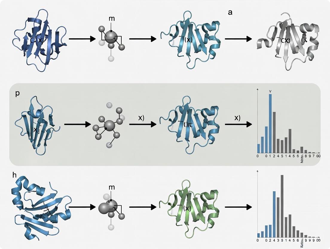
Understanding the FiveFold Ensemble Methodology
What is the FiveFold methodology and how does it address IDP conformational sampling?
The FiveFold methodology is an ensemble-based protein structure prediction framework specifically designed to model conformational diversity, particularly for intrinsically disordered proteins (IDPs). It addresses the critical limitation of single-structure prediction methods by integrating predictions from five complementary algorithms: AlphaFold2, RoseTTAFold, OmegaFold, ESMFold, and EMBER3D [38].
This approach operates on the principle that combining multiple computational strategies creates a more comprehensive predictive framework than any single algorithm can provide. The system strategically pairs multiple sequence alignment (MSA)-dependent methods (AlphaFold2 and RoseTTAFold) with MSA-independent methods (OmegaFold, ESMFold, and EMBER3D) to mitigate individual algorithmic weaknesses while amplifying collective strengths [38]. For IDPs, which comprise approximately 30-40% of the human proteome and often lack sufficient evolutionary information for MSA-based methods, this combination is particularly valuable.
The framework employs two innovative technical components:
- Protein Folding Shape Code (PFSC): A standardized representation system that assigns specific characters to different folding elements (e.g., 'H' for alpha helices, 'E' for extended beta strands), enabling quantitative comparison of conformational differences [38].
- Protein Folding Variation Matrix (PFVM): A systematic framework for capturing and visualizing conformational diversity across the five algorithmic predictions, preserving information about alternative conformational states [38].
Through these components, FiveFold generates multiple plausible conformations rather than attempting to identify a single "correct" structure, making it particularly valuable for drug discovery targeting previously "undruggable" proteins that require strategies accounting for conformational flexibility [38].
How do the component algorithms in FiveFold differ in their approach?
The five algorithms integrated within FiveFold represent complementary methodological approaches to protein structure prediction, each with distinct strengths and limitations for conformational sampling.
Table: Comparison of FiveFold Component Algorithms
| Algorithm | Input Requirements | Key Strengths | IDP Handling Capability |
|---|---|---|---|
| AlphaFold2 | MSA-dependent | Exceptional accuracy for well-folded proteins; captures long-range contacts and complex fold topologies | Limited for highly flexible regions; tends to predict static conformations [38] |
| RoseTTAFold | MSA-dependent | Three-track network analyzing sequence, distance, and 3D structure collectively; good for complex topologies | Similar limitations as AlphaFold2 for disordered regions [38] [39] |
| OmegaFold | MSA-independent | Handles orphan sequences with limited homology; computationally efficient | Improved for proteins lacking evolutionary information [38] |
| ESMFold | MSA-independent | Uses protein language models; fast predictions suitable for high-throughput applications | Effective for sequences with limited homologous information [38] |
| EMBER3D | MSA-independent | Computationally efficient approach; complements other methods | Addresses gaps in conformational sampling [38] |
The consensus-building methodology in FiveFold works by analyzing structural outputs from all five algorithms through several key steps: (1) secondary structure assignment using the PFSC system, (2) alignment and comparison to identify consensus regions and systematic differences, (3) variation quantification through the PFVM, and (4) ensemble generation using probabilistic selection algorithms to sample from consensus and variation data [38].
Troubleshooting Common Experimental Issues
How do I resolve conflicting predictions from different algorithms in the FiveFold ensemble?
Conflicting predictions between algorithms are not necessarily errors but often represent genuine conformational diversity, particularly for IDPs. Follow this systematic troubleshooting approach:
Analyze the Variation Matrix: Examine the Protein Folding Variation Matrix (PFVM) to determine whether conflicts are localized to specific regions or distributed throughout the structure. Regions with high variability may indicate genuine conformational flexibility [38].
Check Input Sequence Quality: Verify that your input protein sequence is complete and correctly formatted. Even small errors in sequence can disproportionately affect predictions, especially for MSA-dependent methods.
Evaluate Evolutionary Coverage: For conflicts between MSA-dependent and MSA-independent methods, check the depth and quality of multiple sequence alignments. Sparse evolutionary information may explain why AlphaFold2 or RoseTTAFold produce low-confidence predictions in certain regions while single-sequence methods perform better [38].
Assess Confidence Metrics: Each algorithm provides confidence estimates (e.g., pLDDT in AlphaFold2). Regions with low confidence across multiple algorithms likely represent genuine disorder rather than algorithmic failure [40].
Prioritize Consensus Regions: Focus initial analyses on regions where multiple algorithms agree, then systematically evaluate areas of disagreement in the context of known biological data or experimental validation.
If conflicts persist, consider the biological context—regions with high conformational diversity may be functionally important for protein-protein interactions or allosteric regulation [38].
What should I do when my ensemble lacks sufficient conformational diversity?
If your generated ensemble appears overly homogeneous and fails to capture expected conformational diversity:
Adjust Sampling Parameters: The FiveFold methodology allows users to define diversity requirements such as minimum RMSD between conformations and ranges of secondary structure content. Increase these thresholds to enforce greater diversity in the output ensemble [38].
Incorporate Experimental Data: Integrate experimental constraints from techniques such NMR chemical shifts or SAXS data to guide the sampling toward biologically relevant states. The maximum entropy reweighting procedure described by Borthakur et al. provides a robust framework for this integration [8].
Supplement with Molecular Dynamics: Use the FiveFold output as starting points for molecular dynamics simulations. All-atom MD simulations with modern force fields can enhance conformational sampling, particularly for disordered regions [8] [41].
Explore Alternative Temperatures in UNRES: If using complementary coarse-grained approaches, try simulations at different temperatures. Research indicates that running UNRES simulations at optimal temperatures (between 270-430 K) can produce comparable results to all-atom force fields for sampling IDP heterogeneity [41].
Verify Input Algorithm Selection: Ensure you're utilizing the full complement of five algorithms, as removing any component reduces the methodological diversity that drives conformational variation in the ensemble.
How can I validate ensemble predictions against experimental data?
Validating conformational ensembles requires different approaches than single-structure validation:
NMR Chemical Shift Comparison: Calculate theoretical chemical shifts from your ensemble and compare with experimental NMR data. The maximum entropy reweighting procedure is particularly effective for this, as it integrates MD simulations with NMR data to determine accurate atomic-resolution ensembles [8].
SAXS Profile Validation: Compute theoretical SAXS profiles from your ensemble and compare with experimental scattering data. Borthakur et al. demonstrate successful integration of SAXS data with MD simulations through their reweighting approach [8].
Radius of Gyration Analysis: Calculate the radius of gyration (Rg) for your ensemble and compare with experimental measurements. UNRES simulations have shown good agreement with experimental Rg values for IDPs when proper temperatures are selected [41].
Paramagnetic Relaxation Enhancement (PRE): If available, PRE data provides distance restraints that are particularly valuable for validating ensemble conformations.
Convergence Assessment: Compare ensembles generated from different initial conditions or force fields. In favorable cases, reweighted ensembles from different MD force fields converge to highly similar conformational distributions after integrating sufficient experimental data [8].
The functional score in FiveFold includes an experimental agreement component (weighted at 40% of the total score) that quantitatively evaluates how well predictions match available experimental structures [38].
Experimental Protocols & Workflows
Protocol: Basic FiveFold Ensemble Generation for IDPs
This protocol outlines the standard workflow for generating conformational ensembles of intrinsically disordered proteins using the FiveFold methodology.
Materials Needed:
- Protein amino acid sequence in FASTA format
- Access to FiveFold implementation (or individual algorithms: AlphaFold2, RoseTTAFold, OmegaFold, ESMFold, EMBER3D)
- Computational resources adequate for running multiple structure predictions
Procedure:
- Input Preparation
- Format your protein sequence as a standard FASTA file
- For MSA-dependent methods (AlphaFold2, RoseTTAFold), prepare multiple sequence alignments using standard databases (UniRef, MGnify)
Parallel Structure Prediction
- Run each of the five algorithms independently using the same input sequence
- Use default parameters initially, as the FiveFold consensus methodology is optimized for standard outputs
PFSC Encoding
- Process each algorithm's output through the Protein Folding Shape Code system
- Assign secondary structure elements to create standardized representations for comparison
Variation Matrix Construction
- Align structural features across all five predictions
- Systematically catalog differences between predictions in the PFVM
- Identify consensus regions and systematic variations
Ensemble Generation
- Set diversity parameters (minimum RMSD: 2-5Å, secondary structure content ranges based on sequence)
- Run probabilistic sampling algorithm to select combinations of secondary structure states from the PFVM
- Apply quality assessment filters to ensure physically reasonable conformations
Validation & Analysis
- Calculate the Functional Score to evaluate conformational utility
- Assess structural diversity, experimental agreement (if data available), and binding site accessibility
Troubleshooting Tips:
- If ensemble lacks diversity, increase the minimum RMSD threshold in step 5
- If specific regions show inconsistent predictions, examine the PFVM for those regions—they may be genuinely disordered
- For proteins with known homologs, compare FiveFold results with AlphaFold Database predictions as a sanity check [42]
Protocol: Integrative Ensemble Refinement with Experimental Data
This protocol describes how to refine conformational ensembles by integrating experimental data using maximum entropy reweighting.
Materials Needed:
- Initial conformational ensemble (from FiveFold or other methods)
- Experimental data: NMR chemical shifts, SAXS profile, and/or Rg measurements
- Software for maximum entropy reweighting (custom implementations based on Borthakur et al.)
- Molecular dynamics simulation capability (optional but recommended)
Procedure:
- Initial Ensemble Preparation
- Generate initial ensemble using FiveFold methodology or molecular dynamics simulations
- For MD, use state-of-the-art force fields (a99SB-disp, Charmm22*, or Charmm36m) with long timescales (≥30μs) [8]
Experimental Data Collection
- Acquire NMR chemical shifts, SAXS data, or other relevant experimental measurements
- Ensure data quality and appropriate error estimation
Forward Model Implementation
- Implement algorithms to predict experimental observables from structural models
- For NMR chemical shifts, use established algorithms like SHIFTX2 or SPARTA+
- For SAXS, use CRYSOL or similar tools to compute theoretical profiles
Maximum Entropy Reweighting
- Apply the maximum entropy principle to introduce minimal perturbation to the computational model while matching experimental data
- Use the Kish ratio (K = 0.10 recommended) to determine the effective ensemble size [8]
- Automatically balance restraint strengths from different experimental datasets based on the desired ensemble size
Convergence Validation
- Compare reweighted ensembles from different initial force fields
- Assess similarity of conformational distributions using appropriate metrics (e.g., Jensen-Shannon divergence)
Final Ensemble Analysis
- Analyze the refined ensemble for biologically relevant features
- Identify key conformational states and their populations
Key Considerations:
- This approach works best when initial ensembles already show reasonable agreement with experimental data
- The method is robust to force field choice when sufficient experimental data is available for reweighting
- For IDPs with little to no residual secondary structure (e.g., Aβ40, α-synuclein), focus validation on global parameters like Rg rather than local structure [8]
FiveFold Ensemble Generation Workflow
Research Reagent Solutions
Table: Essential Computational Tools for Ensemble Prediction of IDPs
| Tool/Resource | Type | Primary Function | Access Information |
|---|---|---|---|
| FiveFold Framework | Ensemble Method | Integrates predictions from 5 algorithms for conformational diversity | Methodology described in Yang et al. [38] |
| AlphaFold2 | Structure Prediction | MSA-based deep learning for accurate single structures | Open source; available via GitHub [39] [40] |
| RoseTTAFold | Structure Prediction | Three-track network for sequence-distance-structure analysis | Open source; available via GitHub [39] |
| OmegaFold | Structure Prediction | MSA-independent method for orphan sequences | Available via GitHub repository |
| ESMFold | Structure Prediction | Protein language model for fast predictions | Available via GitHub repository |
| EMBER3D | Structure Prediction | Computationally efficient complementary method | Research implementation |
| UNRES Web Server | Coarse-Grained MD | Efficient conformational sampling for IDPs | Publicly available web server [41] |
| AlphaFold DB | Structure Database | Over 200 million predicted structures for reference | Publicly accessible at https://alphafold.ebi.ac.uk [42] |
| MaxEnt Reweighting | Validation Method | Integrates MD with experimental data | Implementation described in Borthakur et al. [8] |
Frequently Asked Questions
How does FiveFold compare to molecular dynamics for sampling IDP conformational space?
FiveFold and molecular dynamics (MD) offer complementary approaches for sampling IDP conformational space. FiveFold provides rapid exploration of conformational diversity by leveraging distinct algorithmic biases, making it particularly valuable for initial ensemble generation and when computational resources are limited. MD simulations, particularly with modern force fields like a99SB-disp or Charmm36m, offer physically rigorous sampling of dynamics and thermodynamics but require substantial computational resources for adequate sampling of heterogeneous IDP ensembles [8] [41].
For most applications, an integrative approach is optimal: use FiveFold to generate initial conformational diversity, then refine with MD simulations, and finally validate and reweight using experimental data through maximum entropy methods [8]. The UNRES web server provides a middle ground—a coarse-grained approach that can produce comparable results to all-atom force fields for IDPs with proper temperature selection and requires no investment in computational resources [41].
The computational requirements for FiveFold depend on the implementation strategy:
Minimum Viable Setup:
- Individual algorithm access (AlphaFold2, RoseTTAFold, OmegaFold, ESMFold, EMBER3D)
- Standard workstation with GPU acceleration
- Adequate storage for multiple structure predictions
Optimal Setup:
- Integrated FiveFold implementation with parallel processing capability
- High-performance computing cluster for larger proteins or high-throughput applications
- Storage solution for ensemble data and variation matrices
For reference, running all five algorithms independently for a medium-sized protein (200-300 residues) typically requires 2-8 hours on a well-configured system with GPU support. The PFSC and PFVM steps add minimal additional computational overhead. For researchers without adequate resources, focusing on a subset of algorithms or utilizing coarse-grained alternatives like UNRES may be practical alternatives [41].
Can FiveFold be applied to protein-ligand complexes or multimeric systems?
The standard FiveFold methodology focuses on single-chain protein structure prediction. However, the underlying algorithms have capabilities for complex systems:
- AlphaFold Multimer: Extends AlphaFold2 to protein complexes with multiple chains [39]
- RoseTTAFold All-Atom: Can model assemblies containing proteins, nucleic acids, small molecules, and metals [39]
- AlphaFold3: Supports structural modelling of DNA, RNA, and small molecule ligands [39]
While the full FiveFold ensemble approach hasn't been explicitly validated for complexes, the principles could be extended using these specialized versions of the component algorithms. For protein-ligand systems, ensemble docking approaches that use multiple protein conformations have shown improved performance in drug binding predictions [43].
Decision Workflow for Ensemble Refinement
Specialized Algorithms for Macrocyclic and bRo5 Compound Sampling
Frequently Asked Questions (FAQs)
FAQ 1: Why are specialized algorithms needed for sampling macrocyclic and bRo5 compounds, rather than standard molecular dynamics (MD) tools? Macrocycles and other bRo5 compounds exhibit high conformational flexibility, which is a major determinant of their properties but also makes accurate sampling extremely challenging [44]. Standard MD simulations can be limited by the accuracy of the force fields and the high computational cost required to achieve sufficient sampling, especially for flexible molecules [45]. Specialized algorithms are designed to overcome these hurdles by using enhanced sampling strategies, knowledge-based approaches, or integrating experimental data to efficiently explore the vast conformational space these molecules occupy [46] [47].
FAQ 2: What is "chameleonicity" and why is it important for oral bioavailability in bRo5 compounds? Chameleonicity refers to the capacity of a molecule to alter its conformation and molecular properties based on its environment [48]. A chameleonic compound can adopt open and polar conformations in aqueous environments (favoring solubility) while assuming folded and less polar conformations in nonpolar environments like cell membranes (favoring permeability) [48]. This behavior is crucial for oral bRo5 drugs, as it helps balance the otherwise conflicting requirements of aqueous solubility and membrane permeability, as exemplified by cyclosporin A [48].
FAQ 3: My generated macrocycles are chemically valid but have poor novelty. How can I improve this? Improving the novelty of generated macrocycles involves adjusting the sampling strategy of your generative model. The HyperTemp probabilistic sampling algorithm is designed specifically for this purpose [46]. It works by making fine-grained adjustments to the token probabilities during sequence generation (e.g., of SMILES strings), appropriately reducing the probability of the most optimal tokens while increasing the probability of suboptimal ones [46]. This encourages the exploration of alternative structural pathways, thereby improving novelty while maintaining the validity of the generated macrocycles [46].
FAQ 4: How can I determine if my computational conformational ensemble for a macrocycle is accurate? The most robust approach is to use integrative validation, comparing your computational ensemble against experimental data [8]. Key experimental techniques include:
- Nuclear Magnetic Resonance (NMR) spectroscopy: Provides data on chemical shifts, scalar couplings, and NOEs (nuclear Overhauser effects) that report on local structure and distances [8].
- Small-Angle X-Ray Scattering (SAXS): Provides information on the global shape and size (e.g., radius of gyration) of the molecule in solution [8]. A maximum entropy reweighting procedure can then be used to refine the computational ensemble to achieve exceptional agreement with the experimental data, ensuring its accuracy [8].
FAQ 5: What are the key molecular descriptors to monitor when designing permeable macrocyclic drugs? For bRo5 compounds, a set of simple descriptors can serve as effective guidelines. The following bi-descriptor model can help distinguish oral from parenteral macrocycles [49]:
| Descriptor Combination | Suggests Oral Potential If... |
|---|---|
| HBD (Hydrogen Bond Donors) & MW (Molecular Weight) | HBD ≤ 7 and MW < 1000 Da [49] |
| HBD & cLogP (Calculated Log P) | HBD ≤ 7 and cLogP > 2.5 [49] |
Additionally, the Kier flexibility index (PHI) is a more relevant descriptor for flexibility than the number of rotatable bonds when macrocyclic substructures are present. A value of ≤10 may represent a current upper limit for reasonably accurate 3D prediction of macrocycle cell permeability [45].
Troubleshooting Guides
Issue 1: Poor Coverage of Conformational Space
Problem: Your conformational sampling algorithm produces an ensemble that is too narrow and fails to capture the full range of biologically relevant conformations.
Solutions:
- Combine Multiple Sampling Tools: Use a hybrid approach. Distance-geometry based methods like OMEGA have been shown to yield ensembles spanning larger structure and property spaces than methods like MOE-LowModeMD (MOE) or MacroModel (MC), especially for different environments (polar vs. apolar) [44]. Consider using OMEGA to generate a diverse initial set of conformers.
- Apply Enhanced Sampling with VAEs: For highly flexible systems like Intrinsically Disordered Proteins (IDPs) or complex macrocycles, use deep learning models for enhanced sampling. A Variational Autoencoder (VAE) can be trained on a limited set of conformations from a short MD simulation and can then generate a wider, more diverse conformational landscape that covers the space sampled by much longer simulations [47].
- Monitor Diversity with Polymer Descriptors: Quantify the diversity of your ensemble using polymer physics descriptors. Calculate the radius of gyration (Rg) for size and the instantaneous shape ratio (Rs = Ree²/Rg², where Ree is the end-to-end distance) for shape. Plotting these against each other creates a conformational landscape map that allows you to visually assess the coverage and compare it to reference models [50].
Issue 2: Inaccurate Force Fields Leading to Non-Physiological Ensembles
Problem: The conformational ensemble generated by MD simulation deviates significantly from available experimental data, indicating inaccuracies in the molecular mechanics force field.
Solutions:
- Implement Maximum Entropy Reweighting: Integrate your simulation data with experimental data using a maximum entropy reweighting procedure. This automated method introduces the minimal perturbation to your computational ensemble required to match a set of experimental restraints (e.g., from NMR and SAXS). This corrects for force field bias and yields a force-field independent, accurate conformational ensemble [8].
- Cross-Validate with Multiple Force Fields: Run simulations with different, state-of-the-art force fields (e.g., a99SB-disp, CHARMM36m, CHARMM22*). If reweighting these different initial ensembles with the same experimental data causes them to converge to a highly similar final ensemble, you can have high confidence in the result [8].
Issue 3: Failure to Generate Valid or Novel Macrocyclic Structures
Problem: A generative model produces a high rate of invalid chemical structures or generates compounds that are not novel (i.e., are too similar to the training data).
Solutions:
- Adopt a Progressive Transfer Learning Strategy: To overcome data scarcity for macrocycles, pre-train your model on a large dataset of bioactive linear molecules (e.g., from ChEMBL) to learn general chemical language rules. Then, incrementally transfer this knowledge by fine-tuning on a smaller, specialized dataset of macrocyclic compounds. This approach, as used in the CycleGPT model, effectively adapts the model's knowledge from general chemical space to the macrocyclic domain [46].
- Optimize the Sampling Algorithm: Move beyond standard sampling methods. Implement the HyperTemp algorithm, which has been shown to significantly outperform other models like Char-RNN, MolGPT, and Llamol on the comprehensive metric of "noveluniquemacrocycles," generating more valid and novel macrocycles [46].
Experimental Protocols
Protocol 1: Determining an Accurate Conformational Ensemble via Maximum Entropy Reweighting
Purpose: To determine a physically realistic, atomic-resolution conformational ensemble of a macrocycle or IDP by integrating MD simulations with experimental data.
Materials:
- Software: All-atom MD simulation software (e.g., GROMACS, AMBER), OMEGA conformer generator [44], reweighting code (https://github.com/paulrobustelli/BorthakurMaxEntIDPs_2024/) [8].
- Experimental Data: NMR chemical shifts, J-couplings, and/or SAXS data.
Procedure:
- Generate Initial Ensemble: Perform long-timescale all-atom MD simulations of the molecule using one or more state-of-the-art force fields (e.g., a99SB-disp, CHARMM36m) [8].
- Predict Observables: Use forward models to predict the experimental observables (NMR, SAXS) from every frame of the MD ensemble [8].
- Apply Reweighting: Use the maximum entropy reweighting procedure to calculate new statistical weights for each conformation in the MD ensemble. The goal is to minimize the discrepancy between the predicted and experimental ensemble-averaged observables while maximizing the entropy of the new weights [8].
- Set Ensemble Size: Define a Kish ratio threshold (e.g., K=0.10) to determine the effective number of conformations in the final ensemble. This automatically balances the strengths of restraints from different experimental datasets [8].
- Validate the Ensemble: Analyze the reweighted ensemble to ensure it agrees with the experimental data and possesses expected physical characteristics.
The workflow for this integrative approach is as follows:
Protocol 2: Enhanced Conformational Sampling using Variational Autoencoders (VAEs)
Purpose: To efficiently sample the conformational landscape of a flexible molecule (IDP or macrocycle) using deep learning, reducing reliance on extremely long MD simulations.
Materials:
- Software: MD simulation software, Python with deep learning libraries (e.g., PyTorch, TensorFlow).
- Hardware: GPU-accelerated computing resource.
Procedure:
- Generate Training Data: Run a relatively short MD simulation (e.g., microseconds) of the target molecule. Extract a set of conformations (e.g., 50,000 frames) as training data [47].
- Build and Train the VAE:
- Encoder: Design a 4-layer encoder network (e.g., with 1024, 256, 64, and 16 neurons per layer) to compress the 3D coordinates of a conformation into a low-dimensional latent vector (e.g., 2 dimensions) [47].
- Latent Space: The VAE learns to map inputs to a probability distribution in the latent space, enabling continuous sampling [47].
- Decoder: Design a symmetric 4-layer decoder network to reconstruct the 3D coordinates from the latent vector [47].
- Generate New Conformations: After training, sample random vectors from the latent space and use the decoder to generate new, plausible molecular conformations [47].
- Validate Output: Compare the VAE-generated conformations against a longer, reference MD simulation using metrics like Cα RMSD and Spearman correlation coefficient to ensure they accurately reflect the true conformational diversity [47].
The Scientist's Toolkit: Research Reagent Solutions
Table 1: Key Computational Tools for Conformational Sampling.
| Tool Name | Primary Function | Key Application / Advantage |
|---|---|---|
| OMEGA (OpenEye) | Conformational ensemble generation using distance geometry [44]. | Samples larger structure/property spaces; performance in different dielectric environments [44]. |
| MacroModel (Schrödinger) | Conformational search using low-mode sampling and torsion-based methods [44]. | A standard tool for macrocycle sampling; can generate different ensembles for different environments [44]. |
| MOE-LowModeMD | Conformational search using low-mode molecular dynamics [44]. | A frequently used standard method for macrocycle sampling [44]. |
| CycleGPT | Generative chemical language model for macrocycle design [46]. | Overcomes data scarcity via transfer learning; uses HyperTemp for novel/valid macrocycles [46]. |
| Variational Autoencoder (VAE) | Deep learning model for enhanced conformational sampling [47]. | Generates diverse conformational landscapes from short MD trajectories; reduces computational cost [47]. |
| Maximum Entropy Reweighting | Integrates MD ensembles with experimental data [8]. | Corrects force field inaccuracies; produces accurate, force-field independent ensembles [8]. |
Table 2: Essential Molecular Descriptors for bRo5 Compound Design.
| Descriptor Category | Specific Descriptors | Role in Design and Troubleshooting |
|---|---|---|
| Size & Shape | Molecular Weight (MW), Radius of Gyration (Rgyr), Instantaneous Shape Ratio (Rs) [50] [49]. | Rgyr informs on compactness; Rs (Ree²/Rg²) distinguishes extended (high Rs) from compact (low Rs) shapes [50]. |
| Polarity | Hydrogen Bond Donors (HBD), Topological Polar Surface Area (TPSA) [49] [48]. | Critical for estimating solubility and permeability. HBD ≤7 is a key filter for oral macrocycles [49]. |
| Lipophilicity | Calculated LogP (cLogP) [49] [48]. | Impacts permeability and solubility. cLogP > 2.5, combined with HBD ≤7, suggests oral potential [49]. |
| Flexibility | Number of Rotatable Bonds (NRot), Kier Flexibility Index (PHI) [45] [48]. | PHI is superior for macrocycles. A Kier index ≤10 may be a limit for accurate permeability prediction [45]. |
Algorithm Selection and Application Workflow
The following diagram outlines a logical workflow for selecting and applying specialized sampling algorithms based on your research goal.
Overcoming Sampling Challenges: Force Fields, Hidden Barriers, and Efficiency
Navigating Force Field Limitations for Accurate IDP Representation
Frequently Asked Questions
Why do traditional molecular dynamics force fields often fail to accurately represent Intrinsically Disordered Proteins (IDPs)? Traditional force fields, parameterized for folded proteins with stable tertiary structures, often over-stabilize protein-protein interactions. This leads to an over-population of secondary structures (α-helix and β-sheet) and unnaturally compact conformations in IDPs, which have flatter energy landscapes and fewer hydrophobic residues. The core issue is an imbalance in protein–protein, protein–water, and water–water interactions [51] [52] [53].
What are the primary strategies for improving force fields for IDP simulations? The main strategies involve reparameterizing the force field to better capture the conformational ensemble of disordered states. Key approaches include:
- Adjusting Dihedral Parameters: Refitting backbone dihedral (φ and ψ) parameters to reduce the bias toward folded secondary structures [52].
- Adding CMAP Corrections: Using a grid-based energy correction map (CMAP) to adjust the potential energy surface based on deviations from reference data, such as coil library distributions [51] [52].
- Refining Solvent Models: Combining improved protein force fields with modified water models (e.g., TIP4P-D) to correct for artificial structural collapse caused by oversolvation or undersolvation [53].
My IDP simulations show an unnatural collapse. Is this a sampling or a force field problem? While inadequate sampling can be a factor, an unnatural collapse is frequently a signature of an imperfect force field and water model. Benchmarking studies have shown that some force field/water model combinations (e.g., using TIP3P) lead to artificially compact conformations, whereas others (e.g., with TIP4P-D) produce ensemble properties that align better with experimental data like SAXS and NMR [53].
How can I validate the conformational ensemble generated for an IDP? A robust validation protocol involves comparing multiple predicted observables from your simulation against experimental data. Key metrics include:
- Structural Properties: Radius of gyration (from SAXS) [53] [54].
- Local Structure: Chemical shifts (from NMR) [53] [54].
- Long-Range Contacts: Paramagnetic relaxation enhancement (PRE) from NMR [53].
- Backbone Dynamics: NMR relaxation parameters (R1, R2, heteronuclear NOE), which are highly sensitive to force field inaccuracies [53].
Troubleshooting Guides
Issue: Overly Compact IDP Conformations or Excessive Secondary Structure
Problem: Your simulated IDP ensemble is more compact than experimental data (e.g., from SAXS) suggests, or it shows persistent α-helical or β-sheet content where none is expected.
Solutions:
- Re-evaluate Your Force Field and Water Model Combination Switch to a force field and water model specifically tuned for IDPs. The table below summarizes some recommended options.
Implement an Advanced Sampling Protocol If switching force fields is insufficient, use enhanced sampling techniques to improve conformational sampling and cross energy barriers more efficiently [51].
- Replica Exchange MD (REMD): Run parallel simulations at different temperatures, allowing periodic exchanges to escape local energy minima [51] [52].
- Metadynamics or Bias-Exchange MD: Apply a history-dependent bias potential to encourage exploration of under-sampled regions of conformational space [51].
Validate with a Multi-Observable Approach Do not rely on a single metric. Compare your simulation's predictions for radius of gyration, chemical shifts, and PRE data against experimental results to ensure the ensemble is accurate across multiple dimensions [53] [54].
The following workflow outlines a systematic approach for selecting and validating a force field for IDP simulation:
Issue: Inaccurate Representation of Coupled Folding and Binding
Problem: Simulating the interaction between an IDP and its binding partner fails to reproduce the experimentally observed binding-induced folding or dynamic complex formation.
Solutions:
- Utilize a Multi-Scale Approach For larger systems, consider coarse-grained (CG) models. These models reduce the number of degrees of freedom, allowing you to simulate longer timescales and larger assemblies, such as those involved in liquid-liquid phase separation [51].
- Leverage Experimental Restraints Use experimental data (e.g., PRE-derived distances, chemical shift perturbations, J-couplings) as soft restraints in your simulation to guide the system toward the correct conformational ensemble [54].
- Ensure Adequate Sampling of the Bound and Unbound States The process of binding and folding is complex. Use advanced sampling methods like Hamiltonian replica exchange or bias-exchange metadynamics to ensure all relevant states are sampled adequately [51].
The relationship between key force field parameters and the resulting physical properties of an IDP ensemble is crucial for troubleshooting:
The Scientist's Toolkit: Essential Research Reagents & Materials
The following table details key resources for conducting and validating IDP simulations.
| Item Name | Function / Role | Key Considerations |
|---|---|---|
| IDP-Optimized Force Fields (e.g., CHARMM36m, ff99IDPs, CHARMM22*) | Provides the potential energy function for MD simulations; determines the balance between folded and disordered states. | Must be paired with a compatible water model. Performance can be system-dependent; benchmarking is required [51] [52] [53]. |
| Refined Water Models (e.g., TIP4P-D, TIP3P*) | Defines solvent-solute interactions; critical for preventing artificial chain collapse and achieving accurate solvation of charged/polar residues. | TIP4P-D is specifically designed to work with various force fields to improve IDP dimensions [53]. |
| Advanced Sampling Software (e.g., GROMACS, AMBER, NAMD, OPENMM) | Enables enhanced sampling methods like Replica Exchange MD (REMD) to overcome energy barriers and achieve better convergence. | Necessary for adequate sampling of the heterogeneous IDP conformational landscape within feasible simulation time [51]. |
| Validation Data Suite (NMR Chemical Shifts, PREs, SAXS, RDCs) | Provides experimental benchmarks for validating the simulated conformational ensemble against real-world data. | A multi-pronged validation approach using different data types is crucial for a trustworthy ensemble [53] [54]. |
| Coarse-Grained (CG) Models (e.g., Martini, Gō-models) | Reduces computational cost by grouping atoms into beads; allows simulation of larger systems and longer timescales, such as IDP liquid-liquid phase separation. | Sacrifices atomic detail for scale; often used for initial screening or studying large assemblies [51]. |
Identifying True Reaction Coordinates to Overcome Hidden Energy Barriers
FAQs: Core Concepts and Problem Identification
Q1: What are "true" reaction coordinates (RCs) and why are they critical in disordered protein studies? A1: True reaction coordinates (RCs) are the few essential degrees of freedom in a protein that fully control its functional processes, such as conformational changes or allostery. They are rigorously defined by their ability to predict the committor probability for any given system conformation [55]. In the context of disordered proteins, which sample a vast conformational landscape, identifying these true RCs is crucial because they provide the optimal reduced description of the system's complex dynamics, enabling researchers to understand transition mechanisms and efficiently sample functionally relevant states [55] [51].
Q2: What is the "hidden barrier" problem and how is it diagnosed? A2: The "hidden barrier" problem occurs when the collective variables (CVs) selected for enhanced sampling simulations do not align with the true RCs. This results in an un-accelerated activation barrier remaining in the space orthogonal to the chosen CVs, which prevents efficient sampling of conformational changes [55] [18]. Diagnosis involves:
- Committor Analysis: Testing whether configurations along the putative RC have a committor probability,
pB, of approximately 0.5. A distribution peaked at 0.5 indicates a good RC, whereas distributions skewed towards 0 or 1 suggest a hidden barrier [55]. - Non-Physical Trajectories: Observing if biased simulations produce trajectories that visit high-energy, non-physical regions of the conformational space, indicating the CVs are misdirecting the sampling [18].
Q3: Our enhanced sampling of an IDP fails to converge. Could poor RC choice be the cause? A3: Yes, this is a leading cause. Traditional intuition-based CVs—such as root-mean-square deviation (RMSD), radius of gyration, or principal components—are often insufficient for disordered proteins because their flatter energy landscapes are defined by a complex interplay of many coordinates [55] [51]. Using a CV that misses the true RC will result in the hidden barrier problem, where computational resources are wasted on sampling that does not cross the actual transition state [55] [18].
Q4: How can we validate a proposed reaction coordinate? A4: The gold standard for validation is committor analysis [55] [18].
- From your enhanced sampling, select a set of configurations hypothesized to be near the transition state (e.g., where your proposed RC has an intermediate value).
- For each configuration, launch multiple, short, unbiased MD simulations with random initial velocities.
- Calculate the committor,
pB, as the fraction of trajectories that reach the product state before the reactant state. - A valid true RC will have a
pBdistribution sharply peaked at 0.5 for these configurations. A broad or U-shaped distribution indicates an incorrect RC [55].
Troubleshooting Guides
Table 1: Common Problems and Solutions in RC Identification
| Problem Symptom | Potential Cause | Diagnostic Steps | Recommended Solution |
|---|---|---|---|
| Ineffective Enhanced Sampling | Hidden barriers in orthogonal space due to poor CV choice [55] [18]. | Perform committor analysis on configurations from biased runs. | Shift from geometric CVs (e.g., RMSD) to physics-based methods like Energy Flow Theory or the Generalized Work Functional (GWF) method to identify true RCs [18]. |
Low Committor Probability (pB << 0.5) at Putative Transition State |
The chosen CV does not capture the energy activation process [18]. | Verify the initial state definitions; check if the CV correlates with energy flow. | Use the GWF method to find singular coordinates (SCs) that maximize potential energy flow, as these are candidates for true RCs [18]. |
| Non-Physical Trajectories | Bias potential acts on non-essential coordinates, driving the system into unrealistic conformations [18]. | Visually inspect trajectories for unnatural steric clashes or unrealistic geometries. | Employ methods that compute RCs from a single structure based on energy relaxation, such as the GWF method, to enable predictive sampling [18]. |
| Poor Convergence of IDP Ensembles | Inadequate sampling of the vast conformational space and force field inaccuracies [8] [51]. | Compare ensemble averages (e.g., from SAXS/NMR) with simulations from different force fields. | Integrate simulations with experimental data using maximum entropy reweighting to derive force-field independent, accurate ensembles [8]. |
Table 2: Key Research Reagent Solutions
| Reagent / Method | Function in RC Identification & Sampling | Key Consideration |
|---|---|---|
| Generalized Work Functional (GWF) [18] | Identifies true RCs by generating an orthonormal coordinate system that disentangles the essential degrees of freedom based on energy flow. | Can compute RCs from energy relaxation simulations, requiring only a single protein structure as a starting point. |
| Committor Analysis [55] [18] | The definitive test for validating a proposed reaction coordinate. | Computationally expensive, as it requires many short trajectories for each test configuration. |
| Maximum Entropy Reweighting [8] | Integrates MD simulations with experimental data (NMR, SAXS) to produce accurate, force-field independent conformational ensembles of IDPs. | A fully automated procedure that uses the Kish ratio to balance restraints from multiple experimental datasets. |
| State-of-the-Art Force Fields (e.g., CHARMM36m, a99SB-disp) [8] [51] | Provide a physically accurate baseline for MD simulations, which is essential for any subsequent RC analysis or ensemble generation. | Accuracy for IDPs depends on a balanced treatment of protein-protein, protein-water, and water-water interactions. |
Experimental Protocols
Protocol 1: Identifying True RCs using the Generalized Work Functional (GWF) Method
Principle: This method identifies true RCs as the singular coordinates (SCs) that carry the highest potential energy flow (PEF), which is the energy cost for the motion of a coordinate. These coordinates control both conformational changes and energy relaxation [18].
Workflow:
- System Setup: Start with a single protein structure (e.g., from AlphaFold) solvated in an explicit solvent box.
- Energy Relaxation Simulation: Run a short, standard MD simulation initiated from a non-equilibrium state. This can be a local energy-minimized structure or a thermally perturbed configuration.
- Compute Potential Energy Flows: For each frame of the trajectory, calculate the PEF for all degrees of freedom. The PEF through a coordinate
qiover a time period is defined as:ΔW_i(t1, t2) = - ∫_{qi(t1)}^{qi(t2)} [∂U(q)/∂qi] dqi[18] whereU(q)is the potential energy of the system. - Apply Generalized Work Functional: The GWF algorithm processes the PEF data to generate an orthonormal set of SCs. The SCs are ranked by their associated PEF.
- Identify True RCs: The top-ranked SCs, which carry the largest energy flows, are identified as the candidate true RCs.
- Validation: Perform committor analysis on configurations selected along the candidate RC to confirm its validity (
pB ≈ 0.5).
The following diagram illustrates the logical workflow of this protocol:
Protocol 2: Determining Accurate Conformational Ensembles for IDPs via Maximum Entropy Reweighting
Principle: This integrative approach refines a preliminary conformational ensemble from MD simulations by imposing agreement with experimental data while minimizing the deviation from the simulation's original distribution (maximum entropy principle) [8].
Workflow:
- Generate Initial Ensemble: Perform long-timescale all-atom MD simulations of the IDP using state-of-the-art force fields (e.g., a99SB-disp, CHARMM36m).
- Collect Experimental Data: Acquire extensive ensemble-averaged experimental data, such as NMR chemical shifts, J-couplings, and SAXS profiles.
- Calculate Observables: Use forward models to predict the experimental observables from each conformation in the MD ensemble.
- Set Target Ensemble Size: Choose a target for the Kish effective sample size (e.g., K=0.10), which determines how many conformations from the original simulation will have significant weight in the final ensemble.
- Run Reweighting Algorithm: Apply the maximum entropy reweighting procedure to compute new statistical weights for each conformation in the initial ensemble. The algorithm automatically balances the restraints from all experimental datasets to achieve the best agreement while maintaining the target effective sample size.
- Validate and Analyze: The output is a refined conformational ensemble. Check its agreement with experimental data and analyze its structural properties (e.g., radius of gyration, secondary structure content).
The following diagram illustrates the workflow for this integrative protocol:
Balancing Computational Cost with Sampling Comprehensiveness
FAQs: Navigating Computational Sampling for Disordered Proteins
What is the core challenge when sampling conformational space in Intrinsically Disordered Proteins (IDPs)? The core challenge is that IDPs exist as a dynamic ensemble of rapidly interconverting structures rather than a single, stable fold. Experimentally determining an atomic-resolution ensemble is extremely challenging because techniques like NMR and SAXS provide data that is averaged over the entire ensemble and time. Computationally, Molecular Dynamics (MD) simulations can model these ensembles but achieving sufficient sampling to accurately represent the full breadth of conformations is immensely computationally expensive [8].
Why is balancing computational cost with sampling comprehensiveness so critical in IDP research? Accurate conformational ensembles are vital for understanding IDP function and for rational drug design, as these proteins are implicated in many diseases. However, the computational cost of running a simulation long enough to observe all relevant conformational states is often prohibitive. Without comprehensive sampling, the resulting ensemble may be biased and not reflect the true biological reality, leading to incorrect functional insights or ineffective drug candidates [8].
What are the main computational strategies to improve sampling efficiency? Researchers generally employ two complementary strategies. The first is enhanced sampling methods, which use bias potentials on collective variables (CVs) to accelerate the exploration of conformational space. The second is integrative modeling, which combines shorter, more affordable MD simulations with experimental data to refine and correct the ensemble, ensuring it matches real-world observations [8] [18].
What are "true reaction coordinates" and why are they important for cost-effective sampling? True reaction coordinates (tRCs) are the few essential protein coordinates that fully determine the progression of a conformational change. Using intuition or standard geometric parameters as CVs often leads to inefficient sampling because of "hidden barriers." Biasing simulations along tRCs provides highly efficient acceleration (by factors of 10⁵ to 10¹⁵ have been demonstrated) and ensures the simulated pathways are physically realistic, providing the most cost-effective route to comprehensive sampling [18].
Troubleshooting Guides
Problem: My MD simulation is trapped in a local energy state and won't explore the full conformational landscape.
| Potential Cause | Solution | Key Considerations |
|---|---|---|
| Inadequate simulation time | Extend simulation time if computationally feasible. | Often not practical for complex biomolecules; consider enhanced sampling. |
| Poorly chosen Collective Variables (CVs) | Identify and bias True Reaction Coordinates (tRCs). | tRCs provide optimal acceleration and generate natural transition pathways [18]. |
| High energy barriers | Use advanced sampling methods like metadynamics or umbrella sampling. | Efficacy is entirely dependent on the quality of the selected CVs [18]. |
Recommended Protocol: Identifying True Reaction Coordinates
- Input Structure: Start with a single protein structure.
- Energy Relaxation Simulation: Perform a short simulation to allow the system to relax.
- Calculate Potential Energy Flows (PEFs): Analyze the energy flow through individual coordinates during relaxation. Coordinates with the highest PEFs are critical for driving conformational changes.
- Apply Generalized Work Functional (GWF) Method: This generates an orthonormal coordinate system (Singular Coordinates) that disentangles tRCs from non-essential coordinates.
- Identify tRCs: The Singular Coordinates with the highest PEFs are your tRCs. These can be used as bias coordinates in enhanced sampling simulations to achieve efficient and physically accurate sampling [18].
Problem: My computationally generated ensemble does not match experimental data.
| Symptom | Possible Cause | Corrective Action |
|---|---|---|
| Discrepancies in NMR chemical shifts | Inaccurate force field or insufficient sampling. | Use a maximum entropy reweighting procedure to integrate the simulation with experimental data [8]. |
| Mismatch with SAXS data | Incorrect ensemble compactness or shape. | Apply the same reweighting procedure; this corrects the populations of conformations in the ensemble to match experiment [8]. |
| General poor agreement | The initial simulation model is of low quality. | Ensure the initial unbiased simulation is in "reasonable agreement" with data before reweighting for best results [8]. |
Recommended Protocol: Maximum Entropy Reweighting
- Run Unbiased MD Simulation: Generate an initial conformational ensemble using an all-atom MD simulation.
- Predict Experimental Observables: Use forward models to calculate the expected NMR and SAXS data from every frame of your MD simulation.
- Calculate Optimal Weights: Use a maximum entropy algorithm to determine new statistical weights for each conformation in your ensemble. The goal is to find the smallest perturbation to the original simulation that results in a perfect match with the experimental data.
- Analyze Reweighted Ensemble: The output is a refined ensemble that retains atomic detail from the simulation but now agrees with experimental evidence. The effectiveness can be measured by the Kish ratio, which indicates the fraction of conformations with significant weight in the final ensemble [8].
Problem: The computational cost of generating a comprehensive ensemble is too high for my system.
| Strategy | Implementation | Benefit |
|---|---|---|
| Conformational Sampling (CS) [56] | Use tools like the pucke.rs toolkit to generate a landscape of constraint axes (e.g., torsion angles) for efficient sampling of ring puckering or peptide backbone angles. |
Systematically covers conformational space with fewer optimization steps, reducing resource consumption. |
| Hybrid QM/MM Methods | Employ cheaper semi-empirical quantum mechanical (QM) methods (e.g., HF-3c) for geometry optimizations during initial sampling, reserving higher-level methods (e.g., MP2) for final energy calculations [56]. | Dramatically reduces computation time while maintaining reasonable accuracy for generating potential energy surfaces. |
| Integrative Modeling | Combine shorter MD simulations with experimental data via reweighting, rather than relying solely on ultra-long simulations to achieve convergence [8]. | Leverages experimental data to guide and correct limited simulations, providing an accurate ensemble at a lower computational cost. |
Workflow Visualization
The following diagram illustrates a decision workflow for selecting a sampling strategy based on computational cost and comprehensiveness.
Research Reagent Solutions
The following table details key computational tools and methods used in advanced conformational sampling.
| Tool/Method | Function in Research | Key Application in IDPs |
|---|---|---|
| Maximum Entropy Reweighting [8] | Integrates MD simulations with experimental data to produce accurate conformational ensembles. | Determines force-field independent atomic-resolution ensembles of IDPs by combining NMR/SAXS data with MD. |
| True Reaction Coordinate (tRC) Identification [18] | Identifies the essential coordinates that drive conformational changes for targeted enhanced sampling. | Accelerates sampling of functional processes (e.g., flap opening in HIV-1 protease) by factors up to 10¹⁵. |
pucke.rs Toolkit [56] |
A command-line tool and Python module for conformational sampling of peptides and sugar rings. | Generates constraint axes to map the energy landscape of modified nucleotides (XNA) and amino acids. |
| Semi-empirical Methods (e.g., HF-3c) [56] | Cost-effective quantum mechanical methods for geometry optimization and energy calculations. | Used for rapid generation of potential energy surfaces in conformational sampling benchmarks. |
| Molecular Dynamics Force Fields (e.g., a99SB-disp, C36m) [8] | Physical models defining atom-atom interactions in MD simulations. | Critical for initial ensemble generation; accuracy varies, making integrative validation important. |
Optimizing Parameters for Enhanced Sampling and Meta-Dynamics
Frequently Asked Questions (FAQs) and Troubleshooting
FAQ 1: What are the primary enhanced sampling methods suitable for studying intrinsically disordered proteins (IDPs)?
IDPs require methods that can efficiently sample their vast conformational landscapes. The table below summarizes the most suitable techniques.
Table 1: Enhanced Sampling Methods for IDP Conformational Sampling
| Method | Key Principle | Advantages for IDPs | Key Considerations |
|---|---|---|---|
| Replica-Exchange MD (REMD) [57] | Parallel simulations at different temperatures swap configurations, preventing trapping in local minima. | Efficiently samples different conformational states; good for global folding/unfolding. | High computational cost; performance sensitive to maximum temperature choice [57]. |
| Metadynamics [57] [58] | Adds a history-dependent bias potential to "fill" free energy wells, encouraging exploration. | Excellent for mapping Free Energy Surfaces (FES) along specific Collective Variables (CVs). | Accuracy depends on a low-dimensional set of well-chosen CVs [57]. |
| Parallel Tempering Metadynamics [58] | Combines Metadynamics with replica-exchange across temperatures. | Enhances sampling of both CV space and overall protein conformation. | Even higher computational cost than standard REMD or Metadynamics. |
| Variational Autoencoders (VAEs) [59] | Machine learning model that learns a low-dimensional latent space to generate new conformations. | Can reconstruct diverse conformational ensembles from short MD simulations at low cost. | A "black box" approach; requires initial simulation data for training [59]. |
Troubleshooting Guide: If your simulation is trapped in a limited set of conformations:
- Problem: Inefficient crossing of energy barriers.
- Solution: Consider switching from standard MD to REMD or Parallel Tempering Metadynamics [57] [58]. These methods use elevated temperatures to help the system overcome high-energy barriers.
- Problem: Need to focus sampling on a specific conformational change.
- Solution: Use Metadynamics with a carefully selected CV that describes the process of interest, such as a distance or radius of gyration [57].
FAQ 2: How do I select and optimize Collective Variables (CVs) for metadynamics of disordered proteins?
Choosing the right CVs is critical for successful metadynamics. Poor CVs lead to inaccurate free energy estimates and inefficient sampling.
Table 2: Collective Variables for Disordered Protein Simulations
| CV Category | Example CVs | Best Use Cases | References |
|---|---|---|---|
| Geometric & Physical | Radius of Gyration (Rg), End-to-End Distance (Ree), Solvent Accessible Surface Area (SASA) | Characterizing global compactness and shape; a scatter plot of the instantaneous shape ratio (Rs = Ree²/Rg²) against Rg effectively maps the conformational landscape [50]. | [50] |
| Machine Learning (ML)-Derived | Latent space dimensions from a Variational Autoencoder (VAE) | Generating a broad and diverse set of conformations when the relevant physical CVs are not known a priori [59]. | [59] |
| External Knowledge-Based | AlphaFold-based CV (measures conformity of a structure to AlphaFold's predicted distance map) | Guiding folding simulations or structure refinement; useful when a predicted structure is available [58]. | [58] |
Troubleshooting Guide:
- Problem: My CVs are not capturing the relevant conformational transitions.
- Solution: Employ machine learning to discover relevant CVs directly from simulation data. Methods like VAEs can create a low-dimensional latent space that encapsulates the essential dynamics of the protein [60] [59].
- Problem: I have a predicted structure from AlphaFold, but want to study dynamics.
- Solution: Use an AlphaFold-based CV. This CV scores any conformation based on its agreement with the residue-residue distance probabilities from AlphaFold, allowing metadynamics to explore around the predicted structure [58].
The following workflow diagram illustrates the process of selecting and applying CVs for enhanced sampling of IDPs.
Workflow for CV Selection in IDP Studies
FAQ 3: How can I integrate experimental data to validate and refine my conformational ensembles?
For IDPs, it is crucial to ensure that computational models produce physically realistic and accurate conformational ensembles. Integration with experimental data is the gold standard.
Experimental Protocol: Maximum Entropy Reweighting of MD Ensembles [8]
This protocol is used to refine MD-generated ensembles of IDPs by integrating data from Nuclear Magnetic Resonance (NMR) spectroscopy and Small-Angle X-Ray Scattering (SAXS).
- Perform Long-Timescale MD Simulations: Run multiple, long (e.g., 30 µs) all-atom MD simulations of the IDP using state-of-the-art force fields (e.g., a99SB-disp, Charmm36m).
- Collect Experimental Data: Obtain extensive experimental data for the IDP, such as NMR chemical shifts, J-couplings, and SAXS scattering profiles.
- Predict Observables from Simulation: Use "forward models" (software that calculates experimental observables from atomic coordinates) to predict the experimental data for every snapshot in your MD ensemble.
- Apply Maximum Entropy Reweighting: Use a reweighting algorithm that applies the principle of maximum entropy. This method finds a new set of statistical weights for each MD snapshot such that:
- The reweighted ensemble's averaged observables match the experimental data.
- The perturbation from the original MD ensemble is minimized.
- Validate the Ensemble: The output is a refined conformational ensemble that agrees with experimental data. Convergence can be assessed by checking if ensembles from different starting force fields converge to similar distributions after reweighting [8].
Troubleshooting Guide:
- Problem: My simulated ensemble does not match experimental data.
- Solution: Do not discard the simulation. Apply the maximum entropy reweighting protocol. This corrects for small inaccuracies in the force field by combining the atomic detail of MD with the rigor of experimental data [8].
- Problem: The reweighting process results in overfitting.
- Solution: Use a robust protocol that automatically balances restraints from different experimental datasets and monitors the effective ensemble size (e.g., Kish ratio) to prevent overfitting and ensure statistical robustness [8].
FAQ 4: What are the best practices for running MM/PBSA calculations on dynamic protein systems?
MM/PBSA (Molecular Mechanics/Poisson-Boltzmann Surface Area) is a popular method to estimate binding free energies, but its application to dynamic systems requires careful parameterization.
Table 3: MM/PBSA Protocol Considerations for Dynamic Systems
| Parameter | Standard Practice | Recommendation for Disordered/Dynamic Systems | Rationale |
|---|---|---|---|
| Sampling Approach | Often uses a single, minimized structure. | Use ensemble averaging from explicit-solvent MD simulations [61]. | Captures the dynamic flexibility and multiple conformational states relevant to disordered proteins [61]. |
| Ensemble Generation (1A vs 3A) | 1-average (1A): only samples the complex. | Consider 2-average (2A): samples the complex and the free ligand [61]. | Includes the ligand reorganization energy, which can be significant for flexible molecules [61]. |
| Dielectric Constant (ɛ) | Typically 1-4 for the solute. | May require a higher value (e.g., ɛ=17 has been used) [62]. | A higher constant can partially account for the increased flexibility and electronic polarization in disordered regions [62]. |
| Entropy Estimation | Often omitted or estimated via normal-mode analysis. | Be aware that entropy calculations are computationally expensive and can be a major source of error; trends may be more reliable than absolute values [61]. | Conformational entropy is a large component for IDPs but is notoriously difficult to calculate accurately [61]. |
The logical flow for a reliable MM/PBSA calculation is outlined below.
MM/PBSA Calculation Workflow
The Scientist's Toolkit: Research Reagent Solutions
Table 4: Essential Software and Force Fields for Enhanced Sampling
| Tool Name | Type | Primary Function | Relevance to IDP Research |
|---|---|---|---|
| GROMACS [58] | Software Suite | Molecular dynamics simulation, including enhanced sampling methods. | High-performance engine for running MD, REMD, and metadynamics simulations. |
| PLUMED [58] | Library / Plugin | Defines and analyzes CVs, interfaces with MD codes for enhanced sampling. | Essential for implementing metadynamics, umbrella sampling, and other CV-based methods. |
| AMBER [57] | Software Suite | MD simulation with support for various enhanced sampling algorithms. | Provides implementations of REMD and its variants (H-REMD, M-REMD). |
| Charmm36m [8] | Force Field | Parameters for atomic interactions in MD. | A state-of-the-art force field optimized for folded and intrinsically disordered proteins [8]. |
| a99SB-disp [8] | Force Field | Parameters for atomic interactions, including water model. | Another top-performing force field for IDPs, often used in benchmarking studies [8]. |
| AlphaFold2 [58] | AI Structure Tool | Protein structure prediction. | Provides structural models and can generate CVs for guiding metadynamics simulations [58]. |
| AI2BMD [63] | AI Simulation System | Ab initio biomolecular dynamics with ML force fields. | Offers a path to simulate proteins with quantum chemistry accuracy, potentially overcoming force field limitations [63]. |
Strategies for Efficiently Exploring Large-Scale Conformational Changes
Frequently Asked Questions (FAQs)
FAQ 1: What are the biggest challenges in simulating large-scale protein motions, and how can I overcome them? The primary challenge is the "sampling problem"—the enormous time and size scales (ms-μs and up to 102Å) involved in functional transitions are far beyond what standard atomistic Molecular Dynamics (MD) can typically address [64]. This creates a computational gap of 9–12 orders of magnitude compared to the femtosecond timesteps of MD [64].
- Troubleshooting Guide:
- Symptom: Simulations get trapped in one conformational state and fail to observe the transition to another state.
- Solution 1: Employ Enhanced Sampling Methods. Use techniques like umbrella sampling, metadynamics, or adaptive biasing force that apply bias potentials on collective variables (CVs) to accelerate barrier crossing [18].
- Solution 2: Utilize Coarse-Grained (CG) Models. Reduce computational cost by using models like Elastic Network Models (ENMs), which can predict conformational changes and on-pathway intermediates from the protein's overall shape [64].
- Solution 3: Leverage Advanced Hardware. Run simulations on special-purpose supercomputers like Anton or use GPU acceleration to achieve longer timescales [64].
FAQ 2: How do I choose the right Collective Variables (CVs) for enhanced sampling? Selecting effective CVs is a major bottleneck. Intuition-based CVs (e.g., radius of gyration, RMSD) are often inadequate [18]. The optimal CVs are True Reaction Coordinates (tRCs), which are the few essential coordinates that control the conformational change and determine the committor probability [18].
- Troubleshooting Guide:
- Symptom: Biased simulations do not produce physical transition pathways or fail to accelerate the process.
- Solution: Bias True Reaction Coordinates. A 2025 study demonstrates that tRCs can be computed from energy relaxation simulations, requiring only a single protein structure as input. Biasing these tRCs can accelerate conformational changes and ligand dissociation by factors of 10^5 to 10^15, while generating natural transition pathways [18].
FAQ 3: My molecular simulations disagree with my experimental data. How can I reconcile them? Discrepancies often arise from inaccuracies in the physical models (force fields) used in simulations, especially for flexible systems like Intrinsically Disordered Proteins (IDPs) [8].
- Troubleshooting Guide:
- Symptom: Simulated ensembles are too compact or too extended compared to NMR or SAXS data.
- Solution: Apply Maximum Entropy Reweighting. Integrate MD simulations with experimental data (e.g., from NMR and SAXS) using a maximum entropy reweighting procedure. This method automatically refines the simulated ensemble to achieve exceptional agreement with experiments, converging towards a force-field independent representation of the solution ensemble [8].
FAQ 4: What experimental techniques are best for measuring conformational dynamics in membrane proteins? Traditional structural techniques often cannot capture dynamics in a native membrane environment. Standard FRET can be limited by nonspecific labeling and inaccurate distance measurements [65].
- Troubleshooting Guide:
- Symptom: Inability to measure absolute distances and distance changes for proteins in their native membrane.
- Solution: Implement ACCuRET (Anap Cyclen-Cu2+ Resonance Energy Transfer). This method combines amber codon suppression to introduce a small, specific fluorescent donor with a novel, biocompatible metal chelating acceptor. It provides precise measurements of absolute distances in the 10–20 Å range, ideal for intramolecular rearrangements, and can be used on membrane proteins in unroofed cells [65].
Key Methodologies and Data Tables
| Goal | Methodology | Approach Variants | Key Insight |
|---|---|---|---|
| Transition Ensembles | Molecular Dynamics (MD) | Conventional MD, Long-Timescale MD (Anton, GPUs) | Directly simulates motion but is often limited by time scales. Coarse-graining can help [64]. |
| Enhanced Sampling | Multi-replicate (Replica-exchange), Directed sampling (Essential dynamics), FEL modification (aMD, Metadynamics) | Increases sampling efficiency by focusing on specific degrees of freedom or modifying the energy landscape [64]. | |
| Path Generation | Geometric Morphing | Linear Interpolation, Rigid-body interpolation (MolMovDB, FATCAT) | Generates a path between two known structures without physical simulations [64]. |
| CG-Path Finding | Iterative Normal Mode Analysis (iMODS), simulations (CABS-flex, eBDIMS) | Uses simplified protein representations to efficiently predict large-scale transition pathways [64]. |
Table 2: Experimental Techniques for Validating Conformational Ensembles
| Technique | Measurable Observable | Application in Integrative Modeling |
|---|---|---|
| NMR Spectroscopy | Chemical shifts, J-couplings, Residual Dipolar Couplings (RDCs) [8] | Provides atomic-level structural and dynamic information averaged over the ensemble. Critical for reweighting MD simulations of IDPs [8]. |
| Small-Angle X-ray Scattering (SAXS) | Ensemble-averaged particle size and shape [8] | Provides low-resolution structural information to restrain the global properties of the conformational ensemble [8]. |
| FRET / tmFRET | Interatomic distances and distance changes (10-20 Å range) [65] | Measures sparse distance restraints in solution or native membranes to validate predicted conformational rearrangements [65]. |
Experimental Protocol: Maximum Entropy Reweighting for IDP Ensembles
This protocol is adapted from recent work on determining accurate conformational ensembles of Intrinsically Disordered Proteins (IDPs) at atomic resolution [8].
Objective: To refine an ensemble from an MD simulation to achieve high agreement with experimental NMR and SAXS data.
Procedure:
- Generate Initial Ensemble: Perform long-timescale (e.g., 30 μs) all-atom MD simulations of the IDP using a state-of-the-art force field (e.g., a99SB-disp, Charmm36m) [8].
- Calculate Experimental Observables: Use forward models to predict the experimental data (e.g., chemical shifts, SAXS profile) from every frame of the MD simulation [8].
- Apply Maximum Entropy Reweighting:
- Use a robust, automated procedure to reweight the simulation frames against the experimental data.
- The key principle is to find a new set of statistical weights for each frame in the ensemble that maximizes the entropy of the distribution while minimizing the discrepancy with the experimental data.
- A single free parameter, the desired effective ensemble size (Kish ratio, K), is used to automatically balance the restraints from different experimental datasets and prevent overfitting. A typical threshold is K=0.1, meaning the final ensemble effectively contains 10% of the original frames [8].
- Validation: The reweighted ensemble should show excellent agreement with the input experimental data and, in favorable cases, converge to a highly similar distribution regardless of the initial force field used [8].
Workflow Visualization
Conformational Space Exploration Strategy
The Scientist's Toolkit: Research Reagent Solutions
| Item | Function / Application |
|---|---|
| Specialized Supercomputers (e.g., Anton) | Enables long-timescale MD simulations (microseconds to milliseconds) that are otherwise infeasible on standard hardware [64]. |
| Graphics Processing Units (GPUs) | Dramatically accelerates MD simulations through parallel computing, making enhanced sampling more accessible [64]. |
| Advanced Force Fields (e.g., a99SB-disp, Charmm36m) | Improved physical models for MD simulations that provide more accurate descriptions of IDPs and protein dynamics [8]. |
| Non-canonical Amino Acids (e.g., L-Anap) | A fluorescent amino acid incorporated via amber codon suppression; serves as a small, specific FRET donor for ACCuRET distance measurements [65]. |
| Transition Metal Ions (e.g., Cu²⁺, Ni²⁺) | Act as non-fluorescent FRET acceptors in tmFRET; provide short-range (10-20 Å), orientation-independent distance measurements [65]. |
| Maximum Entropy Reweighting Software | Computational tools to integrate MD simulations with experimental data, refining ensembles to achieve force-field independent accuracy [8]. |
Validating and Comparing Conformational Ensembles: Metrics and Best Practices
FAQs and Troubleshooting Guides
Frequently Asked Questions
Q1: What is the primary goal of maximum entropy reweighting for IDP ensembles? The primary goal is to determine accurate, atomic-resolution conformational ensembles of Intrinsically Disordered Proteins (IDPs) by integrating all-atom molecular dynamics (MD) simulations with experimental data from Nuclear Magnetic Resonance (NMR) spectroscopy and Small-Angle X-ray Scattering (SAXS). This approach aims to produce a force-field independent approximation of the true solution ensemble by applying the minimal perturbation necessary to the computational model to match the experimental data [66].
Q2: My reweighted ensemble shows poor agreement with SAXS data. What could be wrong? Poor agreement with SAXS data can stem from several issues. First, ensure your initial unbiased MD simulation samples a diverse and sufficient conformational space; inadequate sampling is a common culprit. Second, verify the quality of your SAXS data, particularly that contributions from aggregates or interfering components have been properly removed, for instance via size-exclusion chromatography (SEC-SAXS). Finally, check the accuracy of the forward model used to calculate the theoretical SAXS profile from your atomic coordinates [66] [67].
Q3: How do I handle discrepancies between different types of experimental data during reweighting? The maximum entropy reweighting procedure described by [66] uses a fully automated protocol that effectively combines restraints from an arbitrary number of experimental datasets. A key feature is that it automatically balances the strength of restraints from different datasets based on a single free parameter: the desired effective ensemble size (Kish Ratio, K). This minimizes the need for subjective decisions about the importance of different data types [66].
Q4: What is the significance of the Kish Ratio (K) in the reweighting process? The Kish Ratio (K) is a measure of the fraction of conformations in the final ensemble that have statistical weights substantially larger than zero. It defines the effective ensemble size. Setting a threshold for K (e.g., K=0.10, meaning the final ensemble contains about 3000 structures from an initial 30,000) helps produce statistically robust ensembles with excellent sampling of the most populated states and minimal overfitting to the experimental data [66].
Q5: Are there alternative methods if I lack extensive computational resources for all-atom MD? Yes, coarse-grained models can be a viable alternative. For example, the UNRES (UNited-RESidue) web server can be used for Replica Exchange Molecular Dynamics (REMD) simulations of IDPs. This method requires significantly less computational investment and, when run at optimal temperatures, can produce conformational ensembles comparable in accuracy to those from all-atom force fields for many IDPs [41].
Troubleshooting Common Issues
Issue 1: Reweighting fails to achieve a good fit for NMR parameters.
- Potential Cause 1: Inaccuracies in the forward model used to back-calculate NMR observables (e.g., chemical shifts) from the atomic coordinates.
- Solution: Validate and, if necessary, refine the forward model. Ensure it is appropriate for disordered proteins, as their conformational averaging can differ from folded proteins [66].
- Potential Cause 2: The initial MD force field may be generating a population of conformers that is fundamentally incompatible with the experimental data.
- Solution: Try reweighting simulations started from different force fields (e.g., a99SB-disp, Charmm22*, Charmm36m). The study by [66] showed that in favorable cases, reweighting ensembles from different force fields converges to highly similar distributions [66].
Issue 2: The final ensemble is overly narrow or lacks conformational diversity.
- Potential Cause: Over-fitting to the experimental data, often due to restraints that are applied too strongly or an effective ensemble size (K) that is set too high.
- Solution: Reduce the Kish Ratio (K) threshold. This retains a larger number of conformations from the original simulation, preserving more conformational diversity while still improving agreement with data. The goal is to find a balance where the ensemble is consistent with experiments without being unjustifiably narrow [66].
Issue 3: Uncertainty in determining the maximum particle size (Dmax) from SAXS data.
- Potential Cause: The pair distance distribution function, P(r), is sensitive to data quality and the indirect transform methods used to estimate Dmax.
- Solution: Use the Shannon channel approach (e.g., with the program SHANUM) to determine the optimal number of parameters to describe your SAXS profile, which excludes high-angle noisy data that does not contain useful information. This provides a more robust estimate of Dmax [67].
Experimental Protocols and Methodologies
Protocol 1: Maximum Entropy Reweighting for IDP Ensembles
This protocol outlines the procedure for determining atomic-resolution conformational ensembles of IDPs by integrating MD simulations with NMR and SAXS data [66].
Generate Initial Conformational Ensemble:
- Perform long-timescale all-atom MD simulations of the IDP using a state-of-the-art force field (e.g., a99SB-disp, Charmm36m, Charmm22*).
- Recommended Simulation Length: ≥ 30 µs.
- Output: A large set of conformations (e.g., ~30,000 structures) representing the unbiased simulation.
Calculate Experimental Observables from the Ensemble:
Perform Maximum Entropy Reweighting:
- Inputs: The unbiased MD ensemble and the corresponding calculated observables.
- Inputs: The corresponding experimental data for all observables.
- Set the target effective ensemble size using the Kish Ratio (K). A value of K=0.10 is a typical starting point.
- Run the reweighting algorithm to assign new statistical weights to each conformation in the initial ensemble. The algorithm minimizes the discrepancy between calculated and experimental averages while maximizing the entropy of the final weights relative to the initial ones.
Validate the Reweighted Ensemble:
- Check that the recalculated averages from the reweighted ensemble show excellent agreement with the full set of experimental data (NMR and SAXS).
- Assess the conformational properties of the final ensemble, such as radius of gyration (Rg) and secondary structure content.
- Compare ensembles derived from different initial force fields to see if they converge to similar conformational distributions.
Protocol 2: Sample Preparation and SAXS Data Collection for IDPs
This protocol details the steps for obtaining high-quality SAXS data suitable for integrative modeling [67].
Sample Preparation and Characterization:
- Purify the IDP to homogeneity.
- Confirm the sample is monodisperse and free of large aggregates using analytical size-exclusion chromatography (SEC) or dynamic light scattering (DLS).
SAXS Data Collection with SEC-SAXS:
- Use online SEC-SAXS to separate the monomeric IDP from any aggregates or oligomers immediately before the SAXS measurement.
- Pass the purified sample through an SEC column coupled directly to the SAXS flow cell.
- Collect scattering data continuously during elution.
Background Subtraction and Data Processing:
- Select the frame corresponding to the peak of the monomeric protein elution.
- Subtract the scattering from the buffer baseline (measured just before or after the peak) to obtain the subtracted SAXS profile, I(s).
- Process the data to obtain key model-free parameters:
- Radius of Gyration (Rg): From the Guinier plot at low angles (s).
- Maximum Dimension (Dmax): From the pair distance distribution function, P(r).
Data Quality Assessment:
- The dimensionless Kratky plot can be used to assess the folded state of the protein. Disordered proteins typically show a monotonic increase at higher angles, unlike the bell-shaped curve of globular proteins [67].
Key Parameters and Data Tables
Table 1: Key Parameters for Maximum Entropy Reweighting
This table summarizes the crucial parameters involved in setting up a maximum entropy reweighting calculation for IDP ensembles [66].
| Parameter | Description | Typical Value/Range | Purpose and Considerations |
|---|---|---|---|
| Kish Ratio (K) | Effective ensemble size; fraction of conformations with significant weight. | e.g., 0.10 | Primary free parameter. Controls trade-off between data fit and conformational diversity. Lower K retains more diversity. |
| NMR Observables | Experimentally measured parameters. | Chemical shifts, J-couplings, RDCs | Provide local and long-range structural restraints. Require accurate forward models for calculation from atomic coordinates. |
| SAXS Intensity, I(s) | Angular dependence of scattered X-rays. | Scattering vector (s) range: ~0.1-5 nm⁻¹ | Provides global structural restraints on size and shape (Rg, Dmax). Sensitive to aggregation; SEC-SAXS is recommended. |
| Force Field | Physical model for MD simulations. | a99SB-disp, C36m, C22* | Initial conformational sampling is force-field dependent. Reweighting multiple force fields can lead to force-field independent ensembles. |
Table 2: Research Reagent Solutions and Essential Materials
This table lists key computational and experimental resources used in the field of IDP ensemble determination [66] [67] [41].
| Item | Function/Description | Relevance to Experiment |
|---|---|---|
| Molecular Dynamics Software | Software for running all-atom MD simulations (e.g., GROMACS, AMBER, OPENMM). | Generates the initial, unbiased atomic-resolution conformational ensemble for reweighting. |
| Maximum Entropy Reweighting Code | Custom code (e.g., from GitHub repository [66]) | Implements the core algorithm that integrates MD data with experiments to calculate the final ensemble. |
| SAXS Data Processing Suite | Software package for SAXS analysis (e.g., ATSAS suite). | Used for processing raw SAXS data, background subtraction, and calculating model-free parameters like Rg and Dmax. |
| UNRES Web Server | Coarse-grained simulation server for proteins. | Provides an alternative, computationally efficient method for generating initial conformational ensembles of IDPs [41]. |
| Forward Model Calculators | Programs to predict experimental data from structures (e.g., for NMR shifts, SAXS profiles). | Act as a bridge between atomic coordinates and experimental observables, essential for the reweighting process. |
Workflow and Pathway Diagrams
Maximum Entropy Reweighting Workflow
Reweighting Workflow - This diagram illustrates the integrative process of combining molecular dynamics simulations and experimental data to determine an accurate conformational ensemble for an intrinsically disordered protein.
SAXS Data Integration Pathway
SAXS Data Pathway - This diagram shows the flow from SAXS data collection to its integration into the maximum entropy reweighting procedure, highlighting the critical step of data processing.
Benchmarking Against Experimental Structures and Known Conformers
## Frequently Asked Questions (FAQs)
1. What does "benchmarking" mean in the context of disordered proteins? For intrinsically disordered proteins (IDPs), benchmarking refers to the process of validating computational conformational ensembles (sets of structures) by comparing their properties against experimental data, such as NMR spectroscopy and Small-Angle X-ray Scattering (SAXS). The goal is to ensure the calculated ensembles are an accurate, force-field independent representation of the true solution ensemble [8].
2. My molecular dynamics (MD) ensemble doesn't match my experimental data. What should I do? A mismatch suggests the initial MD force field may be biased. Integrative approaches, such as maximum entropy reweighting, can resolve this. This method minimally adjusts the weights of structures in your MD ensemble so that the averaged properties of the reweighted ensemble agree with the experimental data, yielding a more accurate representation without discarding simulation data [8].
3. Can I use AlphaFold2 to generate conformational ensembles for IDPs? Standard AlphaFold2 predictions are limited as they typically output a single, high-confidence structure and are biased toward folded states. However, specialized methods that manipulate AlphaFold2's input, such as clustering the Multiple Sequence Alignment (MSA), can be used to generate diverse conformational states, including for some fold-switching proteins [68].
4. What is the advantage of using an ensemble method like FiveFold? Single-structure prediction methods fail to capture the intrinsic flexibility of IDPs. The FiveFold methodology combines predictions from five different algorithms (AlphaFold2, RoseTTAFold, OmegaFold, ESMFold, and EMBER3D) to generate multiple plausible conformations. This ensemble approach more effectively models the conformational landscape of disordered proteins [69].
5. How can I assess the quality of my generated conformational ensemble? A high-quality ensemble should not only match experimental data (e.g., NMR chemical shifts, SAXS profiles) but also be statistically robust. Metrics like the Kish ratio help ensure the ensemble isn't overfitted by verifying that a sufficient number of conformations contribute significantly to the ensemble's properties [8].
6. What are "true reaction coordinates" and why are they important? True reaction coordinates (tRCs) are the essential few coordinates that control a protein's conformational change. Biasing these coordinates in enhanced sampling simulations can accelerate conformational changes by many orders of magnitude while ensuring the trajectories follow physically realistic pathways, unlike empirically chosen collective variables which can lead to non-physical results [18].
## Troubleshooting Guide
### Low Agreement with Experimental Data
| Problem | Possible Cause | Discussion | Recommendation |
|---|---|---|---|
| Systematic deviation from NMR/SAXS data | Inaccurate physical model (force field) in MD simulations | Different force fields have known biases in describing IDPs, leading to ensembles that may be too compact or too extended [8]. | Apply a maximum entropy reweighting procedure. Integrate your MD simulations with experimental data to reweight the ensemble and achieve exceptional agreement [8]. |
| Inability to sample a key functional state | Ineffective collective variables (CVs) for enhanced sampling | Using intuition-based CVs (e.g., radius of gyration) often fails to overcome "hidden barriers" and does not accelerate the desired conformational change [18]. | Identify and bias true reaction coordinates (tRCs). tRCs control both conformational changes and energy relaxation, enabling predictive sampling from a single input structure and providing up to 10^15-fold acceleration [18]. |
| Ensemble is overly narrow or overfitted | Excessive restraint strength during integrative modeling | Applying experimental restraints too strongly can result in an ensemble that fits the data but lacks conformational diversity and is not physically realistic [8]. | Use an automated reweighting protocol with a single free parameter (e.g., desired effective ensemble size). This balances restraint strengths and minimizes overfitting, preserving conformational diversity [8]. |
### Technical and Methodological Challenges
| Problem | Possible Cause | Discussion | Recommendation |
|---|---|---|---|
| AlphaFold2 predicts a single, fixed structure | Algorithmic bias toward static, folded conformations | AlphaFold2 is trained to predict a single dominant conformation from co-evolutionary signals and struggles with intrinsic disorder and conformational diversity [68]. | Manipulate the MSA input. Use agglomerative hierarchical clustering (AHC) on the MSA to generate sub-alignments. Running AlphaFold2 on these clusters can predict alternative conformations [68]. |
| Lack of knowledge about specific folding conformations for an IDP | Traditional IDP analysis focuses on identifying disordered regions, not structures | Many databases and predictors determine intrinsically disordered regions (IDRs) but provide no knowledge of the specific folding patterns or 3D conformations that the IDP can adopt [70]. | Utilize protein structure fingerprint technology. Employ the FiveFold approach, which uses PFSC and PFVM algorithms to explicitly predict an ensemble of possible 3D conformational structures for an IDP from its sequence [70]. |
| Computational expense of generating ensembles | Running multiple full-length MD simulations or hundreds of AF2 predictions | Generating comprehensive ensembles with traditional methods is computationally prohibitive, and some AF2 ensemble methods require hundreds of runs for limited diversity [68]. | Adopt efficient clustering strategies. For AF2, use AHC with protein language model representations to detect metastable states with fewer, larger clusters, reducing the number of required AF2 runs [68]. |
## Experimental Protocols
### Protocol 1: Maximum Entropy Reweighting for Atomic-Resolution IDP Ensembles
This protocol describes how to refine a molecular dynamics (MD) ensemble of an Intrinsically Disordered Protein (IDP) using experimental NMR and SAXS data to achieve a force-field independent, accurate conformational ensemble [8].
1. Prerequisites
- Initial MD Ensemble: Long-timescale all-atom MD simulation of the IDP (e.g., 30 µs). It is recommended to compare ensembles generated from different force fields (e.g., a99SB-disp, Charmm22*, Charmm36m) [8].
- Experimental Data: Collect extensive experimental data, such as:
- NMR chemical shifts
- NMR scalar couplings (³J(HN-HA))
- Paramagnetic relaxation enhancement (PRE) rates
- SAXS profile
- Software: A robust and fully automated maximum entropy reweighting procedure. Code for such a procedure is available from: https://github.com/paulrobustelli/BorthakurMaxEntIDPs_2024/ [8].
2. Step-by-Step Procedure
- Step 1: Predict Observables. Use forward models to calculate the experimental observables from every frame of your unbiased MD ensemble [8].
- Step 2: Define Uncertainty. Estimate the uncertainty (σ_i) for each experimental datapoint. This can be based on experimental errors or forward model inaccuracies [8].
- Step 3: Set Target Ensemble Size. Choose a target for the effective ensemble size, defined by the Kish ratio (K). A value of K=0.10 means the final reweighted ensemble will effectively contain about 10% of the original frames (e.g., ~3000 structures from an initial 30,000) [8].
- Step 4: Perform Reweighting. Run the maximum entropy reweighting procedure. The algorithm will automatically determine the minimal set of weights for the MD structures that maximizes the entropy of the ensemble while matching the experimental data within the defined uncertainties. The Kish ratio parameter automatically balances the strengths of restraints from different experimental datasets [8].
- Step 5: Validate the Ensemble. Assess the agreement between the reweighted ensemble and the experimental data. Furthermore, compare ensembles derived from different initial force fields; if they converge to highly similar conformational distributions, this indicates a force-field independent, accurate solution ensemble has been achieved [8].
3. Workflow Diagram
### Protocol 2: Generating Conformational Ensembles with AlphaFold2 and MSA Clustering
This protocol uses AlphaFold2 (AF2) to predict multiple conformations for a protein by clustering its Multiple Sequence Alignment (MSA), which is particularly useful for fold-switching proteins or exploring conformational diversity [68].
1. Prerequisites
- Input: A single amino acid sequence of the target protein.
- Software: AlphaFold2 installation. MSA generation tools (e.g., Jackhmmer, HHblits). Python environment with scikit-learn for clustering.
2. Step-by-Step Procedure
- Step 1: Generate the Full MSA. Create a deep and diverse MSA for your target sequence using standard MSA tools [68].
- Step 2: Create MSA Representations. Use a protein language model like the MSA Transformer to generate a structured, continuous latent space representation for each sequence in the MSA. This step helps integrate evolutionary information beyond simple sequence similarity [68].
- Step 3: Cluster the MSA. Perform Agglomerative Hierarchical Clustering (AHC) on the MSA representations. This method creates larger, more cohesive clusters compared to density-based methods like DBSCAN, which is more efficient for downstream analysis [68].
- Step 4: Select Representative Clusters. Choose a manageable number of the largest and most distinct clusters from the AHC result. These clusters represent different evolutionary and potentially structural families [68].
- Step 5: Run AlphaFold2 on Clusters. Use each selected cluster as a separate MSA input for AlphaFold2. Run the structure prediction for each cluster [68].
- Step 6: Analyze Predictions. Compare the predicted structures from different clusters. Calculate RMSD between them and against any known experimental structures. High-confidence (high pLDDT) predictions that are structurally diverse represent the conformational ensemble for your protein [68].
3. Workflow Diagram
## The Scientist's Toolkit: Research Reagent Solutions
| Category | Item / Method | Function in Experiment |
|---|---|---|
| Computational Force Fields & Water Models | a99SB-disp / a99SB-disp water | A protein force field and water model combination shown to provide accurate initial ensembles for IDPs when integrated with experimental data [8]. |
| Charmm36m / TIP3P | Another state-of-the-art force field and water model combination used for benchmarking and generating initial MD ensembles for IDPs [8]. | |
| Integrative Modeling Software | Maximum Entropy Reweighting Code | Fully automated procedure to reweight MD ensembles against experimental data. Available from a public GitHub repository [8]. |
| Enhanced Sampling Coordinates | True Reaction Coordinates (tRCs) | The optimal collective variables for accelerating conformational changes in enhanced sampling simulations, enabling barrier crossing with physical pathways [18]. |
| Ensemble Prediction Platforms | FiveFold Methodology | An ensemble method that combines five structure prediction algorithms (AlphaFold2, RoseTTAFold, OmegaFold, ESMFold, EMBER3D) to model conformational diversity, especially for IDPs [70] [69]. |
| AlphaFold2 Ensemble Tools | MSA Clustering (AHC) | A strategy to generate diverse conformational states with AlphaFold2 by clustering the multiple sequence alignment, enabling prediction of alternative conformations [68]. |
| Experimental Data for Validation | NMR Chemical Shifts & PREs | Nuclear Magnetic Resonance data used as restraints to validate and refine computational conformational ensembles [8]. |
| SAXS Profile | Small-Angle X-ray Scattering data providing low-resolution structural information about the ensemble's overall dimensions, used for validation and refinement [8]. |
### Comparison of Computational Methods for IDP Ensemble Modeling
| Method | Core Principle | Key Metric (Kish Ratio) | Force Field Independence | Best Use Case |
|---|---|---|---|---|
| Maximum Entropy Reweighting [8] | Integrates MD with exp. data via minimal reweighting | K = 0.10 (retains ~3000/30,000 structures) | High - Converges to similar ensembles from different force fields | Determining accurate atomic-resolution ensembles when experimental data is available. |
| True Reaction Coordinate Sampling [18] | Biases essential coordinates controlling conformational change | Acceleration factor: 10⁵ to 10¹⁵ | Not Explicitly Stated | Sampling slow, large-scale conformational changes and transition pathways. |
| FiveFold Ensemble Approach [69] | Consensus from five prediction algorithms | Outputs 10 alternative conformations (user-defined) | Medium - Combines multiple algorithmic biases | Modeling IDPs and conformational diversity without extensive MD or experimental data. |
| AF2 with MSA Clustering [68] | Clusters MSA to input different coevolution signals into AF2 | Identifies 10s of clusters for fold-switching proteins | Low - Dependent on AF2's internal model | Exploring alternative states and fold-switching behavior in proteins with deep MSAs. |
Comparative Analysis of Method Performance Across Diverse IDP Systems
Frequently Asked Questions & Troubleshooting Guides
FAQ 1: My MD simulation of an IDP is producing structures that are too compact compared to experimental data. What could be the cause and how can I fix it?
This is a common issue related to the force field and sampling limitations.
- Potential Cause 1: Outdated or unbalanced force field. Traditional force fields are often parameterized for folded proteins and may over-stabilize protein-protein interactions, leading to overly compact IDP conformations [51].
- Troubleshooting: Switch to a state-of-the-art force field specifically improved for IDPs. Current best practices include using CHARMM36m or a99SB-disp, which have been reparameterized to better balance protein-protein, protein-water, and water-water interactions, resulting in more accurate dimensions for IDPs [8] [51].
- Potential Cause 2: Inadequate sampling. The conformational space of IDPs is vast. Standard MD simulations, even on the microsecond scale, may not sufficiently sample the extended states, biasing the ensemble toward more compact, lower-energy states [51].
- Troubleshooting: Employ advanced sampling techniques (e.g., replica exchange molecular dynamics) to enhance conformational sampling. Alternatively, or in addition, integrate your simulation data with experimental data using a maximum entropy reweighting procedure. This method minimally adjusts the weights of your simulated structures to achieve agreement with experimental data, effectively correcting for force field biases [8].
FAQ 2: How can I determine which computational method is best for characterizing my specific IDP of interest?
The "best" method depends on your IDP's properties and the specific biological question. The table below summarizes the performance of different methodological approaches across various IDP types, which can guide your selection.
| Method Type | Key Features | Best Suited For | Considerations & Limitations |
|---|---|---|---|
| Knowledge-Based Ensemble Methods (e.g., ENSEMBLE, ASTEROIDS) | Generates ensemble from a pool of statistical coil structures; selects and weights structures to match experimental data [71]. | IDPs with little to no residual secondary structure (e.g., Aβ40, α-synuclein) [8] [71]. | Can struggle to reproduce data from ensembles with specific, stable tertiary contacts or complex multi-state equilibria [71]. |
| De Novo Molecular Dynamics (MD) | Uses physics-based force fields to simulate without experimental bias; provides Boltzmann-weighted ensembles and dynamic information [71]. | Studying coupled folding and binding; elucidating detailed mechanistic pathways and kinetics [72]. | Computationally expensive; accuracy is force-field dependent; may require advanced sampling to achieve convergence [51]. |
| Integrative Approaches (MaxEnt Reweighting) | Combines all-atom MD with experimental data (NMR, SAXS) using maximum entropy principle to refine the ensemble [8]. | General purpose: Ideal for determining accurate, atomic-resolution ensembles, especially when initial MD is in reasonable agreement with data [8]. | Requires a substantial set of experimental data for reweighting. The initial simulation must sample the relevant conformational space [8]. |
| AI/Deep Learning Methods | Learns sequence-to-structure relationships from large datasets; can generate diverse ensembles rapidly [73]. | Rapidly generating initial conformational landscapes; exploring sequence-conformation relationships. | Often trained on simulation data; limited by data quality and scalability for larger proteins; may lack physical thermodynamic feasibility [73]. |
FAQ 3: What is the most robust way to combine data from multiple experimental techniques when modeling an IDP ensemble?
The most robust strategy is to use an integrative modeling framework that can objectively balance restraints from different data sources without subjective researcher input.
- Recommended Protocol: Implement an automated maximum entropy reweighting procedure [8]. This approach integrates all-atom MD simulations with multiple experimental datasets (e.g., NMR chemical shifts, J-couplings, and SAXS data).
- Key Advantage: A major strength of the advanced protocol described in [8] is that it automatically balances the strength of restraints from different experimental datasets based on a single, objective parameter: the desired effective ensemble size (Kish ratio). This eliminates the need for manual tuning of restraint weights, which can be a major source of bias and inconsistency [8].
- Workflow: The experimental data are used as ensemble-averaged restraints. The algorithm then reweights the frames from an unbiased MD simulation to find the set of statistical weights that maximizes the entropy of the final ensemble while minimizing the discrepancy with the experimental data [8].
FAQ 4: NMR data for my IDP shows averaged parameters with little structural detail. Can computational methods still provide a structural ensemble?
Yes. The averaged nature of NMR data for IDPs makes computational models essential for interpretation [71].
- Solution: Use the NMR data as ensemble-averaged restraints to guide or validate a computational model. Chemical shifts and J-couplings are useful for validating local backbone dihedral sampling [71].
- Important Consideration: Be aware that some NMR parameters, like chemical shifts, may not be sufficient to distinguish between qualitatively different types of ensembles on their own. For a more reliable ensemble, it is crucial to include multiple types of experimental data that report on different structural features, such as:
Experimental Protocols for Key Methodologies
Protocol 1: Maximum Entropy Reweighting of MD Simulations with Experimental Data
This protocol is used to determine accurate atomic-resolution conformational ensembles by integrating MD simulations with experimental data [8].
- Generate Initial Ensemble: Perform long-timescale, all-atom MD simulations of the IDP using an IDP-optimized force field (e.g., a99SB-disp, CHARMM36m).
- Collect Experimental Data: Acquire extensive experimental data, such as NMR chemical shifts, J-couplings, and SAXS profiles.
- Calculate Theoretical Observables: Use forward models (e.g., SHIFTX for chemical shifts, PALES for RDCs) to predict the experimental observable for every frame in the MD ensemble.
- Define the Reweighting Problem: The goal is to find a new set of weights for each simulation frame that maximizes the entropy of the final ensemble while minimizing the discrepancy (χ²) between the ensemble-averaged predictions and the experimental data.
- Perform Reweighting: Utilize an automated maximum entropy algorithm. A key parameter is the target Kish Ratio (K), which controls the effective ensemble size. A typical threshold is K=0.10, meaning the final ensemble retains ~10% of the original frames with significant weight [8].
- Validate the Ensemble: Cross-validate the reweighted ensemble against experimental data not used in the reweighting process. Analyze the conformational properties (radius of gyration, secondary structure, etc.) of the final, reweighted ensemble.
Protocol 2: Knowledge-Based Ensemble Construction using ENSEMBLE
This protocol uses experimental data directly to derive a structural ensemble from a pool of conformers [71].
- Generate a Conformational Pool: Create a large pool of possible conformations. This is often done using statistical coil generators (e.g., TraDES, Flexible-Meccano) that produce structures based on amino acid-specific propensities, optionally biased for known secondary structure elements.
- Input Experimental Restraints: Load all available experimental data as ensemble-averaged restraints (e.g., chemical shifts, RDCs, PREs, J-couplings, SAXS-derived dimensions).
- Monte Carlo Selection & Weighting: Run a Monte Carlo algorithm to select a subset of structures from the pool and assign them weights. The algorithm minimizes an error function that measures the difference between the calculated (from the weighted ensemble) and experimental values.
- Analyze Output Ensemble: Once the error function is minimized, the output is a weighted ensemble of structures that are collectively consistent with the input experimental data.
Method Performance Across IDP Classes
The table below summarizes how different computational methods perform when applied to different classes of IDPs, based on benchmark studies.
| IDP Class / Example | Residual Structure | De Novo MD Performance | Knowledge-Based Performance | Integrative (Reweighting) Performance |
|---|---|---|---|---|
| Unstructured (e.g., Aβ40, α-synuclein) [8] | Little-to-no secondary structure. | Varies by force field; can be too compact or extended. Improved force fields (a99SB-disp) show good agreement [8]. | Good performance; random coil pools are a reasonable starting point [71]. | Excellent. Reweighted ensembles from different force fields converge to highly similar distributions [8]. |
| Helix-Rich (e.g., ACTR, drkN SH3) [8] | Regions of residual helical structure. | Accuracy depends on force field's ability to model correct helical propensities without over-stabilization [8]. | Performance improves if helical regions are biased during pool generation [71]. | Excellent. Effectively refines the population of helical substates to match experimental data [8]. |
| Stable Elements with Flexible Linker (e.g., PaaA2) [8] | Stable secondary elements connected by flexible linkers. | Can accurately model pre-formed elements but sampling of linker dynamics is key [8] [51]. | Challenging if the pool does not accurately represent the stable elements and their spatial relationships. | Excellent. Can correctly weight the conformations of the flexible linker relative to the stable domains [8]. |
| Complex Multi-State Equilibria | Specific tertiary contacts or transient long-range interactions. | May struggle to sample all relevant states without enhanced sampling [51]. Can reveal mechanisms. | Struggles if the conformational pool lacks the specific tertiary contacts present in the true ensemble [71]. | Good to Excellent. Dependent on the initial MD simulation sampling the correct conformational states. |
The Scientist's Toolkit: Essential Research Reagents & Materials
| Item / Resource | Function / Explanation | Example Use in IDP Research |
|---|---|---|
| IDP-Optimized Force Fields | A set of parameters for MD that accurately balances interactions to model disordered states. | CHARMM36m, a99SB-disp are used in de novo simulations to generate physically accurate initial ensembles [8] [51]. |
| Maximum Entropy Reweighting Software | Code that implements the algorithm to reweight MD ensembles against experimental data. | Used in integrative modeling to refine MD trajectories and achieve force-field independent ensembles [8]. |
| ENSEMBLE Software | A knowledge-based program for building structural ensembles from experimental data. | Generates a weighted ensemble from a random coil pool to match input NMR and SAXS data [71]. |
| NMR Chemical Shifts | NMR parameters sensitive to local backbone and sidechain environment. | Used as experimental restraints for validating or refining computational ensembles, reporting on secondary structure propensity [71] [54]. |
| SAXS Data | Low-resolution scattering data reporting on the global shape and size of a molecule in solution. | Provides a restraint on the overall dimension (e.g., radius of gyration) of the IDP ensemble [8] [71]. |
| Forward Model Software (e.g., SHIFTX, PALES) | Programs that calculate experimental observables from a 3D structure. | Essential for predicting NMR or SAXS data from each frame of an MD simulation for comparison with real experiments [8] [71]. |
Experimental Workflow Visualization
Integrative Modeling with Maximum Entropy Reweighting
Quantifying Ensemble Similarity and Convergence Across Force Fields
Frequently Asked Questions (FAQs)
Q1: What software tools are available to quantitatively compare conformational ensembles from different simulations or experiments? A1: The ENCORE (ENsemble COmparison REsearch) software, integrated with the MDAnalysis toolkit, is specifically designed for this purpose. It implements three distinct methods to quantify the similarity between conformational ensembles by estimating the overlap of their underlying probability distributions [74]:
- Clustering Ensemble Similarity (CES): Combines conformations from all ensembles into a common space which is partitioned into clusters using algorithms like Affinity Propagation or K-Means. The similarity is calculated by comparing the population distributions of the different ensembles across these clusters, typically using the Jensen-Shannon divergence (JSD). A JSD of 0 indicates identical ensembles, while ln(2) indicates maximum dissimilarity [74] [75] [76].
- Dimensionality Reduction Ensemble Similarity (DRES): Projects high-dimensional conformational data into a lower-dimensional space using methods like Stochastic Proximity Embedding (SPE). The ensembles are then compared based on their probability distributions in this simplified space [74] [76].
- Harmonic Ensemble Similarity (HES): A fast method that assumes the fluctuations in each ensemble follow a harmonic (Gaussian) distribution. It compares ensembles based on their mean structures and covariance matrices [74] [76].
Q2: My simulations of an Intrinsically Disordered Protein (IDP) seem over-structured compared to experiments. Is this a force field problem or a sampling problem? A2: This is a classic "combined force field–sampling problem" [77]. Both aspects are critical and interconnected.
- Force Field Limitations: Standard force fields parameterized using folded proteins can have a bias toward overly collapsed and ordered states for IDPs [77].
- Sampling Inadequacy: IDPs have relatively flat energy landscapes with many local minima. Enhanced sampling methods are often necessary to achieve sufficient conformational sampling and correct weighting of sub-populations. Studies have shown that the same force field can yield dramatically different results (e.g., random coil vs. over-structured ensembles) when used with different sampling protocols [77].
- Troubleshooting Steps:
- Test Enhanced Sampling: Employ advanced sampling techniques like Temperature Replica Exchange (T-REMD), Replica Exchange with Solute Tempering (REST), or the Well-Tempered Ensemble (WTE) to improve conformational sampling [77] [78].
- Benchmark Force Fields: Compare results from newer force fields specifically optimized for disordered proteins (e.g., a99SB-disp, CHARMM36m) against your current one [77] [8].
- Integrate Experiments: Use maximum entropy reweighting procedures to integrate your simulation data with experimental data (e.g., from NMR or SAXS). This can correct for inaccuracies in the force field and help identify the most accurate conformational distribution [8].
Q3: How can I assess if my molecular dynamics simulation has converged to a stable conformational distribution? A3: You can use ensemble similarity metrics to monitor convergence by comparing different segments of your simulation trajectory [74].
- Protocol: Split your long trajectory into sequential blocks (e.g., first half vs. second half, or increasingly larger segments versus the final segment).
- Analysis: Calculate the ensemble similarity (e.g., using CES or DRES in ENCORE) between these blocks. As the simulation converges, the similarity between the blocks should increase, and the similarity between a block and the final segment should approach zero [74].
- Error Estimation: The
encore.cesfunction in MDAnalysis supports bootstrapping methods to estimate the error in your similarity analysis, providing average JSD values and standard deviations over multiple resampled datasets [75].
Q4: After reweighting my simulations with experimental data, the ensembles from different force fields are still different. What does this mean? A4: This outcome indicates that the initial unbiased simulations from different force fields were sampling relatively distinct regions of conformational space. In such cases, the maximum entropy reweighting procedure clearly identifies the ensemble with the strongest initial agreement with the experimental data as the most accurate representation of the true solution ensemble [8]. It suggests that for your specific system, the choice of force field remains critical even when integrating experimental data.
Experimental Protocols
Protocol 1: Comparing Force Field Performance using ENCORE
This protocol outlines how to compare structural ensembles generated by different molecular force fields for the same protein [74].
1. Input Preparation:
- Ensembles: Obtain molecular dynamics trajectories for your protein simulated with the different force fields you wish to compare. The trajectories can have different lengths and can be in any of the common formats supported by MDAnalysis (e.g., DCD, XTC, TRR) [74] [75].
- Atom Selection: Typically, the comparison is performed on the Cα atoms (
select='name CA') to reduce computational cost and focus on the protein backbone.
2. Similarity Calculation with CES:
- Software: Use the
encore.cesfunction from the MDAnalysis library [75]. - Basic Command:
- Customization: You can specify different clustering methods and parameters to test the robustness of your results [75].
3. Analysis and Visualization:
- The output
similarity_matrixis a matrix of Jensen-Shannon divergence values between each pair of ensembles. - Visualize the result as a heatmap to easily identify clusters of similar force fields.
- For a higher-level overview, project the pairwise similarities into a 2D plot using a tree-preserving embedding or multidimensional scaling, which helps visualize the relative relationships between all ensembles [74] [79].
Protocol 2: Determining Accurate IDP Ensembles by Integrating Simulations and Experiments
This protocol describes a maximum entropy reweighting procedure to determine accurate, force-field independent conformational ensembles of IDPs [8].
1. Generate Unbiased Simulation Ensembles:
- Run long-timescale or enhanced sampling MD simulations of the IDP using multiple modern force fields (e.g., a99SB-disp, CHARMM36m, CHARMM22*).
2. Collect Experimental Restraint Data:
- Gather extensive experimental data that report on conformational averages. Common data for IDPs include:
- NMR chemical shifts
- NMR J-couplings
- Scalar couplings
- Residual Dipolar Couplings (RDCs)
- Nuclear Overhauser Effect (NOE) data
- Small-Angle X-ray Scattering (SAXS) profiles [8]
3. Perform Maximum Entropy Reweighting:
- Principle: Find a new set of statistical weights for the structures in your simulation ensemble that provide the best agreement with the experimental data while minimizing the deviation from the original simulation distribution (maximum entropy principle) [8].
- Implementation:
- Use forward models to back-calculate the experimental observables from each simulation frame.
- Automatically balance the restraints from different experimental datasets based on a single free parameter: the desired effective ensemble size (Kish ratio, K).
- The algorithm outputs a set of weights for conformations in the original ensemble, effectively creating a refined ensemble that agrees with the experiments.
4. Validate and Compare Reweighted Ensembles:
- Check that the reweighted ensemble maintains agreement with the experimental data used for refinement.
- Use ensemble comparison methods (like ENCORE) to quantify the similarity between reweighted ensembles derived from different initial force fields. Convergence to highly similar distributions suggests a force-field independent, accurate solution ensemble [8].
Table 1: Ensemble Similarity Metrics and Their Characteristics
| Metric | Method | Key Input Parameters | Output Range & Interpretation | Best Use Cases |
|---|---|---|---|---|
| Jensen-Shannon Divergence (JSD) | Core to CES and DRES [75] [76] | Dependent on clustering or projection method | 0.0: Identical ensembles.ln(2) (~0.693): Maximally dissimilar [75]. | General-purpose comparison of ensemble distributions. Symmetric and mathematically well-behaved. |
| Kullback-Leibler Divergence | Underlying principle for HES [76] | Means and covariance matrices of ensembles | 0.0: Identical distributions.>0: Dissimilarity (not symmetric) [76]. | Comparing harmonic ensembles. Theoretical foundation for free-energy differences. |
| Harmonic Ensemble Similarity (HES) | Assumes Gaussian distributions [74] [76] | None (uses covariance directly) | Based on KL-divergence. Lower value = more similar. | Very fast comparison of ensembles with small-scale, near-harmonic fluctuations. |
| Clustering Ensemble Similarity (CES) | Clustering of combined conformations [74] [75] | clustering_method (e.g., Affinity Propagation, K-Means), n_clusters |
JSD between population distributions. | Comparing ensembles with complex, multi-modal distributions. Provides intuitive clusters. |
| Dimensionality Reduction Ensemble Similarity (DRES) | Projection into low-D space [74] [76] | Dimensionality reduction method (e.g., SPE), target dimensions. | JSD between distributions in low-D space. | Visualizing ensemble relationships and comparing very high-dimensional data. |
Table 2: Enhanced Sampling Methods for IDP Conformational Sampling
| Method | Key Principle | Relative Efficiency (vs. T-REMD) | Advantages | Disadvantages |
|---|---|---|---|---|
| Temperature Replica Exchange (T-REMD) | Multiple replicas run at different temperatures are swapped [78]. | Baseline (1x) | Easy to set up, no need to define Collective Variables (CVs) [78]. | Computational cost becomes prohibitive for large systems in explicit solvent [78]. |
| Replica Exchange with Solute Tempering (REST/REST2) | Effectively "heats" only the solute (protein), reducing the number of replicas needed [77] [78]. | ~5-6x more efficient [78]. | High efficiency for explicit solvent simulations; readily applied to part of a system [78]. | Hot replicas sample non-physical potential energy surfaces [78]. |
| Parallel Tempering Well-Tempered Ensemble (PT-WTE) | Biases the potential energy to flatten barriers, increasing exchange probabilities between replicas [78]. | ~5-6x more efficient [78]. | Provides temperature-dependent data; reduces required number of replicas [78]. | More complex setup and analysis. |
| Temperature Cool Walking (TCW) | A non-equilibrium method using one high-T replica to generate trial moves for the target replica [77]. | Converges more quickly than T-REMD at lower computational cost [77]. | High efficiency; can produce qualitatively different and more accurate ensembles for some IDPs [77]. | Non-equilibrium method. |
Workflow and Relationship Diagrams
Ensemble Analysis Workflow
IDP Troubleshooting Logic
Table 3: Essential Software and Computational Tools
| Tool / Resource | Type | Primary Function | Key Features / Notes |
|---|---|---|---|
| ENCORE [74] | Software Library | Quantitative comparison of conformational ensembles. | Integrated with MDAnalysis; implements CES, DRES, HES; works with common trajectory formats. |
| MDAnalysis [75] | Software Library | Molecular object model and analysis toolkit. | Provides the foundation for ENCORE; used for trajectory I/O and standard analyses. |
| UNRES Web Server [41] | Web Server / Coarse-Grained Force Field | Efficient conformational sampling of IDPs. | Good alternative to all-atom simulations when computational resources are limited; requires no prior setup. |
| scikit-learn [75] | Software Library | Machine learning in Python. | Used by ENCORE for clustering (Affinity Propagation, K-Means, DBSCAN) and dimensionality reduction. |
| OpenMM [77] | Software Library | High-performance MD simulation. | Often used for running production simulations, including with enhanced sampling methods like TCW. |
| a99SB-disp [8] | All-Atom Force Field | MD simulations of proteins and IDPs. | Includes compatible water model; shown to perform well for IDPs. |
| CHARMM36m [8] | All-Atom Force Field | MD simulations of proteins and IDPs. | Refined to better model disordered and folded proteins. |
| MaxEnt Reweighting Protocol [8] | Computational Method | Integrates MD simulations with experimental data. | Determines accurate, force-field independent ensembles; minimizes overfitting. |
Establishing Force-Field Independent Reference Ensembles
Intrinsically disordered proteins (IDPs) lack a well-defined tertiary structure and instead populate a conformational ensemble of rapidly interconverting structures. Establishing accurate, force-field independent reference ensembles for IDPs is crucial for understanding their biological functions and for rational drug design. Integrative approaches that combine molecular dynamics (MD) simulations with experimental data are essential to achieve this goal, overcoming limitations inherent to either method alone [8].
Troubleshooting Guides
Common Computational Challenges and Solutions
Issue: My MD simulations produce IDP ensembles that are too compact or too extended compared to experimental data.
- Possible Cause 1: Inaccuracies in the force field. Older or unbalanced force fields may inadequately describe protein-water interactions or side-chain interactions, leading to biased ensembles [80] [81].
- Solution: Switch to a modern, IDP-optimized force field. Consider using force fields like a99SB-disp, Amber ff03ws, Charmm22*, or Charmm36m, which have been shown to yield better agreement with experimental data for many IDPs [8] [80].
- Possible Cause 2: Inadequate conformational sampling. Standard MD simulations may be trapped in local energy minima and fail to explore the full conformational landscape, especially for larger IDPs [80].
- Solution: Employ enhanced sampling methods. Hamiltonian Replica-Exchange MD (HREMD) has proven highly effective in generating unbiased and accurate ensembles that reproduce SAXS and NMR data [80]. Replica Exchange MD (REMD) is another viable strategy [41].
Issue: How do I know if my simulated conformational ensemble has converged?
- Possible Cause: The cumulative simulation time is insufficient, or the sampling method is inefficient, leading to non-converged statistical properties.
- Solution: Monitor the convergence of key observables. Run multiple independent simulations and check if histograms of global parameters, like the radius of gyration (Rg), are consistent across all runs [80]. For RE methods, ensure the replica mixing is efficient. Additionally, track the agreement with a time-independent experimental observable (e.g., SAXS χ²) over the simulation time; convergence is suggested when this value stabilizes [80].
Issue: When integrating experimental data, my reweighted ensemble contains very few conformations with significant weight.
- Possible Cause: The prior MD ensemble (the simulation before reweighting) is a poor representation of the true underlying ensemble. If the simulation is highly inconsistent with the experimental data, the maximum entropy reweighting procedure must assign near-zero weights to most conformations to fit the data [8] [81].
- Solution: The prior ensemble must be reasonable. Ensure your initial simulations use an optimized force field and sufficient sampling. It is difficult to recover a correct ensemble via reweighting if the initial simulated ensemble is fundamentally incorrect [81]. The Kish ratio (K), a measure of the effective ensemble size, should be monitored during reweighting. A very low K indicates this problem [8].
Issue: NMR chemical shifts from my simulation agree with experiment, but SAXS data does not.
- Possible Cause: NMR chemical shifts are sensitive to local structure but are insufficient alone to validate the global properties of an IDP ensemble. A simulation can have accurate local chemical environments but incorrect global chain dimensions [80].
- Solution: Always use multiple, complementary experimental techniques for validation. SAXS and NMR paramagnetic relaxation enhancement (PRE) provide crucial information on global chain dimensions and long-range contacts, respectively, and are essential for testing the validity of IDP ensembles [80] [81].
Force Field Comparison and Selection
The table below summarizes the performance and use cases of several force fields mentioned in the literature for IDP simulations.
Table 1: Comparison of Force Fields for IDP Simulations
| Force Field | Water Model | Key Features / Strengths | Reported Performance on IDPs |
|---|---|---|---|
| a99SB-disp [8] [80] | a99SB-disp / TIP4P-D | Optimized for both structured and disordered proteins; balanced interactions. | Produces comparable results to all-atom force fields; good agreement with SAXS and NMR data. |
| Amber ff03ws [80] | TIP4P/2005s | IDP-optimized by scaling protein-water interactions. | Generates accurate, unbiased ensembles when combined with HREMD. |
| Charmm36m [8] | TIP3P | Adjusted to improve chain compaction properties. | Good initial agreement with experiment for many IDPs; responds well to reweighting. |
| Charmm22* [8] | TIP3P | Correction map applied to backbone torsion potentials. | Reasonable initial agreement with experiment; can be refined via reweighting. |
Frequently Asked Questions (FAQs)
Q1: What does "force-field independent" mean in the context of IDP ensembles? It refers to a conformational ensemble whose structural and dynamic properties are consistent with extensive experimental data and are no longer biased by the specific approximations of the molecular mechanics force field used to generate the initial simulation. When MD simulations started with different force fields are reweighted against the same comprehensive experimental dataset, they can converge to highly similar conformational distributions [8].
Q2: What is the minimum set of experimental data required to refine a force-field independent ensemble? There is no universal minimum, but a combination of data reporting on both local and global structure is crucial. A robust dataset typically includes NMR chemical shifts (reporting on local structure), NMR paramagnetic relaxation enhancement (PREs, reporting on long-range contacts), and SAXS data (reporting on global chain dimensions and shape) [8] [81]. Sparse data can lead to degeneracy, where multiple distinct ensembles explain the data equally well.
Q3: What are the key advantages of maximum entropy reweighting over other integrative methods? The maximum entropy principle ensures the final ensemble is the one that agrees with the experimental data while remaining as close as possible to the prior MD ensemble. This introduces the minimal perturbation needed, helping to avoid overfitting and preserving physically realistic structural features sampled by the force field [8].
Q4: My protein of interest is a long IDP (>100 residues). What special considerations should I take? Longer IDPs require substantially more computational resources for sampling. Enhanced sampling methods like HREMD or REMD are highly recommended over standard MD. Furthermore, convergence checks become even more critical. Using a coarse-grained model like UNRES for initial sampling can be a computationally efficient alternative [41].
Experimental Protocols
Protocol 1: Maximum Entropy Reweighting of MD Ensembles
This protocol describes how to refine an MD-derived ensemble using experimental data via a maximum entropy reweighting procedure [8].
- Generate a Prior Ensemble: Run a long-timescale MD simulation or an enhanced sampling simulation (e.g., REMD, HREMD) of the IDP using a modern, IDP-optimized force field.
- Calculate Experimental Observables: Use forward models to predict the experimental observables (e.g., chemical shifts, SAXS curves, PREs) from every snapshot in your MD trajectory.
- Define the Reweighting Target: Specify the experimental data you wish to fit and their associated uncertainties.
- Perform Reweighting: Apply a maximum entropy algorithm to determine new statistical weights for each conformation in the ensemble. The goal is to minimize the discrepancy between the calculated and experimental ensemble-averaged observables while maximizing the entropy of the weights (minimizing the deviation from the prior).
- Validate the Ensemble: Check the agreement between the reweighted ensemble and the experimental data. Use a cross-validation approach, where some experimental data is held back from the reweighting and used to test the ensemble. Monitor the Kish ratio to ensure the effective ensemble size remains reasonable.
Protocol 2: Hamiltonian Replica-Exchange MD (HREMD) for Unbiased Sampling
This protocol is used to generate a well-sampled, unbiased prior ensemble without the need for subsequent reweighting [80].
- System Setup: Prepare the IDP system in a water box with ions, as in standard MD.
- Replica Setup: Typically, 24-32 replicas are used. The potential energy function for higher-order replicas is scaled (e.g., by scaling the solute-solute and solute-solvent interaction parameters), making the energy landscape smoother and facilitating barrier crossing.
- Simulation: Run the HREMD simulation, allowing exchanges between neighboring replicas at regular intervals based on a Metropolis criterion.
- Analysis: Analyze the lowest replica (with unscaled, physical potentials) for production data. Check for convergence by monitoring properties like Rg and SAXS χ² over time.
- Validation: Directly compare the ensemble-averaged properties (SAXS curve, NMR chemical shifts) with experimental data without any reweighting.
Workflow Visualization
Maximum Entropy Reweighting Workflow
Enhanced Sampling Strategy
The Scientist's Toolkit
Table 2: Essential Research Reagents and Computational Tools
| Item / Resource | Function / Purpose | Example / Note |
|---|---|---|
| IDP-Optimized Force Fields | Provides the physical model for MD simulations; critical for accuracy. | a99SB-disp, Amber ff03ws, Charmm36m [8] [80]. |
| Enhanced Sampling Software | Enables efficient exploration of IDP conformational space. | GROMACS (PLUMED), AMBER, CHARMM for HREMD/REMD [41] [80]. |
| UNRES Web Server | Coarse-grained simulation server for efficient IDP sampling without local computational resources [41]. | Publicly available web server. |
| Forward Calculation Software | Predicts experimental observables from atomic coordinates for validation/reweighting. | SHIFTX2 (NMR chemical shifts), CRYSOL/FOXS (SAXS curves) [80]. |
| Reweighting Software | Integrates simulation and experimental data to refine ensembles. | Custom scripts implementing BME/MaxEnt protocol [8] [81]. |
| Experimental Data | Serves as the ground truth for validating and refining computational ensembles. | NMR chemical shifts, PREs, SAXS/SANS data [8] [80]. |
Conclusion
The field of IDP conformational sampling is rapidly advancing from assessing disparate computational models toward generating accurate, force-field independent atomic-resolution ensembles. The integration of enhanced sampling molecular dynamics, generative AI, and rigorous experimental validation through maximum entropy reweighting now enables researchers to determine biologically realistic conformational landscapes. These advances are critically important for drug discovery, as they provide the structural basis for targeting transient binding sites and allosteric mechanisms in proteins previously considered 'undruggable.' Future progress will depend on developing more efficient sampling algorithms, improving force fields, and creating standardized validation protocols. The ability to accurately model IDP ensembles opens new frontiers for understanding cellular regulation, disease mechanisms, and designing novel therapeutics for a wide range of human disorders, ultimately expanding the druggable proteome and enabling new precision medicine approaches.
References
- 1. Sampling Conformational Ensembles of Highly Dynamic ... [https://pubmed.ncbi.nlm.nih.gov/39984300/]
- 2. The roles of intrinsically disordered proteins in ... [https://www.sciencedirect.com/science/article/pii/S0304416525000170]
- 3. Intrinsically Disordered Proteins in Cellular Signaling and ... [https://pmc.ncbi.nlm.nih.gov/articles/PMC4405151/]
- 4. Computational Tools for Nonnatural Pathway Design [https://www.sciencedirect.com/science/article/pii/S2693125725000421]
- 5. Conformational ensembles [https://en.wikipedia.org/wiki/Conformational_ensembles]
- 6. Conformational Ensembles - an overview [https://www.sciencedirect.com/topics/biochemistry-genetics-and-molecular-biology/conformational-ensembles]
- 7. Sampling Conformational Ensembles of Highly Dynamic ... [https://pubmed.ncbi.nlm.nih.gov/38979147/]
- 8. Determining accurate conformational ensembles of ... [https://www.nature.com/articles/s41467-025-64098-3]
- 9. In vivo conformational space and defects of misfolded ... [https://www.nature.com/articles/s41467-025-63354-w]
- 10. Statistical accuracy of molecular dynamics-based methods ... [https://pubs.rsc.org/en/content/articlelanding/2024/cp/d4cp02564d]
- 11. Diffusing protein binders to intrinsically disordered proteins [https://www.nature.com/articles/s41586-025-09248-9]
- 12. Enhanced Exploration of Protein Conformational Space ... [https://pubmed.ncbi.nlm.nih.gov/PMC12105037]
- 13. Intrinsically disordered proteins: Ensembles at the limits of ... [https://pmc.ncbi.nlm.nih.gov/articles/PMC10903413/]
- 14. Use of AI-methods over MD simulations in the sampling ... [https://www.frontiersin.org/journals/molecular-biosciences/articles/10.3389/fmolb.2025.1542267/full]
- 15. Characterization of partially ordered states in the ... [https://www.nature.com/articles/s41598-020-69322-2]
- 16. Computational Methods to Investigate Intrinsically Disordered ... [https://pmc.ncbi.nlm.nih.gov/articles/PMC11398552/]
- 17. Challenges in describing the conformation and dynamics ... [https://pmc.ncbi.nlm.nih.gov/articles/PMC9382080/]
- 18. Enhanced sampling of protein conformational changes via ... [https://www.nature.com/articles/s41467-025-55983-y]
- 19. Sampling of Protein Conformational Space Using Hybrid ... [https://www.frontiersin.org/journals/molecular-biosciences/articles/10.3389/fmolb.2022.832847/full]
- 20. Protein conformational ensembles in function: roles and ... [https://pmc.ncbi.nlm.nih.gov/articles/PMC10619138/]
- 21. BioEmu: AI‐Powered Revolution in Scalable Protein ... [https://pmc.ncbi.nlm.nih.gov/articles/PMC12643044/]
- 22. The Force Field Frenzy: Choosing the Right One for Your ... [https://drugmarvel.wordpress.com/2023/12/20/the-force-field-frenzy-choosing-the-right-one-for-your-molecular-simulations/]
- 23. Understanding LINCS Warnings in GROMACS [https://parssilico.com/blogs/97-lincs-warnings-in-gromacs]
- 24. Common errors when using GROMACS [https://manual.gromacs.org/2024.4/user-guide/run-time-errors.html]
- 25. Umbrella sampling/weighted histogram analysis method [https://bio-protocol.org/exchange/minidetail?id=6393695&type=30]
- 26. Development of force field parameters for the atomic ... [https://www.nature.com/articles/s42004-025-01722-9]
- 27. Conformational Sampling of Oligosaccharides Using ... [https://pmc.ncbi.nlm.nih.gov/articles/PMC4334390/]
- 28. Simple, yet powerful methodologies for conformational ... [https://pubs.rsc.org/en/content/articlelanding/2015/cp/c4cp05262e]
- 29. Assessing Generative Model Coverage of Protein ... [https://pmc.ncbi.nlm.nih.gov/articles/PMC11761634/]
- 30. PhysicsGen: Can Generative Models Learn from Images to ... [https://arxiv.org/html/2503.05333v1]
- 31. "Introducing Physics-Informed Generative Model for Fluid ... [https://www.linkedin.com/posts/yajie-zhao-820a6784_iccv2025-generativeai-physicssimulation-activity-7386223361070342145-IVg6]
- 32. Sampling of Protein Conformational Space Using Hybrid ... [https://pmc.ncbi.nlm.nih.gov/articles/PMC8855042/]
- 33. Computational strategies for protein conformational ... [https://www.sciencedirect.com/science/article/abs/pii/S0959440X21001299]
- 34. Decoding disorder: Advances in predicting intrinsically ... [https://www.sciencedirect.com/science/article/pii/S157016462500030X]
- 35. Navigating protein landscapes with a machine-learned ... [https://www.nature.com/articles/s41557-025-01874-0]
- 36. Protein structure refinement with a memetic algorithm [https://link.springer.com/article/10.1007/s11047-025-10027-8]
- 37. High-resolution protein modeling through Cryo-EM and AI [https://pmc.ncbi.nlm.nih.gov/articles/PMC12590954/]
- 38. A comprehensive application of FiveFold for conformation ... [https://www.nature.com/articles/s41598-025-17022-0]
- 39. AI Models For Protein Structure Prediction [https://frontlinegenomics.com/ai-models-for-protein-structure-prediction/]
- 40. Highly accurate protein structure prediction with AlphaFold [https://www.nature.com/articles/s41586-021-03819-2]
- 41. Simple and Effective Conformational Sampling Strategy for ... [https://pubmed.ncbi.nlm.nih.gov/36827446/]
- 42. AlphaFold Protein Structure Database [https://alphafold.ebi.ac.uk/]
- 43. Incorporating Protein Dynamics Through Ensemble ... [https://pmc.ncbi.nlm.nih.gov/articles/PMC5961778/]
- 44. Conformational Sampling of Macrocyclic Drugs in Different ... [https://pmc.ncbi.nlm.nih.gov/articles/PMC6173504/]
- 45. Predicting the Permeability of Macrocycles from ... [https://www.sciencedirect.com/science/article/pii/S0022354920306687]
- 46. Exploring the macrocyclic chemical space for heuristic drug ... [https://pmc.ncbi.nlm.nih.gov/articles/PMC12504684/]
- 47. Enhancing Conformational Sampling for Intrinsically ... [https://pmc.ncbi.nlm.nih.gov/articles/PMC10138423/]
- 48. Molecular properties, including chameleonicity, as ... - Hrčak [https://hrcak.srce.hr/322198]
- 49. Macrocycles in Drug Discovery—Learning from the Past for ... [https://pmc.ncbi.nlm.nih.gov/articles/PMC10150360/]
- 50. Map conformational landscapes of intrinsically disordered ... [https://www.sciencedirect.com/science/article/pii/S0006349524002728]
- 51. Advanced Sampling Methods for Multiscale Simulation of ... [https://pmc.ncbi.nlm.nih.gov/articles/PMC8533332/]
- 52. Recent Force Field Strategies for Intrinsically Disordered ... [https://pmc.ncbi.nlm.nih.gov/articles/PMC8256680/]
- 53. Choice of Force Field for Proteins Containing Structured and Intrinsically Disordered Regions - PMC [https://pmc.ncbi.nlm.nih.gov/articles/PMC7136338/]
- 54. Experimental methods to study the structure and dynamics ... [https://www.sciencedirect.com/science/article/pii/S2665928X24000151]
- 55. Reaction Coordinates are Optimal Channels of Energy Flow [https://pmc.ncbi.nlm.nih.gov/articles/PMC12137026/]
- 56. The pucke.rs toolkit to facilitate sampling ... [https://pmc.ncbi.nlm.nih.gov/articles/PMC12007234/]
- 57. Enhanced Sampling Techniques in Molecular Dynamics ... [https://pmc.ncbi.nlm.nih.gov/articles/PMC4339458/]
- 58. Collective Variable for Metadynamics Derived From ... [https://pmc.ncbi.nlm.nih.gov/articles/PMC9234394/]
- 59. Enhancing Conformational Sampling for Intrinsically ... [https://pubmed.ncbi.nlm.nih.gov/37108059/]
- 60. Enhanced Sampling in the Age of Machine Learning [https://arxiv.org/html/2509.04291]
- 61. The MM/PBSA and MM/GBSA methods to estimate ligand- ... [https://pmc.ncbi.nlm.nih.gov/articles/PMC4487606/]
- 62. Protocol for MM/PBSA Molecular Dynamics Simulations of ... [https://pmc.ncbi.nlm.nih.gov/articles/PMC1303073/]
- 63. Ab initio characterization of protein molecular dynamics ... [https://www.nature.com/articles/s41586-024-08127-z]
- 64. Large-Scale Conformational Changes and Protein Function [https://pmc.ncbi.nlm.nih.gov/articles/PMC6848229/]
- 65. Visualizing conformational dynamics of proteins in solution ... [https://pmc.ncbi.nlm.nih.gov/articles/PMC6056233/]
- 66. Determining accurate conformational... | Nature Communications [https://www.nature.com/articles/s41467-025-64098-3?error=cookies_not_supported&code=423a11c8-88bd-4b69-98a5-a324fbd1a34e]
- 67. Methods, development and applications of small-angle X ... [https://pmc.ncbi.nlm.nih.gov/articles/PMC8244429/]
- 68. Evolutionary Constraints Guide AlphaFold2 in Predicting ... [https://pmc.ncbi.nlm.nih.gov/articles/PMC12458692/]
- 69. A comprehensive application of FiveFold for conformation ... [https://pmc.ncbi.nlm.nih.gov/articles/PMC12480659/]
- 70. Expose flexible conformations for intrinsically disordered ... [https://pmc.ncbi.nlm.nih.gov/articles/PMC12270658/]
- 71. Comparison of Structure Determination Methods for ... - PMC [https://pmc.ncbi.nlm.nih.gov/articles/PMC4066902/]
- 72. Atomistic molecular dynamics simulations of intrinsically ... [https://www.sciencedirect.com/science/article/pii/S0959440X25000478]
- 73. Use of AI-methods over MD simulations in the sampling of ... [https://pmc.ncbi.nlm.nih.gov/articles/PMC12011600/]
- 74. ENCORE: Software for Quantitative Ensemble Comparison [https://pmc.ncbi.nlm.nih.gov/articles/PMC4624683/]
- 75. Calculating the Clustering Ensemble Similarity between ... [https://userguide.mdanalysis.org/1.0.0/examples/analysis/trajectory_similarity/clustering_ensemble_similarity.html]
- 76. Similarity Measures for Protein Ensembles | PLOS One [https://journals.plos.org/plosone/article?id=10.1371/journal.pone.0004203]
- 77. The combined force field-sampling problem in simulations of ... [https://pmc.ncbi.nlm.nih.gov/articles/PMC6417907/]
- 78. Sampling conformational space of intrinsically disordered ... [https://www.sciencedirect.com/science/article/pii/S109332631630496X]
- 79. ENCORE: Software for Quantitative Ensemble Comparison [https://journals.plos.org/ploscompbiol/article/figures?id=10.1371/journal.pcbi.1004415]
- 80. Full structural ensembles of intrinsically disordered ... [https://pmc.ncbi.nlm.nih.gov/articles/PMC7902620/]
- 81. Refinement of α-Synuclein Ensembles Against SAXS Data [https://pmc.ncbi.nlm.nih.gov/articles/PMC8100456/]