Mastering NPT Ensemble for Biomolecular Simulations: A Comprehensive Guide from Theory to Clinical Applications
This article provides a comprehensive guide to the NPT (isothermal-isobaric) ensemble for biomolecular simulations in solution, a fundamental technique for studying biological systems under experimentally relevant conditions.
Mastering NPT Ensemble for Biomolecular Simulations: A Comprehensive Guide from Theory to Clinical Applications
Abstract
This article provides a comprehensive guide to the NPT (isothermal-isobaric) ensemble for biomolecular simulations in solution, a fundamental technique for studying biological systems under experimentally relevant conditions. Tailored for researchers, scientists, and drug development professionals, it covers foundational statistical mechanics principles and modern methodological approaches, including machine learning force fields and coarse-grained models. The content delivers practical troubleshooting strategies for common simulation issues and outlines rigorous validation protocols against experimental data such as NMR. By integrating these four core intents, this guide serves as an essential resource for achieving accurate, reliable, and physiologically meaningful simulation results in biomedical research.
The NPT Ensemble Explained: Statistical Mechanics for Biomolecular Stability in Solution
The NPT ensemble, also known as the isothermal-isobaric ensemble, is a cornerstone of molecular dynamics (MD) simulations, particularly in biomolecular research. It defines a system under conditions of constant particle Number (N), constant Pressure (P), and constant Temperature (T). These parameters mirror typical laboratory environments where experiments are conducted at ambient pressure and controlled temperature, making NPT the ensemble of choice for simulating biological systems in solution and for calculating properties relevant to experimental observables [1] [2].
For biomolecular simulations, the NPT ensemble is indispensable for achieving realistic system densities and for studying conformational dynamics under physiologically relevant conditions. It allows the simulation box size and shape to fluctuate, enabling the system to stabilize at its equilibrium density [3]. This is critical for investigations into protein folding, ligand-binding events, and the structural characterization of polymers and membranes, where accurate volume and density are paramount for obtaining meaningful, reproducible results that can be validated against experimental data [1] [2].
Theoretical Foundation and Thermodynamic Relations
In statistical mechanics, the NPT ensemble describes the behavior of a system that is in thermal equilibrium with a heat bath at temperature T and mechanical equilibrium with a pressure reservoir at pressure P. The partition function, Δ, for the NPT ensemble provides a connection between microscopic dynamics and macroscopic thermodynamics and is given by:
Δ = ∑∫∫ (1 / (h³ᴺ N!)) * exp(-β(H(r, p) + P V)) dV dr dp
Where h is Planck's constant, N is the number of particles, β = 1/kᴮT, H is the Hamiltonian of the system, P is the pressure, V is the volume, and r and p represent the positions and momenta of the particles, respectively. From this partition function, all thermodynamic properties, such as Gibbs free energy (G = -kᴮT lnΔ), enthalpy, and volume fluctuations, can be derived.
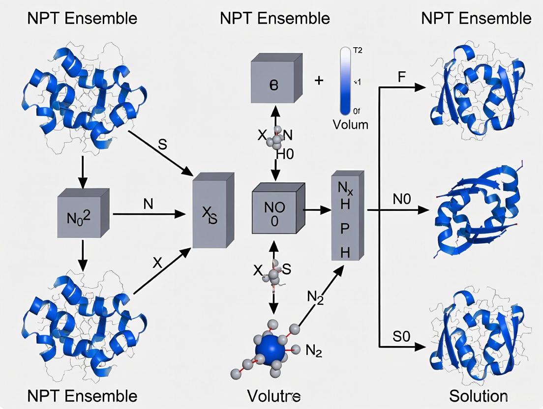
The following diagram illustrates the logical relationship between the defining constants of the NPT ensemble, the algorithms used to maintain them, and the resulting thermodynamic properties and outputs of a simulation.
Coupling Algorithms: Thermostats and Barostats
Maintaining constant temperature and pressure in an MD simulation requires algorithms that couple the system to external thermostats and barostats. The choice of algorithm can significantly influence the quality of the simulation and the physical validity of the generated ensemble [3] [2].
Thermostats control the temperature by scaling particle velocities. Common methods include the Berendsen thermostat, which provides weak coupling to a heat bath, and the Nose-Hoover thermostat, which produces a correct canonical ensemble and is widely used in conjunction with the NPT ensemble for biomolecular simulations [3]. Barostats control the pressure by scaling the simulation box dimensions. The Berendsen barostat offers a simple relaxation scheme, while the Parrinello-Rahman barostat allows for full anisotropic cell fluctuations, which is crucial for simulating crystalline materials or membranes under tension [3]. The Anderson-Hoover NPT ensemble combines a Nose-Hoover thermostat with a barostat for a rigorously correct NPT sampling [3].
Essential Parameters for NPT Simulations in Biomolecular Research
The setup of an NPT simulation requires careful selection of parameters, which are often defined in a molecular dynamics parameter file (e.g., an mdp file in GROMACS) [4]. The tables below summarize the critical parameters and their typical values for biomolecular simulations.
Table 1: Core NPT Control Parameters in MD Software
| Parameter | Description | Common Options & Typical Values |
|---|---|---|
| Integrator | Algorithm for integrating equations of motion. | md (leap-frog), md-vv (velocity Verlet) [4] |
| Ensemble Type | Defines the thermodynamic ensemble. | NPT (selected via imdmet=9,10,11 in ReaxFF; tcoupl and pcoupl in GROMACS) [3] |
| Temperature Coupling | Thermostat algorithm. | Nose-Hoover (NHC), Berendsen [3] |
| Pressure Coupling | Barostat algorithm. | Parrinello-Rahman, Berendsen [3] |
| Compressibility | Isothermal compressibility of the system. | ~4.5e-5 bar⁻¹ for water [4] |
| Coupling Frequency | Interval for applying thermostat/barostat. | Every 1-100 steps (nsttcouple, nstpcouple) [4] |
Table 2: Advanced and System-Specific NPT Parameters
| Parameter | Description | Application Context |
|---|---|---|
| τ_T (tau-t) | Characteristic time constant for temperature coupling. | ~0.5-2.0 ps; slower for production, faster for equilibration [4] |
| τ_P (tau-p) | Characteristic time constant for pressure coupling. | ~1.0-5.0 ps; relates to period of pressure fluctuations [3] |
| Pressure Tensor | Defines which box vectors are allowed to fluctuate. | Isotropic, Semi-isotropic, Anisotropic (e.g., imdmet=10 for fixed-angle cell) [3] |
| Mass Repartitioning | Scales masses of light atoms to enable larger timesteps. | Factor of 3 with constraints=h-bonds for 4 fs timestep [4] |
Experimental Protocol: Equilibration of a Hydrated Protein-Ligand System
This protocol details a robust equilibration procedure for a solvated protein-ligand complex, a common scenario in drug development. The goal is to relax the system from its initial coordinates to a stable state at the target temperature and pressure before beginning production simulation.
The workflow for the NPT equilibration protocol, from system construction to production simulation, is visualized below.
Step-by-Step Methodology
System Preparation
- Obtain the protein structure from a database (e.g., PDB). Prepare the structure using tools like
pdb2gmx(GROMACS) ortleap(AMBER) to assign protonation states, add missing residues, and select an appropriate force field (e.g., AMBER ff99SB-ILDN, CHARMM36) [2]. - Place the ligand in the binding pocket. Generate ligand parameters using tools like
acpypeor the GAFF force field. - Solvate the protein-ligand complex in a periodic box of explicit water molecules (e.g., TIP3P, TIP4P-EW) with a margin of at least 10 Å from the protein to its nearest box image [2].
- Add ions (e.g., Na⁺, Cl⁻) to neutralize the system's net charge and to achieve a physiologically relevant salt concentration (e.g., 150 mM).
- Obtain the protein structure from a database (e.g., PDB). Prepare the structure using tools like
Energy Minimization
- Objective: Remove any bad van der Waals contacts and steric clashes introduced during system building, relaxing the structure to the nearest local energy minimum.
- Protocol: Use a steepest descent or conjugate gradient algorithm for 5,000-50,000 steps until the maximum force is below a specified tolerance (e.g., 1000 kJ/mol/nm for initial steepest descent, then 100 kJ/mol/nm for conjugate gradient) [4] [2].
- Parameters:
integrator=steeporcgemtol= 1000.0nsteps= 50000
NVT Equilibration
- Objective: Gently heat the system to the target temperature (e.g., 310 K) while restraining the heavy atoms of the protein and ligand, allowing the solvent and ions to equilibrate around the solute.
- Protocol: Run a short simulation (100-500 ps) in the NVT ensemble.
- Parameters:
integrator=mddt= 0.002 (ps)nsteps= 50000 (for 100 ps)tcoupl=Nose-Hoover(orBerendsenfor initial heating)tau_t= 0.1-1.0 (ps)ref_t= 310 (K)pcoupl=no
NPT Equilibration
- Objective: Allow the system to reach the target density and stable pressure by releasing the positional restraints and allowing the box volume to fluctuate. This is a critical step for achieving a physiologically realistic system [1].
- Protocol: Run a simulation for 1-5 ns in the NPT ensemble. It is often advisable to run this in two phases: first with weak restraints on protein backbone atoms, and then completely unrestrained.
- Parameters:
integrator=mddt= 0.002 (ps)nsteps= 500000 (for 1 ns)tcoupl=Nose-Hoovertau_t= 1.0 (ps)ref_t= 310 (K)pcoupl=Parrinello-Rahman(for production quality) orBerendsen(for faster equilibration)tau_p= 2.0-5.0 (ps)ref_p= 1.0 (bar)compressibility= 4.5e-5 (bar⁻¹)
Production Simulation
- Objective: Collect data for analysis once the system properties (temperature, pressure, density, total energy, protein RMSD) have stabilized.
- Protocol: Continue the NPT simulation for tens to hundreds of nanoseconds, or longer, using the same parameters as the final NPT equilibration stage but without any restraints. The specific duration depends on the biological process being studied.
The Scientist's Toolkit: Essential Reagents and Software
Table 3: Key Research Reagent Solutions for NPT Biomolecular Simulations
| Item | Function / Description | Example Use Case |
|---|---|---|
| Explicit Solvent Models | Environment for solvating biomolecules; parameterized water molecules. | TIP3P, SPC/E, TIP4P-EW for simulating proteins in aqueous solution [2]. |
| Ion Parameters | Cations and anions for neutralizing system charge and modeling salt concentration. | Na⁺, Cl⁻, K⁺, Mg²⁺, Ca²⁺ parameters compatible with the chosen force field. |
| Biomolecular Force Fields | Empirical potential energy functions defining interatomic interactions. | CHARMM36, AMBER ff99SB-ILDN, GROMOS 54a7 for proteins, lipids, nucleic acids [2]. |
| Small Molecule Force Fields | Specialized parameters for drug-like molecules and ligands. | General Amber Force Field (GAFF), CGenFF for generating parameters for novel ligands. |
| MD Simulation Software | Software packages that perform the numerical integration of the equations of motion. | GROMACS, NAMD, AMBER, Desmond; implement NPT algorithms [2] [5]. |
| System Preparation Tools | Programs for building, solvating, and parameterizing simulation systems. | pdb2gmx (GROMACS), tleap (AMBER), CHARMM-GUI, PackMol. |
| Analysis Suites | Software for processing simulation trajectories to compute properties. | Built-in GROMACS tools, VMD, MDAnalysis, PyTraj for calculating RMSD, RDF, MSD, etc. |
Validation and Analysis of NPT Simulations
A critical final step is to validate that the simulation has reached a state of equilibrium and that the sampled ensemble is physically meaningful and reproducible [2]. Key metrics for validation include:
- Convergence of System Properties: Monitor the potential energy, temperature, pressure, and density over time. These should fluctuate stably around a constant average value after the equilibration phase. The density of a hydrated protein system, for instance, should converge to a value close to that of pure water (~997 kg/m³ at 300 K, depending on the water model used).
- Structural Stability: Calculate the root-mean-square deviation (RMSD) of the protein backbone and the ligand. A stable or converged RMSD indicates that the protein has not undergone large, unphysical conformational changes and is sampling a stable basin in the energy landscape.
- Comparison with Experimental Observables: Whenever possible, validate the simulation against experimental data. This can include comparing calculated radii of gyration with those from small-angle X-ray scattering (SAXS), or validating the structure against NMR-derived observables such as spin-spin coupling constants or residual dipolar couplings (RDCs) [2]. As noted in validation studies, while different simulation packages and force fields may reproduce average experimental observables equally well, the underlying conformational distributions can differ, highlighting the need for careful benchmarking [2].
By rigorously following these protocols and validation steps, researchers can ensure that their NPT simulations provide reliable and meaningful insights for drug development and biomolecular research.
Why NPT is the Gold Standard for Simulating Biological Conditions in Solution
The isothermal–isobaric (NPT) ensemble, where the number of particles (N), system pressure (P), and temperature (T) are kept constant, represents the most physiologically relevant environment for simulating biomolecular processes in solution. Unlike simulations conducted at constant volume (NVT), the NPT ensemble allows the simulation box size to fluctuate, enabling the system to maintain a realistic density that matches experimental conditions. For biomedical researchers and drug development professionals, employing the NPT ensemble is crucial for obtaining quantitatively accurate results that can reliably inform experimental design and interpretation. This protocol outlines the theoretical foundations, practical implementation, and key applications of NPT simulations for biomolecular systems, with a particular focus on achieving and validating proper equilibration.
Theoretical Foundation of the NPT Ensemble
In molecular dynamics (MD), the choice of statistical ensemble directly controls the thermodynamic conditions of the simulation. For biological systems in solution, the NPT ensemble mirrors natural environments where biomolecules experience constant temperature (maintained by a thermostat) and constant atmospheric pressure (maintained by a barostat). The barostat continuously adjusts the simulation box dimensions to maintain the target pressure, allowing the system density to stabilize at its experimental value [6].
This contrasts with NVT simulations, where the fixed box volume can generate an internal pressure that deviates significantly from the desired 1 bar, potentially distorting molecular structures and dynamics. The ability of the NPT ensemble to reproduce correct system densities makes it indispensable for calculating thermodynamic properties, studying conformational changes, and preparing systems for production runs that require physiologically relevant conditions.
Practical Implementation: Protocols for Robust NPT Equilibration
Standard NPT Equilibration Protocol
A typical equilibration protocol for a biomolecular system (e.g., a protein in explicit solvent) involves a two-step process to gradually relax the system before production simulation:
- Step 1: NVT Equilibration. The system is first equilibrated with position restraints applied to the heavy atoms of the biomolecule. This allows the solvent and ions to relax around the fixed solute, typically for 50-100 ps. The temperature is maintained at the target value (e.g., 300 K) using a thermostat such as Nose-Hoover or Berendsen.
- Step 2: NPT Equilibration. All position restraints are removed, and the simulation proceeds in the NPT ensemble. The barostat (e.g., Parrinello-Rahman or Berendsen) is activated to maintain constant pressure. This step typically requires 100-500 ps for the system density to stabilize, though larger systems may require longer.
Addressing the "Leaky Membrane" Effect in Charged Systems
Standard equilibration protocols can encounter specific challenges with chemically complex systems. As noted in assessments of glycolipid membrane simulations, a "leaky membrane effect" occasionally occurs where water molecules improperly enter the hydrophobic region during early NPT equilibration [7]. This artifact stems from very high initial pressure during the first steps of equilibration, which can cause a small box expansion that allows water infiltration.
Recommended Modified Protocol for Charged Glycolipids:
- Short NVT Pre-equilibration: Begin with a brief NVT simulation (e.g., 20-50 ps) at the target temperature.
- Stepwise NPT Equilibration: Implement a stepwise thermalization NPT protocol, gradually increasing the temperature (e.g., 100 K → 200 K → 300 K) under NPT conditions.
- Extended NPT at Target Conditions: Conduct extended NPT equilibration at the final temperature and pressure until density and potential energy stabilize.
This modified approach distributes the pressure adjustment more gradually, preventing the rapid box expansion that can compromise membrane integrity during equilibration [7].
Workflow for NPT Simulation Setup and Equilibration
The following diagram illustrates the logical workflow for establishing a properly equilibrated NPT system, incorporating the specialized protocol for sensitive structures:
Key Applications and Validation
Free Energy Calculations and Protein Folding
The NPT ensemble enables precise free-energy calculations for biomolecular processes. Recent advances in artificial intelligence-based ab initio biomolecular dynamics systems (AI2BMD) demonstrate the power of NPT simulations for studying protein folding and unfolding processes with ab initio accuracy [8]. These simulations can derive accurate 3J couplings that match nuclear magnetic resonance experiments and estimate thermodynamic properties such as melting temperatures of fast-folding proteins that align well with experimental measurements [8].
Calculating Diffusion Coefficients
Accurate calculation of translational diffusion coefficients from MD simulations requires special consideration of trajectory unwrapping in NPT ensembles. Since the barostat constantly rescales particle positions based on box fluctuations, standard unwrapping algorithms can introduce artifacts [6].
Recommended Practice:
- Use the toroidal-view-preserving (TOR) unwrapping scheme rather than heuristic lattice-view approaches
- Apply unwrapping to molecular centers of mass rather than individual atoms
- Ensure molecules are made "whole" before unwrapping to prevent bond stretching artifacts
The TOR scheme preserves the dynamics of the wrapped trajectory by summing minimal displacement vectors within the simulation box, providing more reliable diffusion coefficient estimates from NPT simulations [6].
The Scientist's Toolkit: Essential Research Reagents and Materials
Table 1: Essential computational tools and their functions in NPT biomolecular simulations
| Tool/Reagent | Type/Function | Application in NPT Simulations |
|---|---|---|
| AMOEBA Force Field [8] | Polarizable force field | Models explicit solvent polarization effects for accurate electrostatic interactions |
| Berendsen/Parrinello-Rahman Barostat [7] [6] | Pressure-coupling algorithm | Maintains constant pressure by adjusting box dimensions; Berendsen for equilibration, Parrinello-Rahman for production |
| Nose-Hoover Thermostat [7] | Temperature-coupling algorithm | Maintains constant temperature using extended Lagrangian formalism |
| AI2BMD Potential [8] | Machine learning force field | Provides ab initio accuracy for energy/force calculations with significantly reduced computational cost |
| GROMACS [7] [6] | MD simulation software | Implements various barostats, thermostats, and trajectory analysis tools for NPT ensembles |
| TOR Unwrapping Scheme [6] | Trajectory analysis method | Correctly unwraps molecular trajectories from NPT simulations for accurate diffusion calculations |
Quantitative Comparison of Simulation Ensembles
Table 2: Comparative analysis of NPT versus NVT ensembles for biomolecular simulations
| Parameter | NPT Ensemble | NVT Ensemble |
|---|---|---|
| Controlled Variables | Number of particles, Pressure, Temperature | Number of particles, Volume, Temperature |
| System Density | Fluctuates, converges to experimental value | Fixed, may not match experimental conditions |
| Physiological Relevance | High - mimics lab/physiological conditions | Moderate - constant volume is artificial constraint |
| Pressure Artifacts | Minimal - pressure is controlled | Possible - internal pressure may deviate from target |
| Applications | Protein folding, material properties, solvation studies | Specific studies requiring fixed volume |
| Equilibration Complexity | Higher - requires pressure coupling | Lower - no pressure coupling |
| Diffusion Coefficient Accuracy | High when using proper unwrapping schemes [6] | Generally straightforward but may have density errors |
The NPT ensemble remains the gold standard for biomolecular simulations in solution due to its ability to replicate experimentally relevant thermodynamic conditions. Through careful implementation of equilibration protocols—including specialized approaches for charged systems like glycolipid membranes—and proper trajectory analysis techniques, researchers can obtain quantitatively accurate results that reliably connect simulation data with experimental observables. As force fields and sampling methods continue to advance, particularly with the integration of machine learning approaches like AI2BMD, the NPT ensemble will continue to provide an essential foundation for understanding biological processes at atomic resolution and accelerating drug development efforts.
Statistical Mechanical Theory of the Isothermal-Isobaric Ensemble
The isothermal-isobaric (NPT) ensemble is a cornerstone of molecular simulation, particularly for biomolecular systems in solution. This ensemble maintains a constant number of particles (N), constant pressure (P), and constant temperature (T), thereby closely mimicking the natural experimental conditions under which most biological processes occur. The theoretical foundation of the NPT ensemble derives from statistical mechanics, where the partition function Δ(N,P,T) provides the connection between microscopic molecular behavior and macroscopic thermodynamic observables. For biomolecular simulations, the NPT ensemble is indispensable for reproducing correct densities, solvation environments, and conformational equilibria, as it allows the simulation box size and shape to fluctuate in response to internal and external pressures.
The relevance of NPT simulations in drug development and biomedical research cannot be overstated. Accurate modeling of biomolecules such as proteins, nucleic acids, and lipids in their native aqueous environments enables the study of ligand-binding affinities, conformational dynamics, and solvation effects—all critical factors in rational drug design. The NPT ensemble ensures that the simulated system occupies the appropriate volume and maintains realistic intermolecular distances, providing a reliable platform for predicting properties that can be validated against experimental data [9].
Core Theoretical Framework
Partition Function and Thermodynamics
The isothermal-isobaric ensemble is defined by its partition function, which for a one-component system is given by:
Δ(N, P, T) = C ∫∫ exp[-βH(q, p) - βPV] dq dp dV
where β = 1/kBT, H(q,p) is the system Hamiltonian, V is the volume, and C is a constant that ensures proper normalization. This partition function connects to thermodynamics through the Gibbs free energy:
G(N, P, T) = -kBT ln Δ(N, P, T)
The NPT ensemble thus provides a foundation for calculating equilibrium properties under conditions that mirror most laboratory experiments for systems in solution. The thermodynamic connection enables the extraction of key properties including enthalpy, entropy, and volume fluctuations that provide insights into biomolecular stability and interactions [10].
Equations of Motion and Barostats
In molecular dynamics (MD) implementations of the NPT ensemble, the equations of motion are extended to include volume fluctuations. This is typically achieved through the incorporation of barostats that adjust the simulation cell dimensions to maintain constant pressure. Modern simulation packages employ sophisticated algorithms such as the Parrinello-Rahman barostat, which allows for fully flexible simulation cells, or the Nosé-Hoover Langevin piston for robust control of pressure dynamics.
The resulting volume fluctuations provide direct access to important thermodynamic derivatives, including the isothermal compressibility βT:
βT = -1/V (∂V/∂P)T,N = ⟨(δV)²⟩ / (kBTV)
where ⟨(δV)²⟩ represents the volume fluctuations in the NPT ensemble. This relationship highlights how microscopic fluctuations in the simulation connect to macroscopic material properties [10].
Computational Protocols for NPT Biomolecular Simulations
System Setup and Equilibration
Initial Structure Preparation Begin with a high-quality atomic structure of the biomolecule from experimental sources (e.g., Protein Data Bank) or homology modeling. For the solvated environment, embed the biomolecule in an appropriate water box (typically TIP3P, SPC/E, or TIP4P water models) with a minimum of 10-15 Å padding between the solute and box edges. Add physiological ion concentrations (e.g., 0.15 M NaCl) to mimic biological conditions, ensuring charge neutrality.
Energy Minimization Perform steepest descent or conjugate gradient minimization to remove bad contacts and high-energy configurations. Protocol: 5,000-10,000 steps until the maximum force falls below 100-1000 kJ/mol/nm, preparing the system for stable dynamics.
Stepwise Equilibration
- NVT Equilibration: Run 100-500 ps with position restraints on heavy atoms of the biomolecule (force constant of 1000 kJ/mol/nm²) while allowing solvent and ions to relax. Use a Langevin thermostat or Nosé-Hoover chain to maintain temperature at 300 K.
- NPT Equilibration: Run 1-5 ns with mild position restraints (force constant of 100-500 kJ/mol/nm²) on biomolecule heavy atoms, employing a barostat (e.g., Parrinello-Rahman or Berendsen) to maintain pressure at 1 bar. This allows the solvent density to equilibrate properly.
- Unrestrained NPT Production: Remove all restraints and conduct extended NPT simulation for timescales appropriate to the biological process being studied (typically 50 ns to 1 μs for most biomolecular applications) [11].
Property Calculation and Analysis
Thermodynamic Properties The NPT ensemble enables calculation of various thermodynamic properties through fluctuation formulas and direct averaging:
Table 1: Key Thermodynamic Properties Accessible from NPT Simulations
| Property | Mathematical Expression | Biological Significance |
|---|---|---|
| Thermal expansion coefficient | αP = 1/V (∂V/∂T)P,N = ⟨δVδH⟩ / (kBTV) | Volume changes with temperature relevant for thermal stability |
| Isobaric heat capacity | CP = (∂H/∂T)P,N = ⟨(δH)²⟩ / (kBT²) | Energy requirements for conformational changes |
| Isothermal compressibility | βT = -1/V (∂V/∂P)T,N = ⟨(δV)²⟩ / (kBTV) | Membrane mechanical properties and compressibility |
| Thermal pressure coefficient | γV = (∂P/∂T)V,N = αP/βT | Pressure effects on protein denaturation |
Structural Properties Analyze root-mean-square deviation (RMSD) for structural stability, radius of gyration for compactness, and solvent-accessible surface area for hydration effects. For lipid bilayer systems, calculate electron density profiles and order parameters for comparison with experimental data [9] [12].
Application Case Studies in Biomolecular Research
RNA Structure Refinement
In CASP15 community-wide assessment, NPT-MD simulations were systematically benchmarked for RNA structure refinement using Amber with the RNA-specific χOL3 force field. The study revealed that short NPT simulations (10-50 ns) provided modest improvements for high-quality starting models by stabilizing base stacking and non-canonical base pairs. However, poorly predicted models rarely benefited and often deteriorated, highlighting the importance of initial model quality. The optimal protocol employs NPT-MD as a fine-tuning tool rather than a universal corrective method, with early simulation dynamics (5-10 ns) diagnostic of refinement potential [11].
Table 2: NPT-MD Refinement Outcomes for RNA Models in CASP15
| Starting Model Quality | Simulation Length | Typical RMSD Change (Å) | Key Structural Improvements |
|---|---|---|---|
| High-quality | 10-50 ns | -0.1 to -0.3 | Stabilized stacking, optimized non-canonical pairs |
| Medium-quality | 10-50 ns | -0.05 to +0.2 | Variable improvements, occasional deterioration |
| Low-quality | 10-50 ns | +0.3 to +1.5 | Structural drift, loss of native contacts |
| Any quality | >50 ns | Typically positive | Increased drift, reduced fidelity to experimental data |
Biomembrane Dynamics and Validation
A comprehensive dynamic landscape of fully hydrated palmitoyl-oleoyl-phosphatidylcholine (POPC) bilayers was constructed by combining 13C NMR relaxation data with 8.4 μs NPT-MD simulations. This integrated approach enabled separation of molecular motions by type and timescale, revealing vast differences in motional amplitudes and correlation times depending on molecular position within the bilayer. The NPT ensemble was critical for maintaining appropriate membrane packing and hydration during these simulations, enabling direct validation of simulation results against experimental NMR data through detector analysis methodology [12].
Reactive Simulations for Bond Breaking
The implementation of reactivity in MD simulations using harmonic force fields (Reactive INTERFACE Force Field, IFF-R) enables simulation of bond dissociation and failure in NPT ensembles. By replacing harmonic bond potentials with reactive, energy-conserving Morse potentials, IFF-R maintains compatibility with standard biomolecular force fields (CHARMM, AMBER, OPLS-AA) while enabling bond breaking at approximately 30 times the computational speed of reactive bond-order potentials. This approach is particularly valuable for studying mechanical failure in polymer-protein composites and chemical reactions in biomolecular systems under constant pressure conditions [13].
Workflow Visualization
Diagram 1: NPT Simulation Workflow for Biomolecular Systems. This workflow outlines the standard protocol for setting up and running NPT simulations of biomolecules in solution, progressing from initial structure preparation through production simulation to final analysis.
Diagram 2: NPT Simulation Analysis and Validation Pathway. This diagram illustrates the major analysis pathways for NPT simulations and their connection to experimental validation methods, highlighting the importance of experimental corroboration for simulation results.
Table 3: Essential Tools for NPT Biomolecular Simulations
| Tool/Resource | Type | Function in NPT Simulations | Example Implementations |
|---|---|---|---|
| Molecular Dynamics Engines | Software | Integrates equations of motion with barostats for NPT ensemble | GROMACS, NAMD, AMBER, OpenMM, LAMMPS |
| Force Fields | Parameters | Defines bonded and non-bonded interactions for biomolecules | CHARMM36, AMBER, OPLS-AA, GROMOS |
| Barostat Algorithms | Algorithm | Maintains constant pressure by adjusting simulation cell dimensions | Parrinello-Rahman, Berendsen, Martyna-Tobias-Klein |
| Thermostat Algorithms | Algorithm | Maintains constant temperature by controlling kinetic energy | Nosé-Hoover, Langevin, Velocity Rescaling |
| Solvation Models | Parameters | Represents aqueous environment for biomolecular simulations | TIP3P, SPC/E, TIP4P water models |
| Analysis Tools | Software | Processes trajectory data to extract structural and thermodynamic properties | MDAnalysis, VMD, CPPTRAJ, GROMACS tools |
| Validation Databases | Data Repository | Provides experimental data for validation of simulation results | MolMod Database [10], GPCRmd [14] |
Advanced Methodologies and Emerging Approaches
FAIR-Compliant Data Management
The increasing complexity and volume of NPT simulation data necessitates robust data management strategies aligned with FAIR (Findable, Accessible, Interoperable, Reusable) principles. PostgreSQL-based storage solutions coupled with specialized metadata schemas offer a promising approach for maintaining the essential connection between simulation parameters and resulting trajectories. Such systems enable researchers to efficiently track system compositions, force field assignments, boundary conditions, and thermodynamic ensemble settings—critical information for ensuring the reproducibility and reusability of NPT simulation data [14].
Multi-Scale and Multi-Ensemble Integration
Modern molecular simulation tools like ms2 (release 5.0) demonstrate the trend toward integrated simulation environments that provide access to multiple statistical ensembles, including the NPT ensemble. These platforms implement advanced methodologies such as the Lustig formalism for on-the-fly sampling of thermodynamic properties including isobaric heat capacity, thermal expansion coefficient, and isothermal compressibility directly during NPT simulations. Such capabilities facilitate more efficient calculation of thermodynamic derivatives without requiring multiple separate simulations [10].
The isothermal-isobaric ensemble remains an essential tool in the molecular simulation toolkit for biomolecular research, providing the most physiologically relevant conditions for studying biological processes in solution. The theoretical foundation of the NPT ensemble enables direct connection between microscopic simulations and macroscopic experimental observables, while continued methodological advances improve the accuracy, efficiency, and scope of NPT applications. As force fields continue to refine their parameters for biomolecular systems and simulation methodologies expand to include reactive processes and enhanced sampling, the utility of NPT simulations in drug development and basic biomedical research will continue to grow. Proper implementation of NPT protocols, coupled with rigorous validation against experimental data, ensures that molecular simulations provide reliable insights into biomolecular structure, dynamics, and function.
Comparing NPT with NVT, μVT, and Gibbs Ensembles for Biological Systems
The selection of a statistical ensemble is a foundational step in molecular dynamics (MD) simulations, directly determining the thermodynamic state of the system and the relevance of the simulation to experimental conditions. For biological simulations in solution, researchers must choose an ensemble that not only ensures computational efficiency but also accurately models the realistic environmental conditions under which biomolecules operate. This application note provides a detailed comparison of the NPT (isothermal-isobaric), NVT (canonical), µVT (grand canonical), and Gibbs ensembles, with a specific focus on their theoretical underpinnings and practical applications in biomolecular simulation. The NPT ensemble, which maintains constant particle number (N), pressure (P), and temperature (T), is particularly crucial for simulating biological systems in solution as it most closely replicates laboratory conditions where experiments are conducted at atmospheric pressure and controlled temperature [15] [16]. Within the broader thesis of this work, we establish that proper use of the NPT ensemble is indispensable for obtaining biologically relevant structural, dynamic, and thermodynamic data from molecular simulations of solvated biomolecules.
Theoretical Foundation of Statistical Ensembles
In statistical mechanics, an ensemble is defined as an idealization consisting of a large number of virtual copies of a system, considered simultaneously, each representing a possible state that the real system might be in [17]. The concept was formally introduced by J. Willard Gibbs in 1902 to connect microscopic molecular behavior to macroscopic thermodynamic observables. Different ensembles correspond to different sets of macroscopic constraints, leading to distinct statistical characteristics and applications [17].
Mathematical Definitions of Key Ensembles
Microcanonical Ensemble (NVE): Describes completely isolated systems with fixed particle number (N), volume (V), and energy (E). It forms the foundation of statistical mechanics but has limited practical application for biological systems which typically exchange energy with their environment [17].
Canonical Ensemble (NVT): Characterizes closed systems that can exchange energy with a thermal reservoir at temperature T, maintaining constant particle number (N) and volume (V). The partition function for the NVT ensemble is defined as ( Q(N,V,T) = \sumi e^{-\beta Ei} ), where ( \beta = 1/k_B T ) [17].
Isobaric-Isothermal Ensemble (NPT): Models closed systems that can exchange both energy and volume with a reservoir at constant pressure P and temperature T, with fixed particle number N. The NPT partition function is given by ( \Delta(N,P,T) = \int dV \sumi e^{-\beta (Ei + PV)} ) [16].
Grand Canonical Ensemble (µVT): Describes open systems that exchange both energy and particles with a reservoir at constant chemical potential (µ), volume (V), and temperature (T). This ensemble is particularly valuable for studying systems with fluctuating particle numbers, such as binding processes or phase interfaces [17].
Gibbs Ensemble: A specialized ensemble that enables direct simulation of phase equilibria by maintaining thermal and mechanical equilibrium between two or more distinct regions or phases, with constant total number of particles, total volume, and temperature (NV₂T) or constant temperature and pressure (NPT) for each phase [18].
Table 1: Fundamental Characteristics of Statistical Ensembles
| Ensemble | Fixed Parameters | Fluctuating Quantities | Partition Function | Primary Applications |
|---|---|---|---|---|
| NVE | N, V, E | Temperature, Pressure | ( \Omega(N,V,E) ) | Fundamental studies; isolated systems |
| NVT | N, V, T | Energy, Pressure | ( Q(N,V,T) ) | Structural studies in confined volume |
| NPT | N, P, T | Energy, Volume | ( \Delta(N,P,T) ) | Biomolecules in solution |
| µVT | µ, V, T | Energy, Particle Number | ( \Xi(\mu,V,T) ) | Solvation, adsorption, binding |
| Gibbs | N₁+N₂, V₁+V₂, T (or P) | Energy, Volume distribution, Particle distribution | Specialized forms | Phase equilibria, membrane partitioning |
Comparative Analysis of Ensembles for Biological Applications
NPT Ensemble: The Gold Standard for Biomolecular Simulations
The NPT ensemble is generally considered the most appropriate choice for simulating biomolecular systems in solution, as it accurately replicates standard laboratory conditions where experiments are performed at controlled temperature and atmospheric pressure [15] [16]. In this ensemble, the system can adjust its volume in response to internal forces and external pressure, allowing for natural density fluctuations that are essential for proper biomolecular solvation and hydration. For proteins in aqueous solution, the use of NPT conditions ensures that water molecules maintain appropriate density (approximately 1 g/cm³ for TIP3P and SPC water models at 300 K and 1 bar) and that the simulated system does not develop unrealistic internal pressures that could distort protein structure or dynamics [19].
The theoretical foundation of the NPT ensemble involves an extended Hamiltonian that includes a barostat to regulate pressure and a thermostat to maintain temperature. The definition of instantaneous pressure for microscopic systems can be derived from the minimum energy principle for the Helmholtz free energy [19]. For discrete/continuum approaches like the General Liquid Optimized Boundary (GLOB) model, which is particularly useful for biomolecular simulations, the pressure coupling can be implemented through extended phase-space schemes that account for the boundary between explicit and implicit solvent regions [19].
NVT Ensemble: Applications and Limitations
The NVT ensemble maintains a constant volume, which can be advantageous for specific applications such as comparing directly with experimental data collected under confined conditions or when simulating crystal structures where unit cell dimensions are fixed. However, for biomolecular simulations in solution, the fixed volume constraint presents significant limitations. Without the ability to adjust volume, the system may develop non-physical internal pressures, particularly during equilibration stages or when significant conformational changes occur. This can lead to distorted hydrogen-bonding networks, improper solvation shell structures, and ultimately unrealistic protein dynamics [15].
Despite these limitations, NVT simulations can be useful as part of a multi-stage equilibration protocol, where an initial NVT simulation might precede production NPT simulations to achieve gradual relaxation of the system. Additionally, NVT may be appropriate for short simulations aimed at specific properties that are less sensitive to volume fluctuations.
µVT and Gibbs Ensembles: Specialized Applications
The µVT ensemble is particularly valuable for studying processes involving variable particle numbers, such as ligand binding, ion permeation through channels, or adsorption phenomena. In this ensemble, the chemical potential (µ) of specific components is fixed, allowing particles to enter or leave the simulation volume. This makes µVT ideal for calculating binding affinities, studying competitive solvation, or modeling systems at interfaces [18]. Recent advances in kinetic Monte Carlo (kMC) schemes have improved the accuracy of chemical potential calculations in dense phases where traditional Widom insertion methods fail [18].
The Gibbs ensemble provides a powerful methodology for studying phase equilibria without constructing an explicit interface between phases. By simulating two distinct regions that can exchange particles and volume while maintaining constant total particle number and either total volume (NV₂T) or pressure (NPT), researchers can directly observe phase separation and calculate coexistence properties. This ensemble has been successfully applied to study vapor-liquid equilibria in simple fluids and more complex associating fluids relevant to biological systems [18].
Table 2: Quantitative Performance Metrics of Different Ensembles for Biomolecular Simulations
| Ensemble | Computational Efficiency | Sampling Effectiveness | Accuracy for Aqueous Systems | Ease of Implementation | Recommended Simulation Time |
|---|---|---|---|---|---|
| NPT | High (after equilibration) | Excellent for equilibrium properties | Excellent (density ~1 g/cm³) | Straightforward in most MD packages | ≥100 ns for protein folding |
| NVT | High | Good for structural properties | Good (with careful volume selection) | Very straightforward | 10-100 ns for dynamics |
| µVT | Moderate to Low (depends on insertion success) | Excellent for open systems | Good for solvation thermodynamics | Complex (requires particle exchange moves) | Varies widely with system |
| Gibbs | Moderate | Excellent for phase equilibria | Good for membrane systems | Complex (requires multiple regions) | Varies with phase transition rates |
Practical Protocols for Ensemble Implementation
Standard NPT Simulation Protocol for Proteins in Solution
The following protocol outlines a robust methodology for conducting NPT simulations of biomolecules in explicit solvent, suitable for implementation in common MD packages such as GROMACS, AMBER, or NAMD.
System Preparation
- Initial Structure: Obtain protein coordinates from PDB or predicted structures. Remove crystallographic artifacts and add missing atoms/residues if necessary.
- Solvation: Place the biomolecule in a suitably sized simulation box (typically rectangular or dodecahedral) with a minimum of 1.0-1.5 nm between the protein and box edges. Add explicit water molecules using models such as TIP3P, TIP4P, or SPC, which have been parameterized for use with specific force fields [19] [16].
- Neutralization: Add counterions (typically Na⁺ or Cl⁻) to neutralize the system net charge. Additional ions may be added to achieve specific physiological concentrations (e.g., 150 mM NaCl).
Energy Minimization and Equilibration
- Energy Minimization: Perform steepest descent or conjugate gradient minimization (5,000-10,000 steps) to remove bad contacts and high-energy configurations.
- NVT Equilibration: Equilibrate the system for 100-500 ps in the NVT ensemble to stabilize temperature. Use thermostats such as Berendsen (for initial equilibration) or Nosé-Hoover (for production) with coupling constants of 0.1-1.0 ps.
- NPT Equilibration: Conduct extended equilibration (1-5 ns) in the NPT ensemble to stabilize both temperature and pressure. Use barostats such as Berendsen (equilibration) or Parrinello-Rahman (production) with coupling constants of 1.0-5.0 ps and compressibility set to 4.5×10⁻⁵ bar⁻¹ for water.
Production Simulation
- Parameter Settings: Use a time step of 2 fs with constraints applied to all bonds involving hydrogen atoms (LINCS or SHAKE algorithms). Employ Particle Mesh Ewald (PME) for long-range electrostatics with a real-space cutoff of 1.0-1.2 nm. Apply similar cutoffs for van der Waals interactions.
- Duration: Run production simulations for timescales appropriate to the biological process of interest: typically 100 ns - 1 µs for local conformational changes, and >1 µs for large-scale transitions or folding events [15].
- Data Collection: Save trajectory frames every 10-100 ps for analysis, ensuring a balance between storage requirements and temporal resolution.
Advanced Protocol: µVT Simulations for Ligand Binding Studies
For simulations requiring the µVT ensemble, such as ligand binding or hydration studies, the following specialized protocol is recommended:
System Setup and Preparation
- Simulation Box: Create a solvated system with the protein or binding pocket of interest. Identify the chemical potential reservoir region if using a multi-region approach.
- Parameterization: Define the chemical potential (µ) for the species of interest based on experimental data or preliminary simulations. This may require calibration through iterative simulations.
Monte Carlo/Molecular Dynamics Hybrid Approach
- MD Phase: Perform short molecular dynamics steps (1-100 ps) to sample molecular motions and configurations.
- MC Phase: Implement Monte Carlo moves for particle insertion/deletion and identity changes. The acceptance criteria for identity changes follows: min[1, exp(-βδU + N₂βδμ)] where δU is the energy difference and δμ is the chemical potential difference [18].
- Balancing: Adjust the ratio of MD steps to MC attempts to maintain an acceptance rate of 15-40% for particle exchanges, which typically provides optimal sampling efficiency.
Validation and Analysis Methods
Regardless of the ensemble chosen, rigorous validation against experimental data is essential for establishing simulation reliability:
- Convergence Assessment: Monitor key properties (RMSD, energy, density, radius of gyration) to ensure they have reached stable equilibrium distributions before beginning production analysis.
- Experimental Comparison: Compare simulation results with available experimental data, such as NMR order parameters [20], J-couplings, scattering profiles, or thermodynamic measurements.
- Ensemble Validation: For advanced applications, verify consistency between ensembles by comparing observables that should be ensemble-independent in properly converged simulations [18].
Diagram 1: Standard workflow for biomolecular simulation in the NPT ensemble.
Research Reagent Solutions and Computational Tools
Table 3: Essential Research Reagents and Computational Tools for Ensemble Simulations
| Item | Function/Purpose | Example Implementations | Key Considerations |
|---|---|---|---|
| Force Fields | Describes interatomic interactions | CHARMM, AMBER, OPLS, GROMOS | Select based on target system (proteins, nucleic acids, lipids) |
| Water Models | Solvent representation | TIP3P, TIP4P, SPC, TIP5P | Match to force field parameterization [19] |
| Thermostats | Temperature control | Nosé-Hoover, Berendsen, velocity rescale | Use weak coupling (0.1-1.0 ps) for biomolecular systems |
| Barostats | Pressure control | Parrinello-Rahman, Berendsen, MTK | Compressibility ~4.5×10⁻⁵ bar⁻¹ for aqueous systems [16] |
| MD Software | Simulation engine | GROMACS, AMBER, NAMD, OpenMM | Choose based on hardware compatibility and efficiency |
| Analysis Tools | Trajectory processing | MDAnalysis, VMD, CPPTRAJ | Implement multiple analysis methods for validation |
The selection of an appropriate statistical ensemble is a critical decision point in biomolecular simulation that directly impacts the physical relevance and interpretability of results. For most simulations of biological systems in solution, the NPT ensemble provides the most realistic representation of experimental conditions, allowing natural volume fluctuations that maintain proper system density and hydration. The NVT ensemble serves important but more specialized roles, particularly in systems with constrained volumes or as part of multi-stage equilibration protocols. The µVT and Gibbs ensembles offer powerful capabilities for studying open systems and phase equilibria, respectively, though with increased computational complexity and specialized implementation requirements.
Emerging methodologies, particularly machine learning force fields and advanced sampling techniques, are expanding the horizons of what is possible with each ensemble. Systems like AI2BMD demonstrate how artificial intelligence can achieve ab initio accuracy while maintaining computational efficiency, potentially enabling more sophisticated applications of these ensembles to challenging biological questions [8]. Furthermore, the integration of advanced Monte Carlo schemes with traditional molecular dynamics continues to improve the sampling of complex thermodynamic ensembles, particularly for dense systems and phase interfaces [18].
As the field progresses toward increasingly complex biological questions and multi-scale simulations, the appropriate selection and implementation of statistical ensembles will remain fundamental to generating reliable, experimentally relevant computational data for drug development and basic biological research.
The Critical Role of Pressure Control in Mimicking Physiological Environments
The accurate simulation of physiological conditions is paramount in computational biochemistry for obtaining biologically relevant results. The NPT (isothermal-isobaric) ensemble, which maintains constant particle number (N), pressure (P), and temperature (T), is the cornerstone of molecular dynamics (MD) simulations aimed at replicating these native environments [21]. Within this framework, pressure control emerges as a critical factor, directly influencing system density, volume, and the fundamental energetics of biomolecular interactions [22]. Proper pressure control is not merely a technicality but a physiological necessity; for instance, the electrostatic environment surrounding membrane proteins, characterized by a low dielectric constant, is essential for processes like GPCR activation and ligand binding, and its accurate representation depends on appropriate environmental parameters [23]. This application note details the protocols and considerations for implementing pressure control in MD simulations to faithfully mimic physiological conditions, thereby enhancing the reliability of research in drug development and structural biology.
Theoretical Foundation of Pressure Control
In MD simulations, pressure (P) is a computed property derived from the virial equation, which accounts for both the kinetic energy of particles and the internal forces acting upon them [24]. The role of a barostat, or pressure control algorithm, is to maintain this pressure at a target value by dynamically adjusting the volume of the simulation cell [24].
The choice of barostat significantly influences the quality of the simulation and the physical accuracy of the generated ensemble. Available methods fall into several categories, each with distinct strengths and weaknesses, making them suitable for different stages of research, from system equilibration to production runs.
- Weak Coupling Methods: The Berendsen barostat is a classic example that scales particle coordinates and cell dimensions by an increment proportional to the difference between the instantaneous and target pressure [25] [24]. While highly efficient for rapidly equilibrating a system and driving it to the target pressure, it does not produce a correct thermodynamic ensemble because it suppresses pressure fluctuations [24]. Its use is therefore generally recommended for equilibration only.
- Extended System Methods: These algorithms introduce additional degrees of freedom to represent the system's volume. The Parrinello-Rahman barostat allows for changes in both the size and shape of the simulation cell, making it particularly useful for studying solids under stress or anisotropic systems [22] [26]. The Nosé-Hoover barostat and its refinement, the Martyna-Tuckerman-Tobias-Klein (MTTK) barostat, are other extended system methods that are time-reversible and generate a correct NPT ensemble [24].
- Stochastic Methods: The Langevin Piston method combines the extended system approach with stochastic dynamics. By applying a damping force and random collisions to the piston controlling the volume, it effectively controls oscillations and often converges faster than purely deterministic methods [25] [24]. Stochastic Cell Rescaling is an improved version of the Berendsen barostat that adds a stochastic term, enabling correct fluctuations and making it suitable for production simulations [24].
- Monte-Carlo Methods: These methods involve periodically attempting random changes to the simulation cell volume, accepting or rejecting these changes based on a Metropolis criterion. They do not require the calculation of the virial and are another valid approach for sampling the NPT ensemble [24].
Barostat Selection Guide
The table below provides a comparative overview of common barostats to guide researchers in selecting the most appropriate method.
Table 1: Comparison of Common Pressure Control Algorithms (Barostats)
| Barostat Method | Type | Key Features | Strengths | Weaknesses | Recommended Use |
|---|---|---|---|---|---|
| Berendsen [25] [24] | Weak Coupling | Exponential decay of pressure deviation. | Fast equilibration; numerically stable. | Suppresses fluctuations; incorrect ensemble. | System equilibration only. |
| Parrinello-Rahman [22] [24] | Extended System | Allows full cell shape/size fluctuations. | Correct ensemble; good for anisotropic solids. | "Piston mass" parameter (pfactor) is system-dependent. | Production runs (solids, anisotropic systems). |
| Nosé-Hoover / MTTK [24] | Extended System | Constant enthalpy; correct NPT ensemble. | Correct ensemble; time-reversible. | Can be sensitive to piston mass parameter. | Production runs (general use). |
| Langevin Piston [25] [24] | Stochastic | Damping and random forces on the piston. | Fast convergence; damped oscillations. | Requires setting a damping coefficient. | Production runs (general use, liquids). |
| Stochastic Cell Rescaling [24] | Stochastic | Adds noise to Berendsen's scaling matrix. | Fast and correct fluctuations. | Relatively newer method. | All stages, including production. |
Practical Implementation and Protocols
Successfully implementing pressure control requires careful attention to parameter selection and system setup. The following protocols provide a guideline for configuring barostats in biomolecular simulations.
General Configuration Parameters
Regardless of the barostat chosen, several universal parameters must be defined.
Table 2: Universal Barostat Parameters and Typical Values for Biomolecular Simulations
| Parameter | Description | Typical Values / Considerations |
|---|---|---|
Target Pressure (BerendsenPressureTarget, Parrinello-Rahman Target) |
The desired average pressure of the system. | 1.01325 bar (atmospheric pressure) is standard for mimicking physiological conditions [25]. |
| Pressure Coupling Type | Defines which cell dimensions are allowed to fluctuate. | Isotropic: Uniform scaling in all directions (standard for solution simulations). Semi-isotropic: Different scaling in x-y and z (for bilayers). Anisotropic: Fully flexible cell (for solids) [25] [22]. |
Coupling Frequency (BerendsenPressureFreq) |
How often the barostat is applied. | Typically every 1-20 MD steps. Must be a multiple of other algorithmic frequencies [25]. |
Protocol 1: System Equilibration using the Berendsen Barostat
This protocol is designed for the initial rapid equilibration of a solvated biomolecular system to the target pressure and density.
- System Preparation: Begin with a solvated and neutralized biomolecular system, typically pre-minimized to remove steric clashes.
- Thermostat Configuration: Apply a thermostat (e.g., Berendsen or Nosé-Hoover) to control the temperature. A relaxation time constant (
ttime) of 0.1-1.0 ps is often suitable. - Barostat Configuration:
- Set the barostat to Berendsen.
- Set the
Target Pressureto 1.01325 bar. - Set the
Pressure Relaxation Time(BerendsenPressureRelaxationTime) to 100-500 fs. A shorter time provides stronger coupling and faster equilibration. - Set the
Compressibility(BerendsenPressureCompressibility) to 4.57e-5 bar⁻¹, the experimental value for water [25]. - Use isotropic pressure coupling.
- Simulation Run: Run a short simulation (typically 100-500 ps). Monitor the system density and box volume until they stabilize around equilibrium values.
- Switch to Production Barostat: Once equilibrated, switch the barostat to a method suitable for production (e.g., Parrinello-Rahman or Nosé-Hoover) before beginning the production simulation.
Protocol 2: Production Simulation using the Parrinello-Rahman Barostat
This protocol outlines the setup for a production-level simulation that correctly samples the NPT ensemble, using the Parrinello-Rahman method as an example.
- Initial Equilibration: Ensure the system has been pre-equilibrated using a protocol like the one described above.
- Thermostat Configuration: Use a robust thermostat like Nosé-Hoover or Langevin for temperature control.
- Barostat Configuration:
- Set the barostat to Parrinello-Rahman.
- Set the
Target Pressureto 1.01325 bar. - Define the
Piston Massparameter. In software like ASE, this is often set indirectly via thepfactor(τₚ²B). For a system like fcc-Cu, a value on the order of 10⁶ to 10⁷ GPa·fs² is a good starting point. For biomolecular systems in aqueous solution, extensive testing may be required to find an optimal value that allows for natural fluctuations without causing instability [22]. - Use the appropriate pressure coupling type for your system (e.g., isotropic for a protein in solution).
- Simulation Run: Execute the production simulation. Monitor the pressure and volume to confirm they fluctuate stably around the target values.
The following workflow diagram illustrates the logical decision process for selecting and applying barostats in a typical MD study.
Diagram 1: MD Barostat Selection Workflow
The Scientist's Toolkit: Essential Reagents and Software
Successful NPT simulations rely on a combination of force fields, software tools, and molecular models. The following table lists key resources used in advanced simulation studies.
Table 3: Research Reagent Solutions for NPT Biomolecular Simulations
| Category | Item / Software | Function in Simulation | Example from Literature |
|---|---|---|---|
| Force Fields | OPLS/AA [23], AMBER, CHARMM | Define potential energy functions governing atomic interactions; foundational for accurate dynamics. | Used to parameterize peptides and membrane proteins in low electrostatic environments [23]. |
| MD Software | NAMD [25], GROMACS [24], LAMMPS [27], AMBER | Software engines that integrate equations of motion and implement algorithms for thermostats and barostats. | NAMD used for its implementation of Langevin Piston and Berendsen barostats [25]. |
| Water Models | TIP4P/ϵflex, FBA/ϵ [23] | Solvent molecules parameterized to reproduce key properties like density and dielectric constant. | Reparameterized to create low electrostatic water (LEw) models (FBAmem, TIP4Pmem) for membrane simulations [23]. |
| Analysis Tools | UCSF Chimera [23], OVITO [27], VMD | Visualization and analysis of trajectories; calculation of properties like RMSD, RMSF, and hydrogen bonding. | UCSF Chimera used to build missing residues in peptide structures [23]. OVITO used to visualize atom trajectories [27]. |
| Specialized Scripts | Inflategro [23], LigParGen [23] | Pre-processing tools for setting up complex systems like membrane-protein complexes or generating ligand parameters. | Inflategro used to embed proteins like GPR40 and Rv2617c into a DPPC lipid bilayer [23]. |
Application in Drug Development: A Case Study
The impact of accurate environmental modeling is clearly illustrated in the design of l-amino acid-based alternatives to Ketorolac (KTR), a nonsteroidal anti-inflammatory drug (NSAID) [28]. A primary goal of this research was to design a compound with comparable efficacy but fewer side effects than KTR.
In this study, researchers employed a comprehensive computational workflow, including molecular docking followed by extensive MD simulations in the NPT ensemble. The stability of the proposed drug candidate (AVH) bound to its protein target was assessed through analyses of root-mean-square deviation (RMSD) and root-mean-square fluctuation (RMSF). Crucially, the use of the NPT ensemble ensured that the simulations were conducted under physiologically relevant conditions of constant temperature and pressure. This accurate environmental control was fundamental for reliably evaluating the strength of protein-ligand interactions, which were analyzed through hydrogen bond analysis and calculated binding free energies using the Molecular Mechanics-Poisson-Boltzmann Surface Area (MM-PBSA) method [28].
The results demonstrated that the AVH structure exhibited strong interactions with the protein and, in some cases, improved stability compared to KTR. The binding energy for AVH, while slightly higher than for KTR, remained thermodynamically favorable [28]. This case underscores how precise control over simulation parameters like pressure is not an academic exercise but a practical necessity for obtaining trustworthy data in computer-aided drug design, ultimately guiding the selection of the best candidates for synthesis and experimental testing.
The meticulous implementation of pressure control is a foundational aspect of biomolecular simulations that aspire to model physiological environments with high fidelity. Moving beyond simple equilibration tools like the Berendsen barostat to more advanced, fluctuation-preserving algorithms such as Parrinello-Rahman or Langevin Piston is critical for generating statistically correct ensembles in production simulations. As demonstrated in fields ranging from membrane protein biophysics [23] to rational drug design [28], this rigor directly translates to more reliable predictions of molecular behavior, stability, and binding. By adhering to the detailed protocols and guidelines outlined in this application note, researchers can enhance the credibility of their computational findings and strengthen the bridge between in silico modeling and experimental reality.
Implementing NPT Simulations: From System Setup to Advanced AI-Driven Workflows
Molecular dynamics (MD) simulation serves as a "computational microscope" for life sciences research, enabling the study of dynamic molecular processes that are difficult to observe experimentally [8]. The usefulness of these simulations depends critically on their accuracy and efficiency [8]. For biomolecular simulations in solution, employing the correct thermodynamic ensemble is essential for generating physically meaningful results that can be compared with experimental data.
The NPT ensemble, which maintains constant Number of particles, Pressure, and Temperature, is particularly valuable for modeling biomolecules in their native aqueous environments. This ensemble allows the simulation box size to fluctuate, enabling the system to reach a realistic density before production simulation [29]. Achieving the correct system density is fundamental for meaningful MD results, as skipping proper NPT equilibration or settling for "good enough" density often leads to artifacts in production MD [29]. This protocol outlines a comprehensive workflow from system preparation through production simulation, with particular emphasis on proper NPT equilibration procedures.
Theoretical Framework and Key Concepts
Thermodynamic Ensembles in MD Simulation
MD simulations employ different thermodynamic ensembles to mimic various experimental conditions:
- NVT Ensemble (Constant Number of particles, Volume, and Temperature): Used for initial thermal equilibration, allowing the system to reach the target temperature while keeping the volume fixed.
- NPT Ensemble (Constant Number of particles, Pressure, and Temperature): Used for density equilibration, allowing the system to reach realistic density under constant pressure conditions.
The Berendsen Barostat for NPT Simulations
The Berendsen barostat provides a robust method for pressure control in NPT simulations [30]. It implements weak coupling to a pressure bath and can be used for both isotropic and anisotropic pressure coupling:
- Isotropic Coupling: All cell vectors are rescaled by the same factor, using the isotropic pressure ( P = \frac{1}{3} (P{xx} + P{yy} + P_{zz} ) [30].
- Anisotropic Coupling: Each degree of freedom of the cell vectors is rescaled independently [30].
The barostat is particularly useful for biomolecular systems where maintaining physiological conditions is essential for obtaining meaningful results.
Computational Protocol: A Step-by-Step Workflow
System Preparation
Table 1: System Preparation Steps
| Step | Description | Key Considerations |
|---|---|---|
| Initial Structure | Obtain protein coordinates from PDB or design models | Resolve missing residues/loops; assess protonation states |
| Solvation | Embed biomolecule in explicit solvent box | Ensure sufficient padding (typically 1.0-1.2 nm) from box edges |
| Ion Addition | Add ions to neutralize system charge and achieve physiological concentration | Counterions for neutrality; additional ions for specific concentration |
Energy Minimization
Energy minimization relieves steric clashes and unfavorable geometry introduced during system preparation:
- Method: Steepest descent or conjugate gradient algorithms
- Termination Criteria: Maximum force should typically be below 100-1000 kJ/mol/nm
- Importance: Creates a stable starting configuration for subsequent equilibration phases
NVT Equilibration
The NVT phase stabilizes the system temperature:
- Duration: Typically 50-100 ps, though membrane systems may require longer [31]
- Temperature Coupling: Use algorithms like Berendsen or Nosé-Hoover thermostat
- Monitoring: Confirm temperature stabilization at target value before proceeding
For membrane protein simulations, special considerations apply. The temperature should be above the lipid phase transition temperature (e.g., 323 K for DPPC) [31]. Temperature coupling groups should be defined separately for the protein-lipid complex and aqueous components due to their different diffusion rates [31].
NPT Equilibration
Table 2: NPT Equilibration Parameters for Different System Types
| Parameter | Standard Soluble Protein | Membrane Protein | Notes |
|---|---|---|---|
| Duration | 100 ps (may need extension) | Several nanoseconds | Extend until density stabilizes [31] |
| Pressure Coupling | Isotropic | Semi-isotropic (after membrane formation) | Isotropic recommended initially for membrane self-assembly [32] |
| Barostat Time Constant | 500-1000 fs [30] | 5.0 ps [32] | |
| Reference Pressure | 1.0 bar [30] | 1.0 bar (semi-isotropic: 1.0 bar in x-y and z) [32] | |
| Compressibility | 4.5×10⁻⁵ bar⁻¹ [32] | 4.5×10⁻⁵ bar⁻¹ [32] | System-dependent |
The NPT equilibration phase is crucial for achieving the correct system density. During this phase, the running average of the system density should reach a plateau at the desired value [33]. Pressure tends to fluctuate widely throughout equilibration, so monitoring density stabilization is more informative than watching instantaneous pressure values [33].
For membrane systems in particular, the orientation of the lipid bilayer relative to the pressure coupling directions is critical. When using semi-isotropic pressure coupling, the membrane normal should be aligned with the z-direction to prevent artifacts [32]. If the membrane orientation is unknown (as in self-assembly simulations), begin with isotropic pressure coupling until the membrane forms, then potentially rotate the system for semi-isotropic production simulations [32].
Production Simulation
Once the system is properly equilibrated (with stabilized temperature and density), proceed to production MD:
- Duration: System-dependent, from nanoseconds to microseconds
- Frame Output Interval: Adjust based on analysis needs and storage constraints
- Ensemble: Typically NPT for biomolecular simulations in solution
- Validation: Continuously monitor key system properties (RMSD, energy, density)
Workflow Visualization
Biomolecular Simulation Workflow - This diagram illustrates the sequential steps for setting up and running molecular dynamics simulations of biomolecules, from initial preparation through production simulation and analysis.
Table 3: Essential Tools for Biomolecular MD Simulations
| Tool/Resource | Type | Function | Example Applications |
|---|---|---|---|
| NPT Berendsen Integrator | Algorithm | Pressure and temperature control during simulation | Maintaining constant pressure in biomolecular simulations [30] |
| Polarizable Force Fields | Force Field | More accurate treatment of electronic polarization | Explicit solvent modeling with AMOEBA [8] |
| Machine Learning Force Fields (MLFF) | Force Field | Ab initio accuracy with reduced computational cost | AI2BMD for efficient protein simulations [8] |
| Free Energy Perturbation (FEP) | Method | Calculating relative binding affinities | Drug discovery applications [34] |
| Semi-isotropic Pressure Coupling | Method | Independent pressure control in membrane normal and plane | Membrane protein simulations [32] |
Advanced Applications and Considerations
Special Cases: Membrane Proteins and Self-Assembling Systems
Membrane proteins require special treatment during equilibration. Unlike soluble proteins, these systems contain multiple phases (lipid, aqueous, protein) that equilibrate at different rates [31]. Lipid reorientation around the protein and water reorientation around lipid headgroups can take several nanoseconds [31]. For membrane self-assembly simulations, begin with isotropic pressure coupling until the membrane forms and its orientation is established, then switch to semi-isotropic coupling for production [32].
Advanced Sampling with AI-Driven Approaches
Recent advances in machine learning have enabled new approaches to biomolecular simulation. AI2BMD, for example, uses protein fragmentation and machine learning force fields to achieve ab initio accuracy for proteins comprising more than 10,000 atoms, reducing computational time by several orders of magnitude compared to density functional theory [8]. Such approaches demonstrate potential for efficiently exploring conformational space of peptides and proteins while maintaining high accuracy [8].
Validation and Quality Control
Proper validation is essential for generating reliable simulation results. For NPT equilibration, the simulation should continue until density values stabilize around the expected value [33]. The expected density for water models is approximately 1000 kg/m³ (SPC/E model: ~1008 kg/m³), and deviations from this value after sufficient equilibration may indicate issues with the simulation setup [33].
When applying these methods in drug discovery contexts, Free Energy Perturbation (FEP) calculations can achieve accuracy comparable to experimental reproducibility when careful preparation of protein and ligand structures is undertaken [34]. This makes MD simulations increasingly valuable for prospective drug design applications.
The step-by-step workflow presented here—from energy minimization through NVT and NPT equilibration to production simulation—provides a robust framework for conducting biomolecular simulations in solution. Particular attention to the NPT equilibration phase, with careful monitoring of density stabilization, is crucial for obtaining physically meaningful results. As MD methodologies continue to advance, incorporating machine learning approaches and improved force fields, the accuracy and applicability of these simulations for drug discovery and basic research will further increase.
Practical Guide to NPT Simulation Parameters in Packages like GROMOS and GROMACS
Within the context of biomolecular simulations in solution research, the isothermal-isobaric (NPT) ensemble is a fundamental computational framework. It models a system under constant Number of particles, Pressure, and Temperature, thereby closely mimicking standard experimental conditions [33] [35]. After stabilizing a system's temperature through NVT equilibration, the system may still not have reached the appropriate density [36]. NPT equilibration addresses this by applying pressure to the system until it reaches the correct, stable density, ensuring proper system compactness [33] [36]. This step is critical for the quality of subsequent production simulations, as an improperly equilibrated system can lead to unrealistic densities and simulation artifacts. For researchers in drug development, employing the NPT ensemble is indispensable for simulating realistic biological conditions, such as protein-ligand binding in an aqueous environment, thereby ensuring that derived thermodynamic and kinetic properties are meaningful and reliable.
Core NPT Simulation Parameters
The accuracy and efficiency of an NPT simulation are governed by the parameters defined in the molecular dynamics parameter (.mdp) file. These settings control the integrator, the coupling to temperature and pressure baths, and the treatment of non-bonded interactions.
Integration and Pressure Coupling Parameters
The following table summarizes the key parameters for controlling the integration algorithm and pressure coupling in GROMACS.
Table 1: Key Parameters for Integration and Pressure Coupling in NPT Simulations
| Parameter | Common Setting(s) | Function | Considerations |
|---|---|---|---|
integrator |
md (leap-frog), md-vv (velocity Verlet) |
Algorithm for integrating Newton's equations of motion. | md is typically accurate enough for most production runs [37]. |
dt |
0.001, 0.002 [ps] | Integration time step. | A 2 fs step is common with constraints; 4 fs may be possible with mass repartitioning [37] [38]. |
nsteps |
e.g., 50000 | Number of integration steps to run. | For a 100 ps simulation with dt=0.002, set nsteps=50000 [33]. |
pcoupl |
Berendsen, c-rescale |
Barostat type for pressure coupling. | c-rescale (exponential relaxation) is recommended for equilibration [33] [35]. |
pcoupltype |
isotropic |
Type of pressure coupling for the box shape. | Suitable for standard cubic, octahedral, or dodecahedral boxes. |
tau_p |
5.0 [ps] | Time constant for pressure coupling. | Determines how tightly the system is coupled to the pressure bath [33]. |
ref_p |
1.0 [bar] | Reference pressure for the system. | The target pressure for the simulation [35]. |
compressibility |
4.5e-5 [bar^-1] | Isothermal compressibility of the medium. | For water, this value is typically 4.5e-5 [35]. |
constraints |
h-bonds, all-bonds |
Algorithm for constraining bond lengths. | Allows for a longer dt. h-bonds constrains bonds involving hydrogen only [39]. |
Force Field Specific Parameters: The Case of GROMOS
When using the GROMOS 54A7 force field, particular attention must be paid to non-bonded interaction parameters due to historical differences in parametrization.
Table 2: Recommended Non-Bonded Parameters for GROMOS 54A7 in GROMACS
| Parameter | Recommended Setting | Rationale |
|---|---|---|
cutoff-scheme |
Verlet |
The old group scheme is deprecated; Verlet is required in recent GROMACS versions [40]. |
coulombtype |
PME |
Particle Mesh Ewald provides accurate handling of long-range electrostatics [39] [38]. |
rcoulomb |
1.4 [nm] | Single-range cutoff for electrostatic interactions [39]. |
rvdw |
1.4 [nm] | Single-range cutoff for van der Waals interactions [39]. |
rlist |
1.4 [nm] | Neighbor list cutoff; can be set equal to rcoulomb and rvdw [39]. |
dispcorr |
no |
Long-range dispersion corrections are not typically applied with GROMOS in this setup [39]. |
fourierspacing |
0.18 | Grid spacing for the PME method [39]. |
The GROMOS force field was originally parameterized using a twin-range cutoff scheme, which is no longer supported in modern GROMACS versions [40]. Research indicates that using a single-range cutoff of 1.4 nm with the Verlet neighbor-search scheme yields results that are very close to the original twin-range method, with only minor differences in observed properties [40]. This setup, utilizing PME for electrostatics, is considered a valid and accurate approach for running simulations with the GROMOS 54A7 force field in current versions of GROMACS [39] [40].
Experimental Protocol for NPT Equilibration
This protocol provides a detailed, step-by-step guide for setting up and running an NPT equilibration simulation for a solvated protein system, using common GROMACS workflows and the parameters discussed in the previous section.
The following diagram illustrates the logical sequence of the NPT equilibration protocol, from input preparation to analysis.
Step-by-Step Procedure
Input Preparation
- Input File: Use the final output from a preceding NVT equilibration phase. This is typically a coordinate file (
.gro) and a checkpoint file (.cpt) containing the system state, including velocities [35]. - Topology: Ensure you have the correct topology file (
.top) for your system. - Action: In the GROMACS Wizard or your setup script, specify the NVT output
.grofile as the input coordinates. Often, an auto-fill function can simplify this step [33] [36].
- Input File: Use the final output from a preceding NVT equilibration phase. This is typically a coordinate file (
Parameter File (
.mdp) Configuration- Create an
.mdpfile with the key parameters for NPT equilibration. Below is a template based on recommended settings. Parameters marked with an asterisk (*) are critical for a proper NPT run and were not needed in the same way during NVT. - Position Restraints: These are often applied to the protein backbone during equilibration to allow the solvent to settle without disturbing the protein structure. The relevant options are typically filled in automatically by tools like the GROMACS Wizard based on the input system [33].
- Create an
Generate Binary Input and Execute Simulation
- Use the
grompptool to process the.mdpfile, coordinates, and topology into a binary input file (.tpr). The-tflag is crucial to import the checkpoint file from NVT, which contains the velocities. - Run the simulation using
mdrun. - Execution Options: Computations can be run locally on a PC, on a high-performance computing cluster, or in the cloud, depending on the system size and available resources [33].
- Use the
Analysis and Validation
- Pressure and Density Analysis: After the run, analyze the evolution of pressure and density over time. Use the
energycommand to extract this data from the energy file (.edr). - Success Criteria: The simulation is considered equilibrated when the running average of the density has reached a plateau at a realistic value (e.g., ~1000-1030 kg/m³ for an aqueous system) [33] [35]. Pressure is a quantity that fluctuates widely, so the key is that its average over time is consistent with the reference pressure (
ref_p), within a reasonable statistical uncertainty [35]. - Troubleshooting: If the density has not stabilized, you must extend the NPT equilibration. This is done by using the final output of the current NPT run as the new input and running another NPT simulation [33] [36].
- Pressure and Density Analysis: After the run, analyze the evolution of pressure and density over time. Use the
The Scientist's Toolkit
Table 3: Essential Research Reagents and Computational Tools for NPT Simulations
| Item | Function in NPT Simulation |
|---|---|
| GROMACS | A versatile software package for performing molecular dynamics simulations; it is the engine that executes the NPT protocol [35] [38]. |
| GROMOS Force Field | A family of united-atom force fields (e.g., 54A7, 53A6) providing parameters for bonded and non-bonded interactions for biomolecules [40] [38]. |
| SPC Water Model | A simple point-charge water model, often used with the GROMOS force field to simulate the solvent environment [38]. |
| Particle Mesh Ewald (PME) | An algorithm for the accurate and efficient calculation of long-range electrostatic interactions in periodic systems [38]. |
| C-rescale Barostat | An exponential relaxation barostat used for coupling the system to the reference pressure during NPT simulations; it is robust and suitable for equilibration [33] [35]. |
| Position Restraints | A method to harmonically restrain heavy atoms of the solute (e.g., protein) to their initial positions, allowing the solvent to equilibrate around a stable framework [33]. |
| Thermodynamic Integration (TI) | A method used for force field parameterization and free energy calculations, which involves scaling interaction parameters to determine free energy differences [38]. |
Molecular Dynamics Parameter (.mdp) File |
A plain-text file containing all the settings that control the simulation procedure, from the integrator to the coupling algorithms [37]. |
A properly executed NPT equilibration is a non-negotiable step in achieving reliable and reproducible biomolecular simulations. By understanding the critical parameters—particularly the choice of barostat and force-field-specific non-bonded settings—and following a rigorous protocol for setup and analysis, researchers can ensure their systems exhibit realistic density and pressure. This foundational stability is critical for all subsequent simulation phases, from probing protein folding and conformational changes to accurately calculating binding free energies in drug discovery projects. As methods advance, integrating more accurate, AI-driven potentials like AI2BMD promises to further enhance the predictive power of these computational microscopes [8].
Explicit vs. Implicit Solvent Models and Their Impact on NPT Dynamics
The selection of solvent representation is a critical determinant in the success and outcome of biomolecular simulations within the isothermal-isobaric (NPT) ensemble. This Application Note provides a structured comparison of explicit and implicit solvent models, detailing their theoretical foundations, direct impact on NPT dynamics, and practical implementation protocols. By synthesizing current methodologies—including recent machine learning innovations—we offer a framework for researchers to select appropriate solvation approaches for simulating biomolecules in physiological conditions, with specific application to drug development challenges such as binding affinity prediction and solvation free energy calculation.
In molecular dynamics (MD) simulations of biomolecules, the NPT ensemble is essential for modeling physiological conditions, maintaining constant particle number (N), pressure (P), and temperature (T) [22]. Within this framework, the treatment of the solvent environment—the aqueous solution in which biomolecules are immersed—profoundly influences the accuracy and efficiency of the simulation. Solvent models exist on a spectrum from explicit representations, which model individual solvent molecules, to implicit representations, which replace discrete solvent molecules with a continuous dielectric medium [41] [15].
The core distinction lies in computational demand versus physical completeness. Explicit models capture specific solute-solvent interactions, such as hydrogen bonding and hydrophobic effects, but require simulating many additional degrees of freedom. Implicit solvents offer significant speed advantages by reducing system complexity and smoothing the energy landscape, but may oversimplify critical solvent-mediated effects [41] [42]. This note delineates these trade-offs specifically within the context of NPT simulations, where the solvent model directly influences volume fluctuations, conformational sampling, and the accurate prediction of thermodynamic properties.
Solvent Model Fundamentals and Comparative Analysis
Explicit Solvent Models
Explicit solvent models represent water and ions as discrete molecules with defined atomic coordinates and force field parameters. Common water models include the Simple Point Charge (SPC) model and its variants [43]. In NPT simulations, the explicit solvent surrounds the solute within a simulation box under Periodic Boundary Conditions (PBC). The presence of explicit solvent molecules allows for a natural representation of the system's pressure and volume, as the barostat acts on a realistic molecular fluid [22] [43].
Implicit Solvent Models
Implicit solvent models approximate the average effect of the solvent, replacing explicit water molecules with a potential of mean force (PMF) [41]. The solvation free energy (ΔGsol) is typically decomposed into polar and non-polar contributions.
- Polar Component (ΔGele): Modeled using continuum electrostatics approaches.
- Non-Polar Component (ΔGcav + ΔGvdW): Often modeled as being proportional to the Solvent-Accessible Surface Area (SASA) [41].
Combined approaches, such as GBSA (Generalized Born / Surface Area) and PBSA, are widely used [44]. In NPT simulations, the absence of explicit solvent molecules means the barostat acts directly on the solute, which can significantly alter the dynamics of volume fluctuations compared to an explicit solvent environment.
Table 1: Quantitative Comparison of Explicit and Implicit Solvent Models for NPT Simulations
| Feature | Explicit Solvent | Implicit Solvent |
|---|---|---|
| Theoretical Basis | Discrete molecules with Newtonian dynamics [15] | Potential of Mean Force (PMF); dielectric continuum [41] |
| Computational Cost | High (70-90% of cost on solvent) [42] | Low (dramatically fewer degrees of freedom) [41] |
| Sampling Speed | Slower due to solvent viscosity & friction | Faster (smoother energy landscape) [41] |
| System Size | Large (10,000s - 1,000,000s of atoms) | Small (Solute atoms only) |
| Pressure Control | Natural, barostat acts on molecular fluid [22] | Artificial, barostat acts directly on solute |
| Handling of Solvation | Atomistic, includes specific H-bonds [45] | Mean-field, misses specific interactions [41] |
| Dielectric Response | Explicit, molecular reorientation | Pre-defined, uniform dielectric constant |
| Ionic Strength | Explicit ions | Approximated via Debye-Hückel theory (in PB) |
| Key Strengths | High accuracy; captures specific solvation, viscosity, and diffusion | Computational efficiency; ideal for rapid sampling and screening |
Impact on NPT Dynamics and Biomolecular Applications
The choice of solvent model directly influences structural, dynamic, and thermodynamic properties in NPT simulations.
Conformational Dynamics and Energetics
Implicit solvents, by neglecting the frictional drag and stochastic buffeting of explicit water molecules, can lead to accelerated conformational changes and altered protein folding pathways [41]. While this accelerates sampling, it may produce unrealistic dynamics. Explicit solvents are critical for modeling processes where solvent structure is key, such as ion permeation through channels or water-mediated protein-ligand recognition [45].
Solvation Free Energy and Binding Affinity
Accurate calculation of solvation free energy is the cornerstone of predicting binding affinities—a critical task in drug development. Traditional implicit models (GBSA/PBSA) that use a simple SASA term for the non-polar contribution are prone to significant errors [44]. Recent machine learning-based implicit solvation models, such as the λ-Solvation Neural Network (LSNN), have been trained to match not only forces but also derivatives with respect to alchemical variables, enabling solvation free energy predictions with accuracy comparable to explicit-solvent simulations but at a fraction of the cost [44].
Applicability to Challenging Systems
Implicit solvent models generally perform well for globular proteins in aqueous solution. However, their application to systems with complex, heterogeneous dielectric environments is challenging. This includes nucleic acids (due to their high charge density and specific ion binding) and membranes (due to the low-dielectric lipid bilayer) [41].
Decision Workflow for Solvent Model Selection
The following diagram outlines a logical workflow for selecting an appropriate solvent model for an NPT biomolecular simulation, based on the research objectives and constraints.
Experimental Protocols
Protocol 1: NPT Simulation with Explicit Solvent
This protocol outlines a standard setup for simulating a protein in explicit solvent using GROMACS [43].
System Setup:
- Obtain the protein structure (e.g., from PDB). Place it in the center of a cubic or dodecahedral box.
- Solvation: Fill the box with explicit water molecules (e.g., SPC, TIP3P) using a tool like
gmx solvate. Ensure a minimum distance (e.g., 1.0 - 1.2 nm) between the protein and the box edges. - Neutralization: Add ions (e.g., Na⁺, Cl⁻) to neutralize the system's net charge. Further add ions to match the desired physiological concentration (e.g., 0.15 M NaCl).
Energy Minimization:
- Perform 1500-5000 steps of steepest descent energy minimization to relieve any steric clashes introduced during system building.
Equilibration (NVT and NPT):
- NVT Equilibration: Run a short simulation (e.g., 100 ps) with position restraints on the protein heavy atoms. This allows the solvent and ions to relax around the protein while maintaining the protein's coordinates. Use a thermostat (e.g., velocity-rescaling) to maintain the target temperature (e.g., 300 K).
- NPT Equilibration: Run a second equilibration phase (e.g., 100 ps) with the same position restraints, but now activate a barostat (e.g., Parrinello-Rahman) to maintain constant pressure (e.g., 1 bar). This allows the system density to equilibrate.
Production MD (NPT):
- Release all position restraints and run the production simulation. Use a suitable timestep (e.g., 2 fs), constraining bonds involving hydrogen atoms (e.g., with LINCS). Electrostatic interactions can be handled using the Particle-Mesh Ewald (PME) method. Continue data collection for the required duration (e.g., 100 ns to µs).
Protocol 2: Implicit Solvent Simulation with VESIS Model
This protocol describes an energy minimization and equilibration process using a variational explicit-solute implicit-solvent (VESIS) model, which can be implemented on GPU for efficiency [42].
Initialization:
- Input the atomic coordinates and partial charges of the solute biomolecule. Define the bounded spatial domain
Ωand the initial solute-solvent interfaceΓ.
- Input the atomic coordinates and partial charges of the solute biomolecule. Define the bounded spatial domain
Iterative Free-Energy Minimization:
- The minimization of the free-energy functional
G[Γ,R]is an iterative two-stage process: - Stage 1 (Interface Optimization): Fix the solute atomic positions
R. Minimize the solvation free energy with respect to the dielectric boundaryΓusing a fast binary level-set method. This method approximates the surface area by flipping cells marked -1 (solute) or +1 (solvent) near the interface to lower the energy. - Stage 2 (Solute Relaxation): Fix the optimized interface
Γfrom Stage 1. Minimize the total energyG[Γ,R]with respect to the solute atomic coordinatesRusing an adaptive-mobility gradient descent method. This method uses variable descent steps to navigate the energy landscape efficiently and avoid local minima.
- The minimization of the free-energy functional
Convergence:
- Repeat Stages 1 and 2 until the total free energy and the atomic coordinates converge to within a predefined threshold. The final output is an equilibrium solute-solvent interface and an equilibrium molecular structure.
The Scientist's Toolkit
Table 2: Essential Research Reagents and Computational Tools
| Item / Software | Type | Primary Function in Simulation |
|---|---|---|
| GROMACS | Software Suite | High-performance MD package for running explicit solvent simulations, including energy minimization, equilibration, and production MD [43]. |
| OpenMM | Software Suite | Flexible toolkit for MD simulations with strong support for both explicit and implicit solvent models, often used with Python scripting. |
| VESIS | Model/Algorithm | A variational model that couples explicit solute atoms with an implicit solvent, optimized with fast binary level-set and gradient descent methods [42]. |
| Machine Learning Potentials (e.g., LSNN) | Model | Graph Neural Network-based implicit solvent models trained for accurate solvation free energy and force prediction [44]. |
| Parrinello-Rahman Barostat | Algorithm | A pressure-coupling method used in NPT simulations to maintain constant pressure by allowing all cell dimensions to vary [22]. |
| Generalized Born (GB) Model | Algorithm | A fast implicit solvent model for calculating the electrostatic component of solvation free energy [41]. |
| Solvent-Accessible Surface Area (SASA) | Algorithm | A method to compute the non-polar contribution to solvation free energy, often used in combination with GB or PB [41]. |
Emerging Trends and Future Directions
The field of solvation modeling is being transformed by machine learning. ML-based implicit solvent models, such as the LSNN, are overcoming traditional limitations by providing potentials that are not only fast but also accurate for absolute free energy calculations [44]. Furthermore, ML potentials trained on massive quantum chemical datasets (e.g., Meta's OMol25) are now capable of modeling entire chemical processes in explicit solvent with near-quantum accuracy, bridging the gap between the accuracy of explicit solvent and the speed of implicit models [46] [45]. The integration of these advanced ML approaches into mainstream simulation packages is poised to significantly enhance the predictive power of NPT simulations in structural biology and drug discovery.
Leveraging Coarse-Grained Models (Martini) for Large-Scale NPT Simulations
Within the framework of biomolecular simulation research in solution, the NPT (isothermal-isobaric) ensemble is indispensable for modeling biological processes under physiologically realistic conditions of constant temperature and pressure. For large-scale systems, all-atom simulations often become computationally prohibitive, making coarse-grained (CG) models a vital alternative. The Martini force field is a generic and widely adopted CG framework that utilizes a four-to-one mapping scheme, where on average four heavy atoms and their associated hydrogens are represented by a single interaction center [47]. This reduction in degrees of freedom, combined with a softened energy landscape, allows the Martini model to access microsecond to millisecond timescales and study processes like molecular aggregation, lipid bilayer remodeling, and protein-ligand binding, which are intractable with all-atom detail [47] [48].
The Martini model's parameterization balances physical accuracy with computational efficiency. Non-bonded interactions are primarily derived from experimental partitioning free energies of various chemical compounds between polar and apolar phases, ensuring a thermodynamically consistent representation of solvation and aggregation behavior. Bonded interactions, in contrast, are typically derived from reference all-atom simulations to capture the local flexibility of molecules [47]. The force field defines five main types of interaction sites—polar, non-polar, apolar, charged, and halogen—each with subtypes, enabling a nuanced representation of diverse chemical functionalities [47]. Its ongoing development under the "Martini Force Field Initiative" ensures continuous optimization and extension to new biomolecules and materials, making it a powerful tool for probing the thermodynamic driving forces of biomolecular assembly in solution within the NPT ensemble [47].
Practical Implementation of Martini NPT Simulations
Key Considerations and Parameters
Executing a stable and accurate NPT simulation with the Martini force field requires careful attention to several parameters, as the coarse-graining process intrinsically alters the system's dynamics and thermodynamic properties.
- Thermostat and Barostat Selection: The choice of thermostat and barostat is critical. The velocity-rescale thermostat is often recommended for maintaining a stable temperature due to its stochastic nature, which is suitable for the smoother Martini energy landscape. For pressure control, the Parrinello-Rahman barostat is commonly used for its stability in heterogeneous systems like biomolecules in solution. A typical pressure coupling time constant is around 12 ps [49].
- Timestep and Neighbor Searching: The use of a 20-40 fs timestep is standard practice in Martini simulations, made possible by the simplified potential and softened interactions. The neighbor list, which dictates how often non-bonded interactions are updated, should be refreshed frequently; a neighbor list cutoff of 1.1 nm updated every 20 steps is a common and stable configuration [49].
- Handling Electrostatics: Electrostatic interactions in the current Martini water model are typically treated with the Reaction-Field (RF) method, with a cutoff of 1.1 nm. The recent development of the polarizable water model offers an improved description of dielectric properties, which is particularly important for processes involving ion permeation or interactions at lipid-water interfaces [48].
Table 1: Key Simulation Parameters for Martini NPT Simulations
| Parameter | Recommended Setting | Rationale |
|---|---|---|
| Timestep | 20 - 40 fs | Balances computational efficiency with numerical stability for softened CG potentials. |
| Temperature Coupling | Velocity-rescale thermostat | Stochastic nature ensures proper temperature fluctuations for CG systems. |
| Pressure Coupling | Parrinello-Rahman barostat (semi-isotropic for membranes) | Robust for heterogeneous systems; semi-isotropic coupling is essential for bilayer simulations. |
| Compressibility | System-dependent (e.g., ~3e-4 bar⁻¹ for aqueous systems) | Must be set according to the simulated components to ensure correct box fluctuations. |
| Electrostatics | Reaction-Field (cutoff=1.1 nm) or Particle Mesh Ewald (PME) | RF is standard; PME may be used with specific models for improved accuracy. |
| Van der Waals | Shifted or cut-off (1.1 nm) | Standard treatment for short-range interactions in Martini. |
System Building and Equilibration Protocol
A structured equilibration protocol is crucial for preparing a stable Martini NPT system, especially for complex biomolecular assemblies.
- Energy Minimization: The initial built system should undergo steepest descent energy minimization to remove any steric clashes introduced during the building process.
- Solvent and Ion Equilibration: A short (e.g., 100-500 ps) simulation in the NVT ensemble is performed with position restraints applied to the non-solvent CG beads. This allows the solvent and ions to relax around the fixed solute.
- Full System Equilibration in NPT: Position restraints are released, and the system is simulated in the NPT ensemble. The duration of this step depends on the system size and complexity. For a protein-lipid system, equilibration for 50-100 ns (CG time) is typical to allow for proper lipid packing and relaxation of the biomolecule. Monitoring the convergence of properties like system density, box dimensions, and potential energy is essential to confirm equilibration.
Application Note: Simulating Polymer-Nucleic Acid Complexes for Pulmonary Gene Delivery
Background and Objective
Pulmonary gene delivery is a promising therapeutic strategy for respiratory diseases, but it must overcome several biological barriers, including mucus entrapment and the pulmonary surfactant layer [48]. Polyethylenimine (PEI) and its lipid-functionalized derivatives are efficient non-viral gene carriers. The objective of this application note is to outline how Martini NPT simulations can be used to investigate the structure and interactions of PEI-based nanoparticles with components of the pulmonary surfactant, such as phospholipids and surfactant protein B (SP-B), at a scale relevant to the delivery process [48].
Detailed Protocol
This protocol details the process from model building to production simulation and analysis.
Step 1: Building the Coarse-Grained Models
- PEI and PEI-CA Mapping: A 2 kDa branched PEI is mapped to Martini beads. Each ethylenimine monomer is represented by a P1 bead (uncharged nitrogen) or a Qd bead (charged nitrogen, protonated at ~40% to simulate pH 5-6) [48]. For lipid-substituted PEI (PEI-CA), the caprylic acid tail is mapped to Na (formaldehyde) and C1 (alkane) beads.
- Component Topologies: Obtain or build Martini-compatible topologies for all system components: siRNA (from the Martini biomolecule database), phospholipids like DPPC and DPPG, and SP-B protein.
Step 2: Parameterization and Validation
- Bonded Parameters: For new molecules like PEI-CA, derive bond, angle, and dihedral parameters using an automated bottom-up approach that fits these parameters to distributions obtained from reference all-atom simulations [48].
- Non-Bonded Validation: Validate the non-bonded interactions by calculating the Potential of Mean Force (PMF) between the CG molecule and key partners (e.g., PEI with DPPC, DPPG, SP-B, siRNA). Compare the CG PMF profile with the one obtained from all-atom simulations. The root-mean-square deviation (RMSD) between the AA and CG PMF curves should be comparable to values reported in established Martini literature to ensure compatibility and accuracy [48].
Step 3: System Assembly and Equilibration
- Build the Simulation Box: Place the PEI or PEI-CA complex with siRNA in a solvated box large enough to accommodate subsequent interactions. Add ions to neutralize the system and achieve a physiologically relevant salt concentration (e.g., 150 mM NaCl).
- Staged Equilibration:
- Perform energy minimization (steepest descent, max 5000 steps).
- Run a 100 ps NVT simulation with position restraints (force constant of 1000 kJ mol⁻¹ nm⁻²) on all non-solvent beads.
- Run a 100 ps NPT simulation with the same position restraints.
- Release restraints and perform an extended 50 ns NPT simulation to fully equilibrate the complex. Monitor the root-mean-square deviation (RMSD) of the nanoparticle and the density of the system to ensure stability.
Step 4: Production Simulation and Analysis
- Run Production NPT Simulation: Execute a multi-microsecond Martini NPT simulation to observe the interactions between the PEI nanoparticle and the pulmonary surfactant components. The extended timescale is needed to capture spontaneous processes like adsorption and fusion.
- Analysis Metrics:
- Interfibril Contacts: Quantify the number of contacts between fibers or aggregates over time, as used in studies of cellulose nanofiber aggregation [49].
- Potential of Mean Force (PMF): Use umbrella sampling and the weighted histogram analysis method (WHAM) to calculate the free energy profile of key interactions, such as between a nanoparticle and a lipid bilayer [48].
- Solvent Dynamics: Analyze solvent residence times and mean-square displacement (MSD) around the solute to understand solvation shell stability and mobility, which correlates with aggregation propensity [49].
Diagram 1: Martini NPT Simulation Workflow for Pulmonary Gene Delivery Studies.
Case Study: Solvent-Dependent Aggregation of Cellulose Nanofibers
This case study exemplifies the power of Martini NPT simulations for studying aggregation behavior in multi-component solutions, directly informing biomaterials design.
- Objective: To understand the driving forces behind the irreversible aggregation of cellulose nanofibers (CNFs) in different solvents and to identify solvents that can effectively prevent it [49].
- Method: Martini NPT simulations were set up for systems containing multiple CNF bundles in three solvents: neat water, acetone, and a NaOH-urea-water mixture. The simulations leveraged MARTINI v3.0 parameters for cellulose, with a standard 4:1 mapping for regular beads [49]. Simulations were run for sufficient time to observe the association and dissociation dynamics of the CNFs.
- Results and Validation:
- The CG simulations successfully reproduced results from prior all-atom MD and experiments: CNFs aggregated extensively in neat water and acetone, but remained dispersed in the NaOH-urea-water mixture.
- Analysis revealed that urea and ionic moieties in the effective solvent mixture not only solvated the fibrils directly but also enhanced water confinement around them, as evidenced by longer solvent residence times and reduced water MSD. This created a protective solvation shell that competed with and reduced the driving force for interfiber aggregation [49].
- Conclusion: The study demonstrated that Martini CG MD is a reliable and fast method for screening high-performance solvents for biomaterials processing, accurately capturing the thermodynamic and structural features that dictate aggregation behavior in complex solutions [49].
Table 2: Key Research Reagents and Tools for Martini Simulations
| Reagent/Software Tool | Function in Research | Example/Note |
|---|---|---|
| Martini Force Field | Defines CG interaction parameters. | Core framework; parameters for lipids, proteins, nucleic acids, polymers [47]. |
| GROMACS | Molecular dynamics engine. | Highly optimized for running large-scale CG simulations efficiently [49]. |
| Polyethylenimine (PEI) | Gene delivery carrier (cationic polymer). | Condenses nucleic acids; CG model enables study of nanoparticle formation [48]. |
| DPPC / DPPG | Key phospholipids in pulmonary surfactant. | Used to model the lung surfactant barrier in delivery studies [48]. |
| Cellulose Nanofiber (CNF) | Sustainable biomaterial. | Model system for studying aggregation thermodynamics in solution [49]. |
| Polarizable Water Model | Solvent for Martini simulations. | Improves description of dielectric properties and ion solvation [48]. |
Diagram 2: Logical relationship between solvent type, its molecular-level effect, and the resulting aggregation outcome for cellulose nanofibers, as revealed by Martini NPT simulations [49].
Application Note: AI2BMD for Biomolecular Simulations in the NPT Ensemble
This application note details the use of AI2BMD (AI-based ab initio Biomolecular Dynamics system), a framework designed to perform high-accuracy, full-atom molecular dynamics (MD) simulations of proteins in solution. AI2BMD achieves a previously inaccessible balance, providing ab initio (first-principles) accuracy at a computational cost several orders of magnitude lower than quantum chemistry methods like Density Functional Theory (DFT), making it suitable for simulating large biomolecular systems comprising over 10,000 atoms [8] [50]. Its ability to accurately model kinetic and thermodynamic properties, such as folding free energy and melting temperature, aligns closely with experimental data, offering a powerful tool for drug discovery and biomedical research [8] [50].
Within the context of the isothermal-isobaric (NPT) ensemble, which is crucial for modeling biomolecules in physiological, solvated conditions, AI2BMD integrates a machine learning force field for the protein with a polarizable solvent model (AMOEBA) [8]. This combination allows for the realistic simulation of protein dynamics, including folding and unfolding processes, under controlled temperature and pressure, thereby enabling precise free-energy calculations and exploration of conformational spaces that are inaccessible to classical molecular mechanics (MM) force fields [8] [50].
Quantitative Performance Data
Table 1: Accuracy Assessment of AI2BMD vs. Molecular Mechanics (MM)
Reference values calculated using Density Functional Theory (DFT). Mean Absolute Error (MAE) is shown per atom for energy and per system for force [8].
| Protein (Number of Atoms) | Method | Energy MAE (kcal mol⁻¹ per atom) | Force MAE (kcal mol⁻¹ Å⁻¹) |
|---|---|---|---|
| Chignolin (175) | AI2BMD | 0.038 (avg.) | 1.974 (avg.) |
| MM | ~0.200 (avg.) | 8.094 (avg.) | |
| Trp-cage (281) | AI2BMD | 0.038 (avg.) | 1.974 (avg.) |
| MM | ~0.200 (avg.) | 8.094 (avg.) | |
| Albumin-binding domain (746) | AI2BMD | 0.038 (avg.) | 1.974 (avg.) |
| MM | ~0.200 (avg.) | 8.094 (avg.) | |
| PACSIN3 (1040) | AI2BMD | 0.038 (avg.) | 1.974 (avg.) |
| MM | ~0.200 (avg.) | 8.094 (avg.) | |
| SSO0941 (2450) | AI2BMD | 7.18 × 10⁻³ (avg.) | 1.056 (avg.) |
| MM | 0.214 (avg.) | 8.392 (avg.) | |
| Aminopeptidase N (13728) | AI2BMD | 7.18 × 10⁻³ (avg.) | 1.056 (avg.) |
| MM | 0.214 (avg.) | 8.392 (avg.) |
Table 2: Computational Efficiency of AI2BMD vs. Density Functional Theory (DFT)
Comparison of computation time per simulation step on a desktop with an A6000 GPU card and 32 CPU cores [8].
| Protein | Number of Atoms | AI2BMD (seconds) | DFT (minutes) |
|---|---|---|---|
| Trp-cage | 281 | 0.072 | 21 |
| Albumin-binding domain | 746 | 0.125 | 92 |
| Aminopeptidase N | 13,728 | 2.610 | ~366,000 (est.) |
Experimental Protocol for AI2BMD Simulation
System Setup and Initialization
Objective: To prepare a protein system for ab initio accurate MD simulation within the NPT ensemble. Key Reagents/Materials:
- Protein Structure: Initial coordinates from experimental data (e.g., PDB) or predicted structures.
- AI2BMD Potential: Pre-trained machine learning force field based on the ViSNet architecture [50].
- Solvation Model: Explicit solvent described by the polarizable AMOEBA force field [8].
- Simulation Software: AI2BMD simulation system.
Procedure:
- Structure Preparation: Obtain the protein structure of interest. For comprehensive analysis, utilize multiple initial conformations, including folded, unfolded, and intermediate states, which can be derived from replica-exchange MD simulations [8].
- Solvation: Place the protein in an explicit solvent box using the AMOEBA polarizable force field to model the aqueous environment realistically [8].
- Parameter Assignment: The AI2BMD system automatically handles force field parameters for the protein via its MLFF, eliminating the need for traditional parameterization [8] [50].
AI2BMD Simulation Execution
Objective: To run an MD simulation with ab initio accuracy to explore protein dynamics and thermodynamics. Procedure:
- Force and Energy Calculation: At each simulation step, the AI2BMD potential (ViSNet) calculates the energy and atomic forces for the entire protein system. This model uses atom types and coordinates as input and encodes physics-informed molecular representations with linear time complexity [8] [50].
- Integration: Numerically integrate the equations of motion using the forces computed by AI2BMD to propagate the system through time.
- NPT Ensemble Coupling: Employ a thermostat and barostat (e.g., Nosé–Hoover, Parrinello–Rahman) to maintain constant temperature and pressure throughout the simulation, mimicking physiological conditions.
- Trajectory Propagation: Run the simulation for hundreds of nanoseconds to observe biologically relevant events such as folding, unfolding, and conformational changes [8] [50].
Data Analysis and Validation
Objective: To analyze the simulation trajectory and validate results against experimental data. Procedure:
- Conformational Analysis: Analyze the trajectory to identify key conformations, folding pathways, and metastable states.
- Thermodynamic Property Calculation: Calculate properties such as folding free energy, heat capacity, and melting temperature from the simulation data. AI2BMD has demonstrated strong alignment with wet-lab experiments for these metrics [50].
- Kinetic Property Validation: Derive experimental observables like 3J couplings from Nuclear Magnetic Resonance (NMR) from the simulation and compare them directly to experimental NMR data to validate accuracy [8] [51].
- Comparison with Classical MD: Contrast the explored conformational space and derived properties with those obtained from classical MD simulations to highlight the enhanced capabilities of AI2BMD [8] [50].
Workflow and System Architecture
The Scientist's Toolkit: Essential Research Reagents and Materials
Table 3: Key Research Reagent Solutions for AI2BMD
| Item Name | Function/Description |
|---|---|
| ViSNet Model | A machine learning architecture that serves as the foundation for the AI2BMD potential. It calculates molecular energy and atomic forces from atom types and coordinates with linear time complexity [50]. |
| Protein Fragmentation Scheme | A generalizable method that splits any protein into 21 types of overlapping dipeptide units. This enables the generation of DFT-level training data for manageable molecular fragments and makes simulating large proteins feasible [8]. |
| DFT-Level Dataset | A comprehensively sampled dataset of 20.88 million protein unit conformations used to train the AI2BMD potential. Data was generated using AIMD simulations with the M06-2X functional and the 6-31g* basis set [8]. |
| AMOEBA Force Field | A polarizable force field used to model the explicit solvent environment surrounding the protein in AI2BMD simulations, providing a more accurate representation of electrostatic interactions than non-polarizable models [8]. |
| AI2BMD Simulation System | The integrated software environment that performs the MD simulation, employing the AI2BMD potential for the protein and the AMOEBA force field for the solvent within the NPT ensemble [8] [50]. |
Solving Common NPT Simulation Problems: A Troubleshooting Handbook
Diagnosing and Fixing Temperature Convergence Failures
In biomolecular simulations, the isothermal-isobaric (NPT) ensemble is indispensable for modeling physiological conditions, maintaining constant particle number (N), pressure (P), and temperature (T). This ensemble directly mimics the environment of biological processes in solution, which is critical for research in drug development, such as simulating protein-ligand interactions or membrane dynamics under biologically relevant conditions [2] [22]. However, achieving and maintaining a stable temperature around the target value is a common challenge. Temperature convergence failures can produce non-physical system behavior, compromising the validity of simulation data and leading to erroneous scientific conclusions [52] [53]. This application note details the common causes of these failures and provides structured protocols for their diagnosis and resolution, specifically framed within the context of biomolecular simulation.
Common Causes of Temperature Divergence
Temperature instability in NPT simulations often stems from a few key sources. A primary cause is the inadequate convergence of constraint algorithms, particularly critical in systems containing molecules with highly constrained bonds, such as cholesterol in lipid membranes [53]. The use of coarse-grained force fields or hydrogen mass repartitioning (HMR) schemes, which allow for larger integration time steps, can exacerbate this issue. Suboptimal thermostat parameters, especially inappropriate damping constants (tau_t), represent another frequent source of trouble; a damping parameter that is too large can prevent the thermostat from effectively correcting temperature fluctuations [52]. Furthermore, excessively large integration time steps can lead to instability in the numerical integration of the equations of motion, causing energy drift and temperature spikes [53]. Finally, incorrect system preparation, such as inadequate energy minimization or failure to properly equilibrate the system in the NVT ensemble before switching to NPT, can create initial strains that manifest as temperature control problems [52] [22].
The "Cold Solute-Hot Solvent" Problem and Constraint-Based Artifacts
In heterogeneous biomolecular systems like proteins in solvent or complex lipid bilayers, a known artifact is the "cold solute–hot solvent" problem, where the biomolecule and solvent stabilize at different temperatures despite being set to the same target [53]. A more insidious and recently identified issue arises from non-converged constraints in algorithms like LINCS (Linear Constraint Solver) in GROMACS. When constraints are not solved with sufficient accuracy, energy is not correctly distributed, leading to artificial cooling of highly constrained molecules (e.g., cholesterol) and heating of less constrained neighbors. This can create unphysical temperature gradients that artificially amplify or even induce phenomena like phase separation in lipid membranes [53].
Table 1: Primary Causes and Manifestations of Temperature Convergence Failures
| Cause Category | Specific Example | Observed Symptom |
|---|---|---|
| Constraint Algorithm | Default lincs_iter=1 and lincs_order=4 with a 20-30 fs time step [53] |
Temperature differences between molecule types (e.g., cool cholesterol, warm phospholipids) |
| Thermostat Parameters | Damping constant (Tdamp, tau_t) set too high [52] |
System temperature fluctuates widely and does not settle around the target value |
| Integration Time Step | Time step too large for the chosen force field and constraints [53] | Energy drift, sudden temperature spikes, or simulation crash |
| System Preparation | Insufficient minimization or NVT equilibration before NPT [22] | Poor energy conservation and temperature instability from the start of the NPT run |
Diagnostic Protocol
Follow this step-by-step protocol to systematically identify the root cause of temperature instability in your biomolecular NPT simulations.
Visual Diagnostic Workflow
The following diagram outlines the logical workflow for diagnosing temperature convergence issues, from initial observation to targeted solutions.
Step-by-Step Diagnostic Procedures
Step 1: Interrogate Temperature Coupling Groups Most MD software (e.g., GROMACS, NAMD) allows temperature coupling for different groups (e.g., protein, membrane, solvent). Plot the temperature time series for each group separately. If one group is consistently cooler and another hotter, this indicates poor energy exchange, often due to force constraints or inadequate thermostat coupling [53].
Step 2: Verify Thermostat Parameters
Inspect your simulation input parameters. For a velocity-rescaling thermostat in GROMACS, check the tau_t (or Tdamp) value. A good initial estimate is tau_t = 100 * dt (where dt is the time step). If tau_t is orders of magnitude larger, it is likely the source of poor temperature control [52].
Step 3: Correlate Instability with Constrained Molecules Identify if temperature gradients correlate with the presence of highly constrained molecules. In biomembrane simulations, cholesterol is a classic culprit. In proteins, rigid loops or disulfide bridges can play a similar role. If such molecules are present and are artificially cold, the LINCS algorithm (or its equivalent) is likely not converged [53].
Step 4: Test with Reduced Time Step As a diagnostic test, re-run a short segment of your simulation with the time step reduced by 50%. If the temperature stability improves significantly, the original time step was too aggressive for the system's fastest motions or constraint complexity [53].
Remediation Strategies and Best Practices
Once the likely cause is diagnosed, apply the following targeted solutions.
Optimizing Constraint Algorithms
For simulations in GROMACS using the LINCS algorithm, the lincs_iter (number of iterations) and lincs_order (order of the expansion) are critical. The default settings are often insufficient for systems with molecules like cholesterol.
Table 2: Recommended LINCS Settings for Stable Temperature Control [53]
| Integration Time Step | Minimum lincs_iter |
Minimum lincs_order |
Notes |
|---|---|---|---|
| 20 fs | 2 | 12 | Prevents significant temperature gradients in cholesterol-rich membranes. |
| 30 fs | 3 | 12 | Required for stability with larger time steps; computationally more demanding. |
| Default (Often Inadequate) | 1 | 4 | Will likely cause artificial cooling of constrained molecules. |
Configuring Thermostats and Barostats
The choice and parameterization of thermostats and barostats are crucial for a valid NPT ensemble.
- Thermostat Damping: Use a damping constant
tau_tclose to100 * dt. For example, with a 2 fs time step,tau_t = 0.2 psis a robust starting point [52]. - Multi-Group Coupling: In heterogeneous systems, couple stable phases (e.g., protein, membrane, solvent) to separate temperature baths with the same target temperature. This prevents the "cold solute–hot solvent" artifact [53].
- Barostat Choice: For production runs, use a robust barostat like Parrinello-Rahman [22] [53]. The Berendsen barostat is useful for equilibration due to its efficient damping but does not generate a correct ensemble for production [53].
Ensemble Simulation for Uncertainty Quantification
To ensure the reliability and reproducibility of your results, particularly for calculated properties like binding affinities or material properties (e.g., glass transition temperature, Tg), employ ensemble-based simulation methods. Running multiple independent replicas (e.g., ≥10) with different initial random seeds quantifies the aleatoric (inherent) uncertainty in the simulations. This provides a statistical measure of confidence in your predictions, which is critical for informing drug development decisions [54].
Experimental Protocol: NPT Ensemble for a Protein-Ligand System
This protocol provides a detailed methodology for setting up and running a stable NPT simulation of a protein-ligand complex in solution, incorporating the best practices outlined above.
The Scientist's Toolkit: Essential Research Reagents and Software
Table 3: Key Materials and Computational Tools for Biomolecular NPT Simulations
| Item Name | Function/Description | Example Use Case |
|---|---|---|
| AMBER ff19SB | Protein-specific force field providing accurate dynamics [2]. | Simulating protein folding or conformational changes. |
| CHARMM36 | Comprehensive biomolecular force field for proteins, lipids, and nucleic acids [2] [53]. | Membrane-protein or protein-DNA simulations. |
| TIP4P-EW Water Model | Explicit water model often used with AMBER force fields for improved thermodynamics [2]. | Solvating globular proteins or protein-ligand complexes. |
| GROMACS | High-performance MD software package for NPT simulations [2] [53]. | Running production simulations on CPU/GPU clusters. |
| Parrinello-Rahman Barostat | Algorithm for pressure coupling that generates a correct NPT ensemble [22] [53]. | Maintaining constant pressure in production runs. |
| Nosé-Hoover Thermostat | Extended system thermostat for canonical ensemble generation [22]. | Controlling temperature in solid-state or material simulations. |
Step-by-Step Procedure
Step 1: System Building and Minimization
- Solvate: Place the protein-ligand complex in a truncated octahedral or rectangular box of explicit water (e.g., TIP4P-EW), ensuring a minimum distance of 10 Å between the solute and the box edge [2].
- Add Ions: Add ions to neutralize the system's charge and then to achieve the desired physiological concentration (e.g., 0.15 M NaCl).
- Minimize Energy: Perform multi-stage energy minimization to remove bad contacts.
- Stage 1: Restrain solute heavy atoms with a strong force constant (e.g., 100 kcal/mol/Ų) and minimize the solvent and ions for 500-1000 steps.
- Stage 2: Release restraints and minimize the entire system until the maximum force is below a tolerance (e.g., 1000 kJ/mol/nm) [2].
Step 2: NVT Equilibration
- Initial Velocities: Assign initial velocities from a Maxwell-Boltzmann distribution at the target temperature (e.g., 310 K).
- Thermostat: Couple the entire system to a thermostat (e.g., velocity rescaling) with
tau_t = 0.1 ps(fordt = 2 fs). - Run: Simulate for 50-100 ps with position restraints on solute heavy atoms. This allows the solvent to equilibrate around the solute without the protein undergoing large conformational changes.
Step 3: NPT Production Run
- Thermostat: Use a velocity-rescaling or Nosé-Hoover thermostat. Create multiple temperature coupling groups (e.g., "Protein", "Ligand", "Water-and-Ions"). Set
tau_tto100 * dtfor each group [53]. - Barostat: Use a Parrinello-Rahman barostat for semi-isotropic pressure coupling (for membrane systems) or isotropic coupling (for solution systems). A time constant (
tau_p) of 1-2 ps and a compressibility of 4.5e-5 bar⁻¹ are typical. - Constraints: If using LINCS in GROMACS, apply the settings from Table 2 based on your chosen time step. For a 2 fs time step with a typical protein, defaults may suffice, but for altered dynamics (HMR/Virtual Sites) or coarse-grained systems, stricter settings are needed [53].
- Run Production: Execute the simulation for the required duration (nanoseconds to microseconds). For reliable results, run at least 10 independent replicas to quantify uncertainty [54].
Validation and Data Analysis Workflow
The final diagram illustrates the integrated workflow from system setup to result validation, highlighting key checkpoints to ensure temperature stability and reliable data.
Optimizing Thermostat and Barostat Coupling Parameters (e.g., Tdamp, Pdamp)
In biomolecular simulations, the accurate application of the isothermal-isobaric (NPT) ensemble is fundamental to replicating physiological conditions. The fidelity of these simulations is critically dependent on the precise optimization of thermostat and barostat coupling parameters—namely Tdamp (temperature damping) and Pdamp (pressure damping). These parameters govern the efficiency and physical correctness of the system's coupling to the external temperature and pressure baths. Improper selection can lead to poor control of thermodynamic properties, unphysical dynamics, and non-convergent simulations, ultimately compromising the validity of results in drug development research, such as protein-ligand binding free energy calculations or the study of protein folding. This note details protocols for determining these parameters, grounded in statistical mechanics and tailored for biomolecular systems in aqueous solution.
Theoretical Background and Parameter Significance
The Role of Damping Parameters
In molecular dynamics, thermostats and barostats maintain temperature and pressure by applying a feedback mechanism to the atomic velocities and simulation cell dimensions, respectively. The damping parameters (Tdamp and Pdamp) define the time scale of this feedback.
Tdamp(τ_t): This is the characteristic time constant for the thermostat. It determines how rapidly the system's kinetic energy is scaled to match the target temperature. ATdampthat is too small causes violent, unphysical oscillations in temperature, while one that is too large results in very slow equilibration and an inability to maintain the target temperature [55].Pdamp(τ_p): This is the characteristic time constant for the barostat. It controls the rate at which the simulation cell volume adjusts to match the target pressure. Similar to the thermostat, an excessively smallPdampleads to wild fluctuations in volume and pressure, whereas an overly largePdampprevents the system from reaching the target pressure within a practical simulation time [55].
The physical interpretation of these parameters differs between algorithms. For the Berendsen method, they represent simple relaxation times. For the Nosé-Hoover method, they are related to the period of characteristic oscillations of the thermostat and barostat dynamic variables, influencing their inertial masses [3].
Implications for Biomolecular Simulations
For simulations of proteins, nucleic acids, and their complexes in solvent, the choice of damping parameters is particularly crucial. Biomolecular processes often involve slow, collective motions. A barostat that is too "stiff" (low Pdamp) can artificially interfere with these large-scale conformational changes. Furthermore, the presence of a semi-rigid solute (the biomolecule) surrounded by a mobile solvent (water and ions) creates a system with multiple characteristic timescales. The damping parameters must be chosen to be slower than the fastest physical degrees of freedom to avoid resonances that can destabilize the simulation [56].
Quantitative Parameter Recommendations
The following tables summarize established guidelines and quantitative recommendations for Tdamp and Pdamp from major MD software packages and literature.
Table 1: General Guidelines for Damping Parameters in NPT Simulations
| Parameter | Algorithm | Recommended Value (Time Units) | Recommended Value (Timesteps) | Rationale & Considerations |
|---|---|---|---|---|
Tdamp |
Nosé-Hoover | 0.1 - 1.0 ps | ~100 timesteps [55] | A good initial choice for many models is a Tdamp of around 100 timesteps [55]. |
Pdamp |
Nosé-Hoover (MTTK) | 1.0 - 5.0 ps | ~1000 timesteps [55] | A good choice for many models is a Pdamp of around 1000 timesteps. Pressure relaxation is typically slower than temperature relaxation [55]. |
Table 2: Practical Parameter Selection in Different MD Engines
| Software / Context | Ensemble / Fix Command | Typical Tdamp |
Typical Pdamp |
Key Control Parameters & Notes |
|---|---|---|---|---|
| LAMMPS | fix npt |
temp 300 300 100(100 fs) [55] |
iso 1.0 1.0 1000(1000 fs) [55] [56] |
Tdamp/Pdamp are in time units. The example uses 100 fs and 1000 fs, which correspond to 100 and 1000 steps with a 1 fs timestep. |
| ASE | NPT (Parrinello-Rahman) |
ttime=20*units.fs(20 fs) [22] |
pfactor=2e6(~2,000,000 GPa·fs²) [22] |
pfactor = τ_p² * B, where B is the bulk modulus. This couples the barostat timescale to the system's physical stiffness. |
| ReaxFF (SCM) | imdmet=9 (AH-NPT) |
tdamp (fs) |
pdamp (fs) [3] |
tdamp (τt) determines thermostat mass: Q = N_free * k_B * T_set * τ_t². pdamp (τp) defines barostat mass: W = (N_free + 3) * k_B * T_set * τ_p² [3]. |
| GROMACS | integrator = md-vv(NPT) |
tau_t = 0.1 - 1.0 (ps) |
tau_p = 1.0 - 5.0 (ps) |
Uses tau_t and tau_p parameters for temperature and pressure coupling groups, typically in picoseconds. |
Experimental Protocols for Parameter Optimization
A Systematic Workflow for Parameter Determination
The following diagram outlines a robust, iterative protocol for optimizing Tdamp and Pdamp for a new biomolecular system.
Detailed Protocol Steps
- Initial System Preparation: Begin with a well-equilibrated system in the NVT ensemble. This ensures the system is at the correct density and temperature before introducing the more complex pressure control.
- Initial Parameter Selection: Choose initial parameters based on the system's characteristics. For a typical solvated protein system, a good starting point is
Tdamp = 100 fs(1 ps) andPdamp = 1000 fs(10 ps) [55]. If using the Parrinello-Rahman barostat in ASE, an initialpfactoron the order of 10⁶ - 10⁷ GPa·fs² is recommended for metallic systems and can serve as a starting point for other materials [22]. - Short Simulation and Analysis: Run a short NPT simulation (10-50 ps) and analyze the output:
- Temperature and Pressure: Plot the instantaneous temperature and pressure versus time. The values should fluctuate around the target with a stable mean.
- Density: Monitor the system density. It should converge to a stable value.
- Diagnosis and Iteration:
- If the temperature or pressure oscillates wildly or the simulation becomes unstable, the damping constants are likely too small. Increase
Tdampand/orPdamp(e.g., by a factor of 2) and return to step 2 [55] [57]. - If the temperature or pressure drifts slowly and fails to converge to the target value over tens of picoseconds, the damping constants are likely too large. Decrease
Tdampand/orPdampand return to step 2. - If the fluctuations are stable and the mean values are correct, proceed to a longer production simulation. The fluctuations should be consistent with the expected statistical mechanics for the system size.
- If the temperature or pressure oscillates wildly or the simulation becomes unstable, the damping constants are likely too small. Increase
Advanced Consideration: Stress Fluctuation Analysis
In a well-equilibrated NPT simulation, the stress tensor components should fluctuate around the target values. A significant and sustained drift or an increase in the amplitude of stress fluctuations, as reported in shear deformation studies, can indicate an inappropriate Pdamp value that is close to resonant with the system's internal dynamics [56]. In such cases, adjusting Pdamp to a different value can break this resonance and restore stable dynamics.
The Scientist's Toolkit: Essential Research Reagents and Software
Table 3: Key Software and Computational Tools for NPT Ensemble Simulations
| Item | Function in NPT Simulations | Example Use Case |
|---|---|---|
| LAMMPS | A highly versatile and performant MD engine. Its fix npt command implements Nosé-Hoover style NPT integration [55]. |
Large-scale biomolecular complexes, polymers, and nanomaterials. |
| GROMACS | A high-performance MD package optimized for biomolecular systems. Offers multiple integrators and coupling schemes, including velocity Verlet with Nosé-Hoover and Parrinello-Rahman coupling [58]. | Standard protein, lipid, and nucleic acid simulations in solution. |
| ASE (Atomic Simulation Environment) | A Python library for atomistic simulations. Provides NPT dynamics object combining Nosé-Hoover thermostat and Parrinello-Rahman barostat [59] [22]. |
Custom simulation workflows, hybrid simulations, and rapid prototyping. |
| AMBER, NAMD, CHARMM | Specialized biomolecular simulation suites with robust implementations of NPT ensemble dynamics. | Academic and industrial drug discovery research. |
| Nosé-Hoover Chain Thermostat | An extension of the Nosé-Hoover thermostat that uses a chain of thermostats to ensure proper ergodicity [3]. | Systems with stiff bonds or where accurate canonical sampling is critical. |
| Parrinello-Rahman Barostat | An extended system barostat that allows for fully flexible simulation cells, essential for simulating anisotropic materials or structural phase transitions [3] [22]. | Simulations where the box shape may change, such as membrane proteins or crystal structure prediction. |
| Berendsen Thermostat/Barostat | A weak-coupling algorithm that provides efficient and robust relaxation to the target temperature and pressure [3] [58]. | Often preferred for the initial equilibration stages due to its strong damping properties. |
Managing Volume Drift and Unstable Pressure Control
Within the framework of biomolecular simulation research, the isothermal-isobaric (NPT) ensemble is indispensable for modeling physiological conditions, as it maintains constant particle number (N), pressure (P), and temperature (T). A paramount challenge in employing the NPT ensemble for simulations of biomolecules in solution is combating volume drift and unstable pressure control. These instabilities manifest as non-converging system density or volume, potentially leading to simulation collapse and non-physical results, thereby compromising the thermodynamic validity of the simulation data [60]. The core of this issue often lies in the improper selection of barostats, inadequate system equilibration, or the use of incorrect simulation parameters. This Application Note delineates detailed protocols and quantitative analyses to manage these instabilities, ensuring robust and reliable NPT simulations for drug development and biological research.
Understanding Pressure Control in the NPT Ensemble
In molecular dynamics, a barostat is the algorithm responsible for maintaining constant pressure by dynamically adjusting the simulation box size. The choice of barostat and its parameters critically influences the stability of the simulation and the correct sampling of the NPT ensemble. The NPT ensemble aims to replicate experimental conditions where biomolecules, such as proteins in solution, interact within a defined volume that can fluctuate under a constant external pressure [60].
Some barostats, while computationally stable, do not rigorously reproduce the correct physical ensemble. For instance, the Berendsen barostat is known to effectively suppress pressure oscillations but does not generate the correct phase-space distribution, making it suitable primarily for equilibration stages rather than production runs [60]. For production simulations, stochastic and extended-ensemble barostats are recommended. The NPT Bernetti Bussi method, a stochastic variant, rescales the unit cell to properly sample the NPT ensemble even for very small unit cells. Similarly, the NPT Martyna-Tobias-Klein algorithm is a reliable choice for production simulations [60].
The Barostat time scale parameter is a critical setting that determines how quickly the system pressure approaches and oscillates around the target pressure. An optimal value ensures stable control without introducing artificial dynamics [60].
Quantitative Analysis of Barostat Performance
The performance and stability of a barostat are governed by its underlying algorithm and specific parameters. The following table summarizes key barostat types and their operational characteristics, providing a guide for selection based on the simulation phase.
Table 1: Characteristics and Applications of Common Barostats in Biomolecular Simulations
| Barostat Name | Algorithm Type | Key Control Parameter | Ensemble Fidelity | Recommended Application Phase |
|---|---|---|---|---|
| Berendsen | Coupling to pressure bath | Barostat time scale | Approximate, suppresses fluctuations [60] | System equilibration [60] |
| Martyna-Tobias-Klein | Extended Lagrangian | Barostat time scale / Chain length | Correct (Deterministic) [60] | Production simulation [60] |
| Bernetti Bussi | Stochastic rescaling | Barostat time scale | Correct (Stochastic) [60] | Production simulation (especially for small cells) [60] |
The Barostat time scale parameter, common to these methods, dictates the coupling strength to the pressure bath. A tighter coupling (smaller time scale value) forces the system pressure to the target more rapidly but can interfere with the system's natural dynamics. A looser coupling (larger time scale value) allows for more natural pressure fluctuations but may correct deviations more slowly [60].
Experimental Protocols for Stable NPT Simulations
Achieving a stable NPT simulation requires a meticulous, multi-stage approach to system preparation and equilibration. The workflow below outlines the critical steps to minimize volume drift and ensure stable pressure control.
System Preparation and Energy Minimization
Objective: Relieve severe atomic clashes and strained bonds in the initial structure that can cause simulation failure.
Detailed Protocol:
- Solvation: Place the biomolecule (e.g., a protein) in an appropriate simulation box (e.g., dodecahedron or triclinic) using a tool like
gmx solvate, ensuring a sufficient minimum distance (e.g., 1.2 nm) between the solute and the box edges [61]. - Neutralization: Add counter-ions (e.g., Na⁺, Cl⁻) to neutralize the system's net charge using a tool like
gmx genionorgmx insert-molecules[61]. - Energy Minimization: Run an energy minimization algorithm (e.g., steepest descent) until the maximum force is below a reasonable threshold (e.g., 1000.0 kJ/mol/nm). This step is required to resolve bad contacts introduced during system building [61]. It is often advisable to minimize the solute structure in vacuo before introducing solvent.
Equilibration in the NVT Ensemble
Objective: Gently raise the system to the target temperature while holding the biomolecular structure in place.
Detailed Protocol:
- Thermostat Selection: Use a thermostat like Nose-Hoover or Bussi-Donadio-Parrinello for accurate sampling.
- Position Restraints: Apply harmonic position restraints to the heavy atoms of the solute (protein and ligands). This allows the solvent and ions to relax and equilibrate around the fixed solute.
- Simulation Duration: Run for a sufficient duration (e.g., 100-500 ps) until the temperature stabilizes and fluctuates around the target value.
Equilibration in the NPT Ensemble
Objective: Allow the system density to reach the correct value and the pressure to stabilize.
Detailed Protocol:
- Barostat Selection: For this equilibration stage, the Berendsen barostat is recommended due to its robust and stable coupling, which effectively suppresses oscillations [60].
- Release of Restraints: Initially, maintain the position restraints on solute heavy atoms. Optionally, these can be gradually released in a step-wise fashion.
- Parameter Settings: Set the
Barostat time scaleparameter (e.g., 2-5 ps) and the target pressure (e.g., 1.0 bar for isotropic coupling). - Simulation Duration: Run until the system density and potential energy plateau, typically for 1-5 ns. Monitor the volume for signs of drift.
Production Simulation in the NPT Ensemble
Objective: Conduct the main simulation with correct ensemble sampling for data collection.
Detailed Protocol:
- Barostat Selection: Switch to a barostat that correctly samples the NPT ensemble, such as the Bernetti Bussi (stochastic) or Martyna-Tobias-Klein (deterministic) algorithm [60].
- Parameter Settings: Use a
Barostat time scaleparameter consistent with the natural fluctuations of the system. For biological systems in water, a value of 5-10 ps is often a good starting point. - Removal of Restraints: All position restraints must be removed for a valid production simulation.
- Velocity Generation: Do not re-generate velocities at the start of the production run to maintain a continuous thermodynamic trajectory [61].
The Scientist's Toolkit: Essential Research Reagents and Materials
A successful biomolecular simulation relies on a combination of software tools, force fields, and parameters. The following table details the essential components of the simulation workflow.
Table 2: Key Research Reagent Solutions for Biomolecular NPT Simulations
| Item Name | Function / Role | Example / Specification |
|---|---|---|
| Molecular Dynamics Engine | Software that performs the numerical integration of the equations of motion. | GROMACS [61], QuantumATK [60], Desmond [5] |
| Force Field | A set of empirical potentials describing interatomic interactions. | CHARMM36 [62], AMBER, OPLS-AA [5] |
| Water Model | Explicit solvent model representing water molecules. | SPC/E, TIP3P, TIP4P; Flexible models may be considered for minimization [61] |
| Barostat Algorithm | Regulates system pressure by adjusting the simulation box volume. | Berendsen (equilibration), Bernetti Bussi (production) [60] |
| Barostat Time Constant | Determines the coupling strength to the pressure bath. | 1-10 ps (adjust based on system size and observed stability) [60] |
| System Building Tools | Utilities for solvation, ion placement, and topology generation. | gmx solvate, gmx insert-molecules [61], CHARMM-GUI [61] |
Troubleshooting Common Instability Issues
Despite careful setup, simulations can exhibit instability. The decision diagram below provides a logical pathway for diagnosing and resolving common issues related to volume drift and pressure control.
Specific Corrective Actions:
- Check the Equilibration Foundation: Ensure the NVT and NPT equilibration stages were run for a sufficient duration. The potential energy, temperature, pressure, and density should all have plateaued before starting the production run [61].
- Validate the Time Step: A time step that is too large can cause a catastrophic energy increase. For systems with light atoms (e.g., hydrogen) or strong bonds, a smaller time step (e.g., 1-2 fs) is necessary [59]. Monitor the conservation of total energy in an NVE simulation to assess the time step.
- Verify Topology and Force Field: Incorrectly defined molecules, missing atoms, or problematic force field parameters can lead to instabilities. Use tools like the Automated Topology Builder or CHARMM-GUI to ensure topology correctness [61]. For polarizable force fields, ensure compatibility with the chosen barostat [8].
- Address Finite-Size Effects: If the simulation box is too small, self-interaction of the biomolecule with its periodic images can cause artificial forces and instability. Ensure the box size is larger than twice the cut-off distance used for non-bonded interactions [60].
Managing volume drift and unstable pressure control is a critical, achievable objective in biomolecular NPT simulations. By understanding the mechanics of barostats, adhering to a rigorous multi-stage equilibration protocol, and systematically troubleshooting common pitfalls, researchers can establish stable and thermodynamically valid simulation conditions. This robustness is fundamental for obtaining reliable insights into protein folding, ligand binding, and other dynamic processes in solution, thereby strengthening the computational pillar of modern drug development and life sciences research.
Addressing System Instabilities in Complex Biomolecular Complexes
System instabilities, such as simulation collapse, unrealistic conformational sampling, and inaccurate free energy estimations, present significant challenges in biomolecular simulations. These instabilities often originate from inaccuracies in the underlying force fields, which may lack chemical accuracy, or from the inefficient sampling of complex energy landscapes, particularly within the isothermal-isobaric (NPT) ensemble commonly used for simulating biomolecules in physiological solution conditions [8] [62]. The NPT ensemble is crucial for biomedical research as it models biomolecules in their native aqueous environment, allowing for realistic volume fluctuations and density properties. However, achieving both accuracy and stability in these simulations has been a persistent hurdle. Recent advances integrating artificial intelligence (AI) with physics-based simulations and novel sampling methods are providing robust solutions to these problems, enabling more reliable studies of drug binding, protein folding, and allosteric regulation [63] [64].
Results
AI-Driven Force Fields for Ab Initio Accuracy and Stability
The AI2BMD (AI-based ab initio Biomolecular Dynamics) system represents a transformative approach to overcoming the accuracy limitations of classical force fields. This method utilizes a generalized protein fragmentation scheme, decomposing proteins into 21 types of dipeptide units. A machine learning force field (MLFF) trained on a comprehensively sampled dataset of 20.88 million conformations calculated at the density functional theory (DFT) level with the M06-2X/6-31g* model then provides energy and force calculations [8].
Table 1: Performance Comparison of AI2BMD against Traditional Methods
| Metric | AI2BMD | Classical MM Force Field | Quantum Chemistry (DFT) |
|---|---|---|---|
| Energy MAE (per atom) | 0.038 kcal mol⁻¹ (avg.) | ~0.2 kcal mol⁻¹ (avg.) | Reference |
| Force MAE | 1.056 - 1.974 kcal mol⁻¹ Å⁻¹ | 8.094 - 8.392 kcal mol⁻¹ Å⁻¹ | Reference |
| Computational Time (vs. DFT) | Up to 10⁶ times faster | N/A | Reference (e.g., 21 min for 281 atoms) |
| System Scalability | >10,000 atoms | Large systems | Limited to small systems |
As shown in Table 1, AI2BMD achieves a remarkable reduction in computational time—by several orders of magnitude compared to DFT—while maintaining ab initio accuracy. It demonstrates superior performance in energy and force calculations compared to classical molecular mechanics (MM), which shows significantly broader error distributions [8]. This enhanced accuracy directly addresses system instabilities related to poor force field parametrization, enabling reliable simulation of large biomolecules over hundreds of nanoseconds, including the observation of protein folding and unfolding processes.
Robust Force Field Parameterization via Bayesian Inference
Force field instability is often linked to non-optimal, non-transferable parameters. A Bayesian learning framework has been developed to create more physically grounded and robust force fields. This method learns parameters, such as atomic partial charges, directly from ab initio molecular dynamics (AIMD) data of solvated molecular fragments, thereby inherently capturing condensed-phase environmental effects like electronic polarization [62].
The core of this approach uses Markov Chain Monte Carlo (MCMC) sampling to learn the posterior distribution of force field parameters. Local Gaussian Process (LGP) surrogate models are employed to efficiently map trial parameters to simulated quantities of interest (QoIs), such as radial distribution functions (RDFs) and hydrogen-bond counts, avoiding the need for costly molecular dynamics simulations at each step [62]. When applied to 18 biologically relevant molecular motifs, this Bayesian framework yielded optimized partial charges that systematically improved agreement with AIMD reference data across nearly all species and QoIs. Hydration structure errors (RDFs) remained below 5% for most species, and significant improvements were observed, particularly for charged systems like anions [62]. This method provides not only an optimal parameter set but also confidence intervals, offering a systematic and transparent strategy for deriving predictive molecular models and enhancing confidence in simulations.
Enhanced Conformational Sampling for Complex Energy Landscapes
Proteins often function by toggling between distinct conformations, but sampling these large-scale transitions is computationally challenging and can lead to simulations getting trapped in local energy minima. Self-Guided (SG) simulation methods, such as Self-Guided Molecular Dynamics (SGMD) and Self-Guided Langevin Dynamics (SGLD), address this by enhancing low-frequency motions that are critical for conformational changes like folding and allosteric transitions [64].
These methods incorporate local averages of momenta or forces into the equation of motion to guide the system along low-frequency modes. Key parameters include the momentum guiding factor (λ) and the force guiding factor (μ). A reformulation of the method led to the SGLD-Generalized Langevin Equation (SGLD-GLE), which rigorously enhances sampling while maintaining the canonical ensemble [64]. Furthermore, combining SG methods with a replica-exchange scheme in Replica-Exchange Self-Guided Langevin Dynamics (RXSGLD) significantly improves sampling efficiency for large systems compared to traditional temperature-based replica exchange. This enhanced sampling capability allows for more efficient and robust exploration of complex energy landscapes, reducing instabilities related to insufficient sampling.
Discussion
The integration of AI, robust statistical parameterization, and enhanced sampling algorithms is paving the way for a new generation of stable and predictive biomolecular simulations. These developments are particularly impactful within the NPT ensemble, where accurately modeling biomolecules in solution is paramount for drug discovery. For instance, understanding small-molecule partitioning into biomolecular condensates—an emerging therapeutic target—requires stable simulations that can capture the complex interplay between the compound and the condensate environment [65]. Similarly, accurately predicting the structures of autoinhibited proteins, which toggle between active and inactive states, remains a challenge for AI structure-prediction tools like AlphaFold, highlighting the continued need for dynamic simulation approaches [66].
The methods discussed create a synergistic framework: AI-driven force fields like AI2BMD provide a accurate and scalable potential energy surface, Bayesian learning ensures the force field parameters are grounded in high-fidelity data and come with uncertainty quantification, and SG methods enable efficient navigation on this energy surface. This multi-faceted approach significantly mitigates the root causes of system instabilities in complex biomolecular simulations.
Experimental Protocols
Protocol 1: Running a Stable Simulation with the AI2BMD Potential
This protocol outlines the steps for performing a stable biomolecular dynamics simulation with ab initio accuracy using the AI2BMD framework [8].
System Preparation:
- Obtain the initial protein structure from a database like the PDB or from a prior simulation.
- Place the protein in an explicit solvent box (e.g., TIP3P water) using a program like
pdb2gmxortleap. Add ions (e.g., Na⁺/Cl⁻) to neutralize the system's charge.
Energy Calculation Setup:
- The AI2BMD system will automatically fragment the protein into its constituent dipeptide units.
- The pre-trained ViSNet-based MLFF potential is used to calculate the energy and atomic forces for the entire protein by assembling the contributions from the individual fragments.
Simulation Execution (NPT Ensemble):
- Employ a simulation engine that integrates the AI2BMD potential. The polarizable AMOEBA force field is recommended for the solvent [8].
- Energy Minimization: Use the steepest descent algorithm (e.g.,
mdrunin GROMACS) to minimize the energy of the system. Set an initial step size of 0.01 nm and minimize until the maximum force is below a threshold (e.g., 1000 kJ/mol·nm). - NVT Equilibration: Heat the system from 0 K to the target temperature (e.g., 310.15 K) over 100 ps.
- NPT Equilibration: Conduct a 100 ps simulation using a barostat (e.g., Berendsen) to equilibrate the pressure at 1 bar.
- Production Run: Run the production MD simulation for the desired duration (e.g., 100 ns or more). Use a 2 fs integration time step. Constrain all hydrogen bonds with the LINCS algorithm. Calculate electrostatic interactions using the Particle-Mesh Ewald (PME) method with a cutoff of 1.2 nm.
Protocol 2: Bayesian Parameterization of Partial Charges
This protocol describes the workflow for refining partial charge distributions using a Bayesian framework to improve force field accuracy and stability [62].
Reference Data Generation:
- Perform ab initio MD (AIMD) simulations for the target molecular fragment solvated in explicit water. Use DFT with a suitable functional and basis set.
- From the AIMD trajectory, extract reference structural QoIs: Radial Distribution Functions (RDFs) between solute and solvent, hydrogen bond counts, and ion-pair distance distributions (if applicable).
Surrogate Model Training:
- Define a prior distribution for the partial charges of the fragment atoms (e.g., a truncated normal distribution based on typical force field values).
- Sample thousands of charge sets from this prior.
- For each sampled charge set, run a short classical MD simulation of the solvated fragment and calculate the corresponding QoIs.
- Train a Local Gaussian Process (LGP) surrogate model to map the partial charges to the QoIs. This model will emulate the MD simulation at a fraction of the computational cost.
Bayesian Inference via MCMC Sampling:
- Define the likelihood function, which quantifies the agreement between the QoIs from a trial charge set (predicted by the LGP surrogate) and the reference AIMD QoIs.
- Use an MCMC sampler (e.g., Metropolis-Hastings) to explore the parameter space. The sampler will draw samples from the posterior distribution of the partial charges, which is proportional to the product of the likelihood and the prior.
- Run the MCMC until convergence is achieved. The resulting posterior distribution provides the optimized charge sets along with their confidence intervals.
The Scientist's Toolkit
Table 2: Essential Research Reagents and Software Solutions
| Item Name | Function/Application | Key Features/Notes |
|---|---|---|
| AI2BMD | ML-driven MD system for large biomolecules | Uses fragmentation & MLFF for ab initio accuracy; >10⁶ speedup vs. DFT [8] |
| GROMACS | MD simulation software package | Used for running all-atom MD with AMBER/CHARMM force fields; highly optimized [67] |
| CHARMM/AMBER | Empirical force fields | Include parameters for proteins, nucleic acids, lipids; AMOEBA is polarizable [8] [62] |
| Bayesian FF Framework | Force field parameterization | Learns parameters from AIMD data; provides confidence intervals [62] |
| SGLD/SGMD | Enhanced conformational sampling | Promotes low-frequency motion; available in CHARMM & AMBER [64] |
| HDOCK / RosettaDock | Protein-protein docking | For rigid & flexible docking of complexes (e.g., nanobody-antigen) [67] |
| FoldX / gmx_MMPBSA | Binding energy analysis | Calculates protein-protein interaction energy & binding free energy (MM/GBSA) [67] |
| AlphaFold2/3 | Protein structure prediction | Benchmarked on autoinhibited proteins; performance varies with conformational state [66] |
Workflow Diagrams
Best Practices for Equilibration and Ensuring Ergodicity in NPT
Within biomolecular simulation, the NPT (isothermal-isobaric) ensemble is indispensable for replicating realistic experimental conditions where systems interact with their environment at constant temperature and pressure. Achieving a properly equilibrated system in NPT is a critical precursor to obtaining reliable production simulation data. This process entails allowing the system's density to stabilize at the target value, ensuring that the structural properties are representative of the thermodynamic state point, and confirming that the simulation has sampled a sufficient volume of phase space to be considered ergodic. This application note details established and emerging protocols for efficient NPT equilibration, with a particular focus on methodologies relevant to biomolecular systems in solution.
Foundational Concepts and Challenges
The Critical Role of Equilibration
Equilibration in molecular dynamics (MD) is the process whereby an initially constructed, and often non-equilibrium, system evolves until its properties no longer exhibit a systematic drift and fluctuate around stable average values. In the context of NPT simulations, the primary metric for successful equilibration is the stabilization of the system's density at the desired value [33]. Pressure, being a quantity that can fluctuate widely, is also monitored, but the convergence of its running average is the key indicator [33]. The equilibration state of a simulation cell holds significant importance in MD simulations, as it ensures that subsequent trajectory analysis yields statistically meaningful results reflective of the true thermodynamic ensemble [1].
The Problem of Non-Ergodicity
Ergodicity, a cornerstone of statistical mechanics, posits that the time average of a system's property should equal its ensemble average. In practical MD, non-ergodicity arises when the simulation becomes trapped in a localized region of phase space, failing to sample other energetically accessible states. This is a significant concern in biomolecular simulations featuring complex energy landscapes with multiple metastable states and high energy barriers [68]. For drug-protein systems, such as the ABL-imatinib complex, complex landscapes with orthogonal barriers can lead to parallel pathways that complicate convergence and sampling [68]. Consequently, properties calculated from a non-ergodic trajectory may be severely biased and inaccurate.
Established Equilibration Workflows and Protocols
A robust equilibration protocol typically precedes the NPT stage with initial energy minimization and NVT (canonical ensemble) equilibration. The following workflow and detailed protocols outline this process.
The diagram below illustrates the standard multi-step pathway to prepare a system for production simulation under NPT conditions.
Step-by-Step NPT Equilibration Protocol
This protocol assumes the system has been energy-minimized and underwent NVT equilibration to stabilize temperature.
Input Structure Preparation
- Use the output structure from the previous NVT equilibration step (typically a
.grofile in GROMACS) [33]. - Ensure that the topology and coordinate files are consistent.
- Use the output structure from the previous NVT equilibration step (typically a
Parameter Selection
- Integration Time Step: Commonly 2 fs when using bond constraints (e.g., LINCS) for bonds involving hydrogen atoms [15].
- Simulation Duration: A typical starting point is 100–500 ps, but this is system-dependent. The run should be continued until the running average of the density plateaus at the desired value [33].
- Pressure Coupling: Use an exponential relaxation barostat (e.g.,
c-rescalein GROMACS) with a time constant of ~5 ps. The reference pressure is typically set to 1 bar for biomolecular simulations in solution [33]. - Temperature Coupling: Maintain the same temperature coupling parameters (thermostat and reference temperature) used during the NVT equilibration step [33].
- Position Restraints: These are often automatically applied to heavy atoms or the solute during the initial NPT stage based on the input system, and can be gradually released in subsequent steps [33].
Execution
- Launch the simulation using your preferred MD engine (e.g., local cluster, cloud computing) [33].
Analysis of Results
- Primary Check: Plot the system's density and pressure over simulation time. The density must have stabilized (reached a plateau) around the experimental or target value [33].
- Secondary Checks: Monitor the potential energy and Root-Mean-Square Deviation (RMSD) of the protein backbone for stability.
- Action: If the density has not stabilized, extend the NPT equilibration using the final frame as the new starting structure and run for additional time [33].
Quantitative Comparison of Equilibration Methods
A 2025 study on ion exchange polymers provides a quantitative comparison of equilibration efficiencies, offering valuable insights for biomolecular simulations [1].
Table 1: Performance comparison of molecular dynamics equilibration methods for a polymer system [1].
| Method | Key Characteristics | Computational Efficiency (Relative to Lean Method) | Reported Efficacy |
|---|---|---|---|
| Proposed Ultrafast Method | Novel, robust algorithm | ~600% more efficient | Variation in diffusion coefficients reduces with more chains; significantly reduced errors in 14- and 16-chain models. |
| Conventional Annealing | Iterative cycling between NVT/NPT over a wide temperature range (e.g., 300–1000 K) | ~200% more efficient | Widely used but computationally expensive; can require many cycles to achieve target density. |
| Lean Method | Two-step process: extended NPT followed by NVT | Baseline | Less computationally efficient than modern alternatives. |
The Scientist's Toolkit: Essential Research Reagents and Materials
Table 2: Key software and tools for performing and analyzing NPT equilibration.
| Item Name | Function/Brief Explanation | Example Use Case |
|---|---|---|
| GROMACS | A high-performance MD software package for simulating Newtonian equations of motion. | Performing the entire simulation workflow: minimization, NVT, NPT, and production MD [33]. |
| VMD | A visual molecular dynamics program for displaying, analyzing, and visualizing biomolecular systems. | Visualizing the equilibrated trajectory and checking for structural stability [68]. |
| PLUMED | An open-source library for enhanced sampling algorithms and data analysis. | Implementing advanced sampling techniques to improve ergodicity and calculate free energies [68]. |
| PyRETIS | A software package for path sampling simulations. | Studying rare events like ligand dissociation where ergodicity is a major challenge [68]. |
| chemtrain | A Python framework for learning neural network potential models via automatic differentiation. | Creating custom training routines to develop accurate machine-learning force fields [69]. |
Advanced Strategies for Ensuring Ergodicity
For systems with complex energy landscapes, standard equilibration may be insufficient. The following advanced strategies can help promote ergodic sampling.
Enhanced Sampling and Path Sampling Methods
When spontaneous transitions between states are rare on the timescale of a simulation, enhanced sampling techniques are required.
As illustrated, methods like Replica Exchange Transition Interface Sampling (RETIS) and its variants offer a bias-free alternative to approaches requiring predefined reaction coordinates [68]. However, these methods can face significant challenges in achieving full convergence for high-dimensional systems like drug-protein dissociation, where multiple metastable states and orthogonal barriers lead to parallel pathways [68].
Machine Learning-Enhanced Simulations
Machine learning (ML) is revolutionizing MD by enabling more accurate and efficient simulations. Machine-learning interatomic potentials (MLIPs), trained on first-principles quantum mechanics data, can achieve near-quantum accuracy while retaining the speed of classical MD [70] [69]. This is particularly valuable for simulating solid-solution systems and capturing subtle electronic effects that influence structure and dynamics. Frameworks like chemtrain facilitate the development of such models by providing customizable training routines that can combine multiple algorithms, potentially incorporating both simulation and experimental data [69]. Using MLIPs can lead to a more accurate underlying potential energy surface, which in turn promotes more realistic sampling and helps address ergodicity problems stemming from inaccuracies in classical force fields.
A methodical approach to NPT equilibration is fundamental to the integrity of biomolecular simulations. Adherence to a structured protocol—initial minimization, NVT temperature stabilization, and careful NPT density equilibration with continuous monitoring—forms the foundation for producing reliable data. For systems plagued by slow dynamics or rugged energy landscapes, practitioners must be prepared to employ advanced path sampling and machine-learning techniques to break the barriers of non-ergodicity. As methodologies continue to advance, particularly with the integration of machine learning, the community moves closer to robust and predictive simulations of complex biological processes in their native-like environments.
Validating Your NPT Ensemble: Ensuring Accuracy Against Experimental Data
Within the context of biomolecular simulations in solution, the isothermal-isobaric (NPT) ensemble is critically important as it most closely mimics standard laboratory conditions, where experiments are conducted at constant temperature and pressure [71]. The fidelity of molecular dynamics (MD) simulations performed in this ensemble is ultimately judged by their ability to reproduce experimental observables. Benchmarking against these observables is therefore not merely a validation step but a fundamental practice for ensuring that the simulated model captures the essential physics of the real biological system. This application note details the protocols for using Nuclear Magnetic Resonance (NMR) spectroscopy, density, and thermodynamic properties as key experimental benchmarks for biomolecular simulations conducted under NPT conditions. We provide a structured guide for researchers to bridge the gap between computational models and empirical data, thereby enhancing the reliability of simulations for drug development and basic research.
Theoretical Framework: Linking the NPT Ensemble to Experiment
The NPT ensemble is defined by a constant number of particles (N), constant pressure (P), and constant temperature (T) [71]. In this ensemble, the system can exchange heat with its surroundings and adjust its volume to maintain constant pressure, typically through the action of a barostat [71]. This flexibility makes NPT the ensemble of choice for simulating biomolecules in solution, as it allows the system to find its natural density at a given temperature and pressure, just as in a real experiment.
A robust benchmarking workflow involves a direct comparison between properties extracted from the NPT-MD simulation trajectory and those measured experimentally. The following diagram illustrates the cyclical process of simulation and experimental validation.
Diagram 1: The iterative process of benchmarking and validating an NPT molecular dynamics simulation against experimental data.
The key to this framework is that the experimental observables are not arbitrary but are directly linked to molecular-level properties and interactions that the simulation aims to model. NMR chemical shifts are exquisitely sensitive to the local electronic environment, hydrogen bonding, and molecular conformation [72] [73] [74]. The system density is a direct, bulk output of a well-equilibrated NPT simulation. Thermodynamic properties, such as those derived from chemical exchange processes (e.g., free energy, enthalpy, entropy of activation), provide a stringent test of the energy landscape sampled by the simulation [75].
NMR Spectroscopy as a Benchmarking Tool
Core Principles Relevant to Simulation
NMR spectroscopy provides a rich set of parameters that report on the structure and dynamics of biomolecules in solution. For simulation benchmarking, the most relevant parameters are:
- Chemical Shift (δ): The resonant frequency of a nucleus, expressed in parts per million (ppm), which is highly sensitive to the local chemical environment and electron density [72] [73]. For example, the 1H chemical shift of water exhibits a strong, non-linear dependence on temperature due to the weakening of hydrogen bonding, a phenomenon that must be reproducible by simulation [74].
- J-Coupling (ⁿJ): The indirect through-bond spin-spin coupling constant, which provides information on dihedral angles and molecular conformation [76].
- Relaxation Times (T₁, T₂): Parameters that report on the dynamics and mobility of molecular bonds and segments on various timescales [76] [74].
The quantitative nature of NMR spectroscopy makes it ideal for following dynamic processes, such as conformational changes or chemical exchange events, under well-defined experimental conditions [75].
Protocol: Determining Thermodynamic Parameters from NMR Kinetics
A powerful application of NMR is the determination of kinetic and thermodynamic parameters for molecular processes, which can be directly compared to free energy profiles from simulation.
1. Experiment Overview: This protocol describes how to extract thermodynamic activation parameters for a reversible chemical exchange process (e.g., protein folding, ligand binding, conformational change) using variable-temperature NMR spectroscopy [75].
2. Materials & Equipment:
Table 1: Key Research Reagents and Equipment for NMR Kinetics
| Item | Function/Brief Explanation |
|---|---|
| NMR Spectrometer | High-field spectrometer (e.g., 400 MHz, 500 MHz) for acquiring high-resolution spectra [75] [72]. |
| Temperature Control Unit | Precise control of sample temperature is critical for kinetic and thermodynamic measurements [75]. |
| NMR Tube | Standard (e.g., 5 mm) sample tube; material must be compatible with solvent and temperature range. |
| Deuterated Solvent | Provides a signal for the spectrometer lock system and allows for solute signal observation without interference [72]. |
| Reference Compound | Tetramethylsilane (TMS) or other suitable compound for chemical shift referencing [72]. |
| Sample | The biomolecule of interest (e.g., protein, nucleic acid) in a solution that facilitates the exchange process under study. |
3. Step-by-Step Procedure:
- Sample Preparation: Dissolve the biomolecule in an appropriate deuterated solvent. Add a small amount of TMS (e.g., 0.03% v/v) or use the residual solvent peak as an internal chemical shift reference [72].
- Data Acquisition: Acquire a series of one-dimensional ¹H NMR spectra (or two-dimensional EXSY spectra for complex systems) at a minimum of four different temperatures, ensuring the system remains at equilibrium at each temperature [75].
- Kinetic Analysis:
- For a two-state exchange (A ⇌ B), measure the change in signal intensities of states A and B over time or analyze the line-broadening and coalescence phenomena as a function of temperature.
- Extract the observed exchange rate constant, kex, at each temperature. For a first-order reversible reaction, kex = k₁ + k_₋₁ [75].
- Thermodynamic Analysis:
- Using the Eyring equation, plot ln(kex/T) versus 1/T (an Eyring plot) [75].
- The slope of the resulting line is equal to -ΔH‡/R, and the intercept is ln(kB/h) + ΔS‡/R.
- From the linear fit, calculate the enthalpy of activation (ΔH‡) and the entropy of activation (ΔS‡).
- Calculate the Gibbs energy of activation (ΔG‡) at a specific temperature T using the equation: ΔG‡ = ΔH‡ - TΔS‡ [75].
4. Data Interpretation: The determined activation parameters (ΔG‡, ΔH‡, ΔS‡) describe the energy barrier of the molecular process. These experimental values serve as a direct benchmark for the same parameters calculated from an NPT-MD simulation, for instance, from the free energy profile along a reaction coordinate or from transition state theory applied to simulated dynamics.
Density as a Fundamental Benchmark
Protocol: Measuring and Comparing System Density
The system density is one of the most straightforward and critical benchmarks for an NPT simulation.
1. Experimental Measurement: The density of the biomolecular solution can be measured experimentally using an oscillating U-tube densitometer. This method provides high-precision density values by measuring the resonance frequency of a tube filled with the sample solution.
2. Computational Measurement from NPT-MD: In an NPT simulation, the density is not an input but an output. After the system has reached equilibrium, the average density is calculated from the simulated volume, V, and the total mass of the system: ρ = m_total / V.
3. Benchmarking: The simulated average density is directly compared to the experimentally measured value. A significant discrepancy suggests that the force field may not accurately capture the intermolecular interactions, potentially due to issues with solvation, partial charges, or van der Waals parameters.
A Practical Workflow for Biomolecular Simulation Benchmarking
The following protocol integrates the benchmarking of NMR, density, and thermodynamic properties into a standard workflow for NPT-MD simulations of biomolecules.
1. System Setup & Equilibration:
- Build System: Construct the initial coordinates of the biomolecule and solvate it in a water box with appropriate ions to neutralize the system.
- Energy Minimization: Remove any steric clashes using steepest descent or conjugate gradient algorithms.
- NVT Equilibration: Perform a short simulation (e.g., 100 ps) in the canonical ensemble to stabilize the system temperature [71].
- NPT Equilibration: Perform a simulation (e.g., 100 ps to 1 ns) in the isothermal-isobaric ensemble to adjust the system density to the target temperature and pressure [71]. Monitor the potential energy, temperature, pressure, and density to confirm stability.
2. Production Simulation:
- Run NPT-MD: Execute a long-timescale production simulation (tens to hundreds of nanoseconds) in the NPT ensemble. The specific time required depends on the size of the system and the dynamics of interest.
- Data Saving: Save the atomic coordinates and velocities at regular intervals (e.g., every 10-100 ps) for subsequent analysis.
3. Benchmarking & Validation:
- Calculate Density: From the production trajectory, compute the average volume and therefrom the system density. Compare to the experimental value.
- Calculate NMR Observables:
- Chemical Shifts: Use empirical or quantum mechanics-based programs (e.g., SHIFTX2, SPARTA+) to predict NMR chemical shifts from the MD trajectory. Compare the root-mean-square deviation (RMSD) and correlation coefficient (R²) between predicted and experimental chemical shifts.
- J-Couplings: Calculate ³J-coupling constants from the simulated dihedral angles and compare to experimental values.
- Ensemble Properties: Compare simulated order parameters or relaxation rates to those measured by NMR, if available.
- Compare Thermodynamics: If applicable, compute the free energy profile for a conformational change or binding event from the simulation (e.g., using umbrella sampling or metadynamics) and compare the barrier heights and relative free energies to those determined from NMR kinetics (see Protocol 3.2).
The relationships between the simulation ensemble, the calculated properties, and the experimental benchmarks are summarized below.
Diagram 2: The logical relationship between properties extracted from an NPT simulation trajectory and the corresponding experimental observables used for benchmarking.
The Scientist's Toolkit: Essential Reagent Solutions
Table 2: Key Research Reagent Solutions for Biomolecular NMR and Simulation
| Item | Function/Brief Explanation |
|---|---|
| Deuterated Solvents (D₂O, CD₃OD, etc.) | Allows for locking and shimming of the NMR spectrometer while minimizing the intense background signal in ¹H NMR spectra [72] [74]. |
| Internal Reference Standard (TMS, DSS) | Provides a universal reference point (0 ppm) for chemical shift measurement, ensuring consistency and reproducibility across experiments [72]. |
| Buffer Salts (Deuterated or at low concentration) | Maintains physiological or otherwise relevant pH conditions for the biomolecule without interfering with NMR signals. |
| Paramagnetic Relaxation Agents | Used in NMR to study solvent accessibility or to shorten experimental times by reducing relaxation times [76]. |
| Force Field Parameters (e.g., CHARMM, AMBER, OPLS) | A set of mathematical functions and constants defining bonded and non-bonded interactions; the foundational model determining the accuracy of the MD simulation. |
| Water Model (e.g., TIP3P, SPC/E, TIP4P) | Computational representation of water molecules; critical for simulating solvation effects and hydrogen bonding accurately in NPT simulations. |
The rigorous benchmarking of NPT biomolecular simulations against experimental observables is a non-negotiable practice for producing scientifically valid and reliable models. NMR spectroscopy provides a multifaceted set of data, from chemical shifts that report on local structure to kinetic parameters that define the energy landscape of conformational changes. When combined with the fundamental benchmark of system density, these methods form a powerful toolkit for validating and refining force fields and simulation protocols. By adhering to the detailed protocols and workflows outlined in this application note, researchers in drug development and basic science can significantly increase the predictive power and interpretive value of their computational studies.
The accuracy of energy and force calculations is a cornerstone of reliable molecular dynamics (MD) simulations, which are an indispensable tool in structural biology and computational drug discovery. The predictive power of these simulations is fundamentally governed by the underlying molecular mechanics force field. In the context of the NPT (isothermal-isobaric) ensemble—essential for modeling biomolecules in physiological, solvated conditions—the force field must accurately balance inter-molecular interactions, protein-solvent forces, and internal conformational energies. Even state-of-the-art force fields exhibit distinct strengths and weaknesses, and a systematic assessment of their accuracy is paramount for obtaining trustworthy insights into biomolecular function and interactions.
Quantitative Comparison of Force Field Accuracy
The assessment of force field accuracy spans multiple dimensions, from the reproduction of high-level quantum mechanical data to the prediction of experimental observables. The following tables summarize key performance metrics across different force fields and systems.
Table 1: Performance of Machine-Learned and Bayesian-Refined Force Fields
| Force Field / Method | System | Key Metric | Reported Accuracy | Reference for Comparison |
|---|---|---|---|---|
| GEMS (MLFF) [77] | Polyalanine, Crambin (>25k atoms) | Energy/Force RMSE vs. DFT | Energy: 0.450 meV/atom; Forces: 36.704 meV/Å | Density Functional Theory |
| SpookyNet (MLFF) [77] | General Biomolecules | Non-local effects (e.g., charge transfer) | Explicitly modeled | Physical Model |
| Bayesian FF [62] | 18 Biologically Relevant Fragments | Hydration Structure (RDF) Error | Typically <5% | Ab Initio MD (AIMD) |
| Bayesian FF [62] | 18 Biologically Relevant Fragments | Hydrogen Bond Count Deviation | Typically 10-20% | Ab Initio MD (AIMD) |
| ByteFF (Data-driven MMFF) [78] | Drug-like Molecules | Torsional Energy Profiles | State-of-the-art | B3LYP-D3(BJ)/DZVP QM |
Table 2: Performance of Classical Force Fields on Specific Biomolecular Systems
| Force Field | System | Performance Assessment | Experimental/Native Reference |
|---|---|---|---|
| OPLS-AA / TIP3P [79] | SARS-CoV-2 PLpro | Superior in maintaining native fold and catalytic domain stability over long simulations. | Native crystal structure |
| AMBER ff99SBws [80] | Ubiquitin, Villin HP35 | Maintained structural integrity (RMSD <0.2 nm). | X-ray crystal structure (e.g., PDB: 1D3Z) |
| AMBER ff03ws [80] | Ubiquitin, Villin HP35 | Exhibited significant instability and local unfolding. | X-ray crystal structure |
| AMBER ff99SB-disp [80] | Aβ16-22, Ubiquitin | Overestimates protein-water interactions; underestimates aggregation/self-association. | Aggregation & dimerization assays |
| CHARMM36m [80] | Aβ16-22, Ubiquitin | Correctly predicts Aβ16-22 aggregation but overestimates ubiquitin self-association. | Aggregation & dimerization assays |
| AMBER ff19SB-OPC [80] | Aβ16-22, Ubiquitin | Intermediate behavior; weak ubiquitin dimerization predicted accurately. | Aggregation & dimerization assays |
| Multiple (OL3, DES-AMBER) [81] | RNA-Ligand Complexes | Generally stabilize RNA but show instability in RNA-ligand contacts. | Crystal/NMR structures (e.g., HARIBOSS DB) |
Experimental Protocols for Force Field Assessment
A rigorous assessment of force field accuracy requires well-defined protocols that benchmark simulation outcomes against reliable reference data, either from high-level theory or experiment.
Assessment Against Ab Initio Molecular Dynamics (AIMD)
Principle: This protocol uses structural data from AIMD simulations as a quantum-mechanically rigorous ground truth for optimizing and validating classical force fields, particularly for solvated systems [62].
Workflow:
- Reference Data Generation: Perform AIMD simulations of the target molecular fragment (e.g., a carboxylate or phosphate group) in explicit solvent.
- Quantity of Interest (QoI) Extraction: From the AIMD trajectory, calculate key structural observables, such as:
- Radial distribution functions (RDFs) between solute and solvent atoms.
- Hydrogen bond counts and lifetimes.
- Ion-pair distance distributions.
- Classical Simulation and Emulation: Run classical MD (FFMD) simulations with trial force field parameters. To reduce computational cost, a local Gaussian process (LGP) surrogate model is trained to map force field parameters directly to the QoIs [62].
- Bayesian Inference: Using the LGP emulator, apply Markov chain Monte Carlo (MCMC) sampling to learn the posterior distribution of force field parameters (e.g., partial charges) that maximize the likelihood of the AIMD-derived QoIs.
- Validation: The optimized force field is validated by comparing its QoIs directly against the original AIMD reference data, typically reporting errors as Normalized Mean Absolute Error (NMAE) [62].
Assessment of Folded Protein and IDP Stability
Principle: This protocol evaluates a force field's ability to simultaneously maintain the stability of folded proteins while reproducing the expanded dimensions of intrinsically disordered proteins (IDPs), a key challenge for modern force fields [80].
Workflow:
- System Setup:
- Folded Proteins: Simulate well-characterized proteins like Ubiquitin (PDB: 1D3Z) and the Villin headpiece (HP35, PDB: 2F4K) in explicit solvent under NPT conditions.
- IDPs: Simulate disordered peptides (e.g., the RS peptide) in explicit solvent.
- Simulation Parameters: Run multiple independent microsecond-scale simulations at physiological temperature (310 K) and pressure (1 atm), using a suitable water model (e.g., TIP4P-D, OPC, modified TIP3P).
- Trajectory Analysis:
- For Folded Proteins: Calculate the backbone root-mean-square deviation (RMSD) and root-mean-square fluctuation (RMSF) relative to the native crystal structure. A stable force field will maintain low RMSD (<0.2-0.3 nm) without significant unfolding.
- For IDPs: Calculate the radius of gyration (Rg) or compare simulation snapshots to Small-Angle X-Ray Scattering (SAXS) data to ensure the ensemble is not overly collapsed.
- Balanced Performance Assessment: A force field is considered "balanced" if it successfully passes both tests: preserving the native fold and reproducing the experimentally observed dimensions of IDPs [80].
Assessment of RNA-Ligand Binding Stability
Principle: This protocol tests the ability of force fields to model the stability of interactions in RNA-small molecule complexes, an area where many force fields struggle [81].
Workflow:
- Structure Curation: Select RNA-ligand complexes from a curated database (e.g., HARIBOSS). Preprocess the structures by adding hydrogens and determining correct protonation states for ligands.
- System Preparation: Solvate the complex in an appropriate water model (OPC for AMBER force fields, TIP4P-D for DES-AMBER), add neutralizing and physiological ions (e.g., K+, Cl− to 150 mM).
- Simulation and Analysis: Run multiple, microsecond-long, unrestrained MD simulations. Key analyses include:
- LoRMSD: Ligand-only RMSD after aligning on the RNA backbone, quantifying ligand drift.
- Contact Map Analysis: Calculate the occupancy and its variation (σ) of specific ligand-RNA heavy-atom contacts (distance < 4.5 Å) over the trajectory. A stable complex will show high occupancy and low variation for native contacts [81].
Workflow Visualization
The following diagrams illustrate the logical relationships and key workflows described in the protocols above.
Bayesian Force Field Optimization from AIMD Data
Balanced Force Field Assessment for Proteins
The Scientist's Toolkit: Essential Research Reagents and Materials
Table 3: Key Computational Reagents for Force Field Development and Assessment
| Reagent / Resource | Function / Role in Assessment | Specific Examples |
|---|---|---|
| Ab Initio MD (AIMD) | Generates quantum-mechanically accurate reference data for solvated molecular structures. | Density Functional Theory (DFT) with dispersion corrections (e.g., PBE-D2, PBE-D3) [62] [82]. |
| Classical Force Fields | Provides the empirical potential energy functions being evaluated and optimized. | AMBER (ff19SB, ff99SB-disp), CHARMM (CHARMM36m), OPLS-AA, GROMOS [79] [83] [80]. |
| Specialized Water Models | Critical for balancing protein-solvent interactions in the NPT ensemble; part of the force field definition. | Four-site models: TIP4P2005, TIP4P-D, OPC [80]. Three-site models: TIP3P (modified) [79] [80]. |
| Machine Learning Potentials | Offers a path to near ab initio accuracy with greater computational efficiency than direct QM. | GEMS [77], SpookyNet [77], Gaussian Approximation Potential (GAP) [82]. |
| Bayesian Inference Tools | Enables robust parameter optimization with uncertainty quantification. | Markov Chain Monte Carlo (MCMC) sampling coupled with Local Gaussian Process (LGP) emulators [62]. |
| Analysis Software & Packages | Used to compute key observables and compare simulation trajectories to reference data. | VMD (visualization, RMSD), MDAnalysis (trajectory analysis), PLUMED (enhanced sampling), Contact Map Explorer (interaction analysis) [81]. |
| Validated Benchmark Systems | Standardized molecular systems for testing specific force field properties. | Folded proteins (Ubiquitin, Villin HP35), IDPs (RS peptide), RNA-ligand complexes (from HARIBOSS) [81] [80]. |
The accuracy of biomolecular simulations in the isothermal-isobaric (NPT) ensemble is fundamentally constrained by the underlying energy functions used to describe atomic interactions. For decades, the field has been divided between the high accuracy but low scalability of quantum chemical methods and the high scalability but limited accuracy of classical molecular mechanics (MM) force fields. The recent emergence of machine learning force fields (MLFFs), particularly the AI-based ab initio biomolecular dynamics system (AI2BMD), represents a paradigm shift, promising to bridge this long-standing gap. This Application Note provides a quantitative comparison of these methodologies, detailing protocols for their application and benchmarking within the context of biomolecular simulations in solution.
Quantitative Performance Comparison
The table below summarizes the key performance metrics for AI2BMD, classical force fields, and quantum chemistry benchmarks, highlighting the trade-offs between accuracy and computational efficiency.
Table 1: Quantitative Performance Benchmarking of Simulation Methods
| Performance Metric | AI2BMD | Classical Force Fields (MM) | Quantum Chemistry (DFT) |
|---|---|---|---|
| Energy MAE (per atom) | 0.038 kcal mol⁻¹ (on 1,040-atom protein) [8] | 0.214 kcal mol⁻¹ (on 2,450-atom protein) [8] | Reference Value |
| Force MAE | 1.974 kcal mol⁻¹ Å⁻¹ (average across 5 proteins) [8] | 8.094 kcal mol⁻¹ Å⁻¹ (average across 5 proteins) [8] | Reference Value |
| Computational Time (vs. DFT) | 6 orders of magnitude faster (for 13,728-atom protein) [8] | N/A (Typically faster than AI2BMD) | Baseline (Prohibitive for large proteins) |
| System Size Scalability | >10,000 atoms [8] | >100,000 atoms (practical limit) | ~100s of atoms (practical limit for dynamics) |
| Accuracy in J-couplings | Matches NMR experiments [8] | Varies; often requires re-parameterization | Reference Quality |
| Protein Folding/Unfolding | Accurately simulates process and free energy [8] | Possible but accuracy depends on force field [8] | Theoretically accurate but not feasible |
The performance gap is further illustrated by specific benchmarks. AI2BMD demonstrated a force mean absolute error (MAE) of 1.974 kcal mol⁻¹ Å⁻¹ across several proteins, a substantial improvement over the 8.094 kcal mol⁻¹ Å⁻¹ error of a classical force field, using Density Functional Theory (DFT) as the reference [8]. In terms of computational efficiency, AI2BMD required only 2.61 seconds per simulation step for a large protein (Aminopeptidase N, 13,728 atoms), a task estimated to take DFT over 254 days [8].
Methodologies and Experimental Protocols
AI2BMD Simulation Workflow
AI2BMD employs a novel fragmentation strategy to achieve generalizable ab initio accuracy across diverse proteins. The following protocol outlines its core workflow for running an NPT ensemble simulation.
Protocol 1: AI2BMD Simulation with NPT Ensemble
System Preparation:
- Obtain the initial protein structure from a PDB file or a previous simulation.
- Solvate the protein in an explicit solvent model. AI2BMD uses a polarizable force field (AMOEBA) for water [8].
- Add counterions to neutralize the system and salt to achieve physiological ionic concentration (e.g., 0.15 mol∙L⁻¹ NaCl).
Initial Equilibration (NPT Ensemble):
- Perform energy minimization to remove steric clashes.
- Gradually heat the system to the target temperature (e.g., 300 K) over 300 ps.
- Equilibrate the system in the NPT ensemble for at least 700 ps to achieve stable density. Use a Berendsen or Nosé-Hoover thermostat and a barostat (e.g., Berendsen) to maintain constant temperature and pressure (1 atm) [84].
AI2BMD Production Run:
- The AI2BMD potential, based on the ViSNet architecture, calculates the energy and atomic forces at each step [8] [50].
- The force calculation leverages a pre-trained model on a dataset of 20.88 million protein unit conformations, generated at the DFT level (M06-2X/6-31g*) [8].
- The integration time step is typically 1 fs. Configure the simulation to write trajectories and energy data at regular intervals for subsequent analysis.
Analysis:
- Analyze trajectories to compute properties such as root-mean-square deviation (RMSD), radius of gyration (Rg), radial distribution functions (RDFs), and J-coupling constants for comparison with NMR data [8].
- Perform free energy calculations using methods like umbrella sampling or Markov state models to estimate thermodynamic properties such as protein folding free energy [8].
Benchmarking Against Quantum Chemistry
To validate the ab initio accuracy of a new method like AI2BMD or a classical force field, benchmarking against quantum chemistry calculations is essential. The protocol below uses a small protein like Chignolin, which is feasible for DFT.
Protocol 2: Quantum Chemistry Benchmarking for Proteins
Conformational Sampling:
- Perform extensive conformational sampling of the target protein (e.g., Chignolin) using classical replica-exchange MD (REMD) and long-timescale MD simulations (e.g., 100 μs aggregate time) to generate a diverse set of structures covering folded, unfolded, and intermediate states [84].
Anchor Selection:
- Reduce the dimensionality of the simulation data using time-lagged independent component (tIC) analysis to identify slow modes and key conformational states [84].
- Cluster structures on the free energy landscape and select thousands of representative "anchor" structures for subsequent ab initio simulation [84].
Ab Initio MD Simulations:
- Perform ab initio MD simulations for each anchor structure using a quantum chemistry software package (e.g., ORCA).
- Employ a functional and basis set suitable for biomolecules, such as M06-2X/6-31G*, which models dispersion and weak interactions well [84].
- Run short simulations (e.g., 225 fs) for each anchor at the target temperature (e.g., 340 K) using a thermostat [84].
Data Compilation and Benchmarking:
- Extract coordinates, potential energies, and atomic forces from the ab initio trajectories to create a reference dataset.
- Compare the energies and forces predicted by the method being evaluated (e.g., AI2BMD or a classical force field) against this DFT reference on the same set of conformations to calculate quantitative error metrics like MAE [8].
Workflow Visualization
The following diagram illustrates the key steps and logical flow for establishing a benchmark and using it to validate a simulation method, integrating the protocols described above.
Diagram 1: Benchmarking workflow for force field validation.
This section details essential computational tools and data resources required for conducting and benchmarking high-accuracy biomolecular simulations.
Table 2: Essential Research Reagents and Resources
| Resource Name | Type | Function & Application | Key Features / Notes |
|---|---|---|---|
| AI2BMD | AI Simulation System | Enables full-atom protein MD simulations with ab initio accuracy at a fraction of the cost of DFT [8] [50]. | Uses ViSNet-based potential; incorporates polarizable AMOEBA solvent [8]. |
| ViSNet | Machine Learning Architecture | Serves as the force field potential in AI2BMD; models molecular geometry and many-body interactions [8]. | Physics-informed; calculates four-body interactions with linear time complexity [8]. |
| AIMD-Chig Dataset | Reference Data | Provides 2 million conformations of Chignolin with DFT-level energies/forces for MLFF training and benchmarking [84]. | Sampled with M06-2X/6-31G*; covers full conformational space [84]. |
| M06-2X/6-31G* | DFT Functional/Basis Set | High-level quantum chemistry method for generating reference data and running AIMD for biomolecules [8] [84]. | Models dispersion and weak interactions well; balances accuracy and cost [84]. |
| AMOEBA | Polarizable Force Field | Models explicit solvent molecules in AI2BMD simulations, improving the description of electrostatic interactions [8]. | More advanced electrostatics compared to fixed-charge models like TIP3P. |
| L-OPLS-AA | Classical All-Atom FF | A refined classical force field shown to accurately predict density and viscosity of long-chain molecules in tribological studies [85]. | Recommended over united-atom models for accurate friction and structural properties [85]. |
The quantitative data and protocols presented herein establish a clear framework for selecting and validating force fields for biomolecular simulation. AI2BMD emerges as a transformative technology that effectively bridges the divide between quantum accuracy and molecular mechanics scalability. Its demonstrated ability to reproduce experimental observables and explore conformational space more effectively than classical force fields makes it a powerful tool for critical applications in drug discovery and protein engineering, such as modeling protein-drug interactions and enzyme catalysis. As the field progresses, rigorous benchmarking against standardized quantum chemistry datasets and experimental data will remain crucial for driving further innovations in force field development.
Integrating MD Ensembles with Experimental NMR Relaxation Data for Validation
The integration of Molecular Dynamics (MD) simulations with experimental Nuclear Magnetic Resonance (NMR) relaxation data represents a powerful approach for validating and refining dynamic conformational ensembles of biomolecules in solution. This integration is crucial for moving beyond static structural models to achieve a time-resolved, four-dimensional understanding of protein function, which is essential for applications in structural biology and rational drug design. The paradigm has shifted from viewing proteins as rigid, single-conformation structures to recognizing them as dynamic systems that sample multiple conformational states; their functional properties are often defined by the entire energy landscape and the transitions between these states [86]. Within the context of biomolecular simulations in an NPT ensemble (constant Number of particles, Pressure, and Temperature), which mimics physiological solution conditions, MD simulations generate theoretical dynamical ensembles. However, validating these theoretical models against experimental data remains a significant challenge. NMR relaxation experiments provide a unique and powerful set of observables that report on molecular motions across a wide range of timescales, offering an ideal dataset for this validation [86] [87]. This Application Note details the methodologies and protocols for the synergistic combination of MD ensembles and NMR relaxation data to produce accurate, biologically relevant conformational ensembles.
Background and Significance
Proteins are inherently dynamic, and their conformational heterogeneity is essential for function, including substrate binding, catalysis, and allosteric regulation [86]. Traditional structural biology methods often produce a single, time-averaged structure, which can obscure the dynamic information critical for understanding mechanism. MD simulations can model these dynamics by calculating the trajectories of atoms over time, providing an atomic-level view of conformational fluctuations. Nevertheless, the accuracy of an MD ensemble is limited by the force field used, the sampling time, and other computational parameters [86].
Solution-state NMR spectroscopy serves as a powerful companion to MD by providing experimental insights into protein dynamics. Key observables include longitudinal (R1) and transverse (R2) relaxation rates, and the heteronuclear Nuclear Overhauser Effect (NOE), which are sensitive to motions on the pico- to nanosecond timescale [86] [88]. The model-free (MF) analysis of this data yields the generalized order parameter (S²), which quantifies the spatial restriction of internal motions, and the effective correlation time (τₑ), which reflects their rate [86]. Therefore, by comparing back-calculated relaxation parameters from an MD trajectory with experimental NMR data, researchers can validate, refine, and select the most physically accurate segments of the simulation, leading to a validated 4D conformational ensemble [86] [89].
Table 1: Key NMR Relaxation Parameters for MD Ensemble Validation
| Parameter | Timescale Sensitivity | Structural/Dynamic Insight |
|---|---|---|
| Longitudinal Relaxation Rate (R₁) | Picosecond-Nanosecond | Rates of fast internal motions; overall molecular tumbling. |
| Transverse Relaxation Rate (R₂) | Picosecond-Nanosecond, Microsecond-Millisecond | Rates of fast internal motions; chemical exchange processes (R₂ₑₓ). |
| Heteronuclear NOE | Picosecond-Nanosecond | Degree of rigidity/flexibility of bond vectors. |
| Order Parameter (S²) | Picosecond-Nanosecond | Amplitude of internal motions (0 = disordered, 1 = rigid). |
| Cross-Correlated Relaxation (ηₓᵧ) | Picosecond-Nanosecond | Provides complementary dynamics information, less sensitive to exchange broadening than R₂ [86]. |
The general workflow for integrating MD ensembles with NMR relaxation data involves a cyclic process of simulation generation, experimental data acquisition, comparison/validation, and ensemble refinement. Recent advances have incorporated AI-based structure prediction tools like AlphaFold to generate high-quality starting structures for simulations [86] [89].
The following diagram illustrates the core workflow for integrating MD and NMR data.
Experimental Protocols
NMR Relaxation Data Acquisition
This section provides a detailed protocol for acquiring standard backbone amide ¹⁵N relaxation data, a cornerstone for validating MD simulations of proteins [86].
4.1.1 Key Materials and Equipment
- Protein sample: Uniformly ¹⁵N-labeled protein in an appropriate aqueous buffer (e.g., 20 mM phosphate, pH 6.5, 50 mM NaCl).
- NMR Spectrometer: High-field spectrometer (e.g., 600 MHz and above) equipped with a cryogenically cooled probe.
- NMR Software: Pulse sequence programming and data acquisition software (e.g., Topspin, Bruker).
4.1.2 Longitudinal Relaxation (R₁) Measurement The inversion-recovery experiment is the standard method for determining R₁ rates.
- Sample Preparation: Ensure the protein sample is stable and concentrated sufficiently for a good signal-to-noise ratio. The sample volume and shimming must be optimized.
- Pulse Sequence: Use a standard ¹⁵N inversion-recovery pulse sequence like
hsqcetf3gpsior its equivalent, which incorporates water flip-back pulses for stability. - Data Acquisition:
- Set the recovery delay array (D7 or T) to a series of values (typically 8-12 values) that span a range from shorter than to longer than the expected T₁. A common set is 10, 50, 100, 200, 400, 600, 800, 1000, 1500, 2000 ms (adjust based on pilot experiments).
- The relaxation delay (D1) between transients should be set to at least 5 times the longest T₁ of interest to ensure full magnetization recovery between scans for quantitative accuracy [90].
- Acquire data with an adequate number of transients and points in the indirect dimension to achieve sufficient resolution.
4.1.3 Transverse Relaxation (R₂) Measurement
- Pulse Sequence: Use a Carr-Purcell-Meiboom-Gill (CPMG)-based pulse sequence (e.g.,
hsqcetf3gpsicpmg). - Data Acquisition:
- The CPMG delay (T) is varied (e.g., 8-12 values) while keeping the number of pulses constant. A typical set might be 10, 30, 50, 70, 90, 110, 130, 150, 170, 190 ms.
- The same advice for D1 and resolution applies as for the R₁ experiment.
4.1.4 ¹⁵N{¹H} Heteronuclear NOE Measurement
- Pulse Sequence: Use a standard steady-state NOE experiment.
- Data Acquisition:
- Acquire two interleaved datasets: one with proton saturation applied during the recovery delay and one without.
- The proton saturation is typically achieved using a train of 120° pulses.
- The recovery delay (D1) should be long (e.g., 5-6 seconds) to establish the steady-state NOE.
NMR Data Processing and Analysis
The following protocol outlines the steps for processing inversion-recovery (R₁) data in Topspin, which can be adapted for R₂ analysis.
4.2.1 Processing R₁ Data in Topspin
- Fourier Transform: Open the dataset and process the direct dimension (F2) with appropriate window functions (e.g., exponential line broadening). Then, process the indirect dimension.
- Phasing: Perform a manual 2D phase correction. Phase the spectrum with the shortest recovery delay to have negative peaks and the spectrum with the longest delay to have positive peaks [90].
- Baseline Correction: Apply an automatic baseline correction in both dimensions.
- Chemical Shift Referencing: Calibrate the chemical shifts using a known reference signal (e.g., DSS or water peak).
- Peak Integration and T₁ Fitting:
- Use the dedicated T₁/T₂ analysis tool in Topspin (found in the "Applications/Dynamics" menu in Topspin 4) [90].
- Select the last spectrum (longest recovery delay) and manually define integration regions for all well-resolved peaks.
- Enter the "Relaxation Window" and confirm the list of recovery delays (D7 array) is correctly recognized.
- In the fitting parameters, select "Area" as the measurement type.
- Execute the fitting routine. The software will fit the recovery of each peak's intensity (I) to the equation: ( I(t) = I_0 \cdot (1 - 2 \cdot e^{-t / T₁}) ), where ( t ) is the recovery delay.
- Quality Control: Check the fitted T₁ values for reasonableness and inspect the fitting curves for each peak to ensure a good fit. Generate a report and save the results [90].
4.2.2 Model-Free Analysis
Once R₁, R₂, and NOE values are extracted for each residue, they can be analyzed using the Model-Free approach, implemented in software like relax, to extract the order parameter (S²) and internal correlation time (τₑ) [86].
Computational Protocols
Generating the MD Ensemble
The computational workflow begins with generating a dynamic ensemble via MD simulation under NPT conditions.
5.1.1 Starting Structure Preparation
- A high-quality initial structure is critical. An AlphaFold2-predicted structure has been shown to be an excellent starting point for MD simulations of various proteins [86] [89].
- The structure should be protonated and placed in a solvation box (e.g., TIP3P water) with appropriate counterions to neutralize the system.
5.1.2 MD Simulation Parameters
- Ensemble: NPT (Constant Number of particles, Pressure, and Temperature).
- Temperature: 300 K or 310 K, maintained with a thermostat (e.g., Nosé-Hoover).
- Pressure: 1 bar, maintained with a barostat (e.g., Parrinello-Rahman).
- Force Field: Choose a modern, validated force field (e.g., CHARMM36, AMBER ff19SB) [86].
- Simulation Time: Perform a long, unconstrained simulation (hundreds of nanoseconds to microseconds) to achieve adequate sampling of conformational space. Multiple shorter replicates can also be used.
Integration and Validation of MD with NMR
The core of the methodology is the quantitative comparison between the MD trajectory and experimental NMR data.
5.2.1 Back-Calculation of NMR Parameters
- Trajectory Analysis: From the MD trajectory, the motions of the N-H bond vectors are analyzed. This typically involves calculating the time-autocorrelation function of the bond vector orientation.
- Parameter Calculation:
- The generalized order parameter (S²) can be directly computed from the plateau of the autocorrelation function.
- Alternatively, relaxation rates (R₁, R₂, NOE) can be back-calculated from the trajectory using spectral density functions [86]. This approach can more directly incorporate effects like chemical exchange.
5.2.2 Ensemble Validation and Selection
- Comparison: The back-calculated parameters (S² or relaxation rates) are compared with the experimental values. A common metric is the root-mean-square deviation (RMSD) or χ² between calculated and experimental datasets.
- Trajectory Selection: Instead of reweighting the entire trajectory, one robust method is to select specific segments of the MD trajectory (identified as "RMSD plateaus") that show the best agreement with the experimental relaxation data [86] [89]. This helps identify and isolate the most biologically relevant conformational states sampled during the simulation.
- Refinement (Optional): In some integrative approaches, the ensemble can be refined using methods like Bayesian inference or maximum entropy (MaxEnt) to minimally adjust the weights of conformations in the ensemble to achieve better agreement with experiment [86].
Table 2: Essential Research Reagent Solutions for MD-NMR Integration
| Category | Item / Software | Function / Description |
|---|---|---|
| Sample Preparation | Uniformly ¹⁵N/¹³C-labeled Protein | Enables specific detection of protein signals in NMR experiments. |
| NMR Buffer Components | Provides a stable, physiologically relevant environment for the protein. | |
| NMR Software | Topspin (Bruker), VnmrJ (Agilent) | Software for operating the spectrometer, acquiring, and processing raw NMR data. |
| NMRFAM-SPARKY, CCPN Analysis | Specialized software for NMR spectral analysis and resonance assignment. | |
| MD Software | GROMACS, AMBER, NAMD | Molecular dynamics simulation packages used to generate conformational ensembles. |
| AlphaFold2 | AI system for generating accurate initial protein structures for MD simulations. | |
| Analysis Tools | relax |
Software for model-free analysis of NMR relaxation data. |
MDTraj, cpptraj |
Libraries for analyzing MD trajectories (e.g., calculating RMSD, S²). | |
| Custom Scripts (Python/MATLAB) | For back-calculation of NMR parameters from MD trajectories and validation. |
Case Study and Application
A practical application of this integrated approach is illustrated in a 2025 study on the extracellular region of Streptococcus pneumoniae PsrSp [86] [89]. The researchers began with an AlphaFold-predicted structure and conducted long, unconstrained MD simulations. They then acquired experimental ¹⁵N NMR relaxation data (R₁, NOE, and cross-correlated relaxation ηₓᵧ). By comparing back-calculated relaxation parameters from the entire MD trajectory with the experimental data, they found that only specific segments of the simulation were consistent with experiment. The resulting validated conformational ensemble uncovered two regions with elevated flexibility, both of which were implicated in important biological functions, demonstrating the power of the method to reveal dynamics-function relationships that would be missed by either technique alone [86].
Another study on cyclic Lys48-linked diubiquitin (Ub2) combined coarse-grained MD simulations with NMR relaxation dispersion experiments [88]. This integration helped visualize how protein cyclization slowed down intrinsic domain motion, explaining the unexpected NMR relaxation results and characterizing the structural stabilization conferred by cyclization [88].
Workflow Visualization
The complete integrated process, from initial setup to final validated ensemble, is summarized in the following workflow.
Molecular dynamics (MD) simulations conducted in the isothermal-isobaric (NPT) ensemble are indispensable for modeling biomolecular processes in a physiologically relevant environment, maintaining constant temperature and pressure. Accurate prediction of protein folding and binding free energies within this ensemble remains a central challenge and goal in computational biophysics and drug discovery. This case study examines two advanced methodologies—AI2BMD, an artificial intelligence-based ab initio biomolecular dynamics system, and QresFEP-2, a hybrid-topology free energy perturbation (FEP) protocol—that have demonstrated significant breakthroughs in achieving chemical accuracy and computational efficiency for biomolecular simulations in solution [91] [8]. These methods enhance the predictive power of simulations, providing researchers with robust tools for protein engineering and drug design.
Key Methodologies and Performance Benchmarking
AI2BMD for Ab Initio Accuracy
The AI2BMD framework overcomes the traditional trade-off between the chemical accuracy of quantum mechanics and the scalability of classical molecular dynamics. Its innovation lies in a generalizable protein fragmentation approach, which decomposes proteins into 21 types of manageable dipeptide units. A machine learning force field (MLFF) based on the ViSNet architecture is then trained on a massive dataset of 20.88 million conformations calculated with density functional theory (DFT) [8]. This allows AI2BMD to perform full-atom simulations of proteins exceeding 10,000 atoms with ab initio fidelity at a fraction of the computational cost.
- Performance Data: The accuracy and efficiency of AI2BMD are quantified in the table below.
Table 1: Performance Benchmarking of AI2BMD and QresFEP-2
| Method | Key Innovation | Benchmark System | Accuracy Metric | Performance Result | Computational Efficiency |
|---|---|---|---|---|---|
| AI2BMD [8] | AI-driven force field; protein fragmentation | 9 proteins (175 - 13,728 atoms) | Energy Mean Absolute Error (per atom) | 0.038 kcal mol⁻¹ (vs. 0.214 kcal mol⁻¹ for MM) | >1,000,000x faster than DFT for a 746-atom protein |
| Force Mean Absolute Error | 1.056 - 1.974 kcal mol⁻¹ Å⁻¹ (vs. ~8.4 kcal mol⁻¹ Å⁻¹ for MM) | Near-linear scaling with system size | |||
| QresFEP-2 [91] | Hybrid-topology FEP | 10 protein systems (~600 mutations) | Protein Stability Prediction | Excellent correlation with experimental data (R² = 0.79-0.92) | Highest computational efficiency among FEP protocols |
| GB1 domain (400+ mutations) | Domain-wide Mutagenesis Scan | Robust accuracy across a wide range of mutations | Suitable for high-throughput virtual screening |
QresFEP-2 for High-Throughput Free Energy Calculations
For precise quantification of mutational effects on protein stability and binding affinity, the QresFEP-2 protocol offers a robust solution. It employs a hybrid-topology approach, which combines a single-topology representation for the conserved protein backbone with a dual-topology representation for the mutating side chains. This design avoids the transformation of atom types or bonded parameters, enhancing the robustness and convergence of the alchemical transformations [91]. The protocol has been extensively validated on nearly 600 mutations affecting protein stability and has also been successfully applied to predict the effects of site-directed mutagenesis on protein-ligand binding in a GPCR target and protein-protein interactions [91].
Experimental Protocols
Protocol 1: AI2BMD Simulation for Protein Folding
This protocol details the steps for setting up and running a protein folding simulation with AI2BMD to achieve ab initio accuracy [8].
- System Preparation:
- Obtain an initial protein structure (from PDB or predicted).
- Fragment the protein into its constituent dipeptide units according to the AI2BMD fragmentation scheme.
- Force Field Calculation:
- For each simulation step, the AI2BMD potential (ViSNet model) calculates the energy and atomic forces for the entire protein by processing the coordinates of the fragmented units.
- Solvation and Simulation:
- Solvate the protein in an explicit solvent environment modeled using the polarizable AMOEBA force field [8].
- Employ the NPT ensemble, using a thermostat (e.g., Nosé-Hoover) and a barostat (e.g., Parrinello-Rahman) to maintain constant temperature and pressure, mimicking physiological conditions.
- Trajectory Analysis:
- Analyze the resulting trajectory to compute observables such as 3J couplings for validation against NMR data, root-mean-square deviation (RMSD) to monitor folding, and free-energy landscapes via methods like Markov state models.
Protocol 2: QresFEP-2 for Protein Mutational Stability Scan
This protocol outlines the procedure for using QresFEP-2 to calculate the change in protein stability (ΔΔG) upon mutation [91].
- System Setup:
- Start with a crystal structure or a high-quality predicted structure of the wild-type protein.
- Prepare the mutant structure by modifying the side chain of the residue of interest. Both wild-type and mutant systems must be solvated in the same manner.
- Hybrid Topology Construction:
- Create a hybrid topology file where the backbone atoms are shared (single topology), while the non-equivalent side-chain atoms of the wild-type and mutant exist simultaneously (dual topology) but in non-interacting states.
- Alchemical Transformation:
- Define a λ schedule (typically 12-24 windows) that gradually decouples the wild-type side chain from its environment while concurrently coupling the mutant side chain.
- Run a series of MD simulations in the NPT ensemble at each λ window. Spherical boundary conditions can be used to maximize computational efficiency without sacrificing accuracy [91].
- Free Energy Analysis:
- Use the Bennett Acceptance Ratio (BAR) or Multistate BAR (MBAR) method to compute the free energy difference from the collected data across all λ windows.
- The final ΔΔG is calculated by performing the transformation in both the folded protein and the unfolded state (often approximated in solution), completing the thermodynamic cycle.
The following diagram illustrates the logical workflow and key decision points common to both protocols:
The Scientist's Toolkit: Essential Research Reagents and Solutions
Table 2: Key Research Reagents and Computational Tools
| Item | Function in Research | Application in NPT Simulations |
|---|---|---|
| AI2BMD Potential [8] | Machine learning force field providing energy/forces with DFT-level accuracy. | Enables ab initio accuracy for protein folding and dynamics in the NPT ensemble. |
| QresFEP-2 Software [91] | Open-source FEP implementation for calculating free energy changes from mutations. | Used for high-throughput prediction of protein stability and binding affinity changes. |
| Polarizable Force Fields (e.g., AMOEBA) [8] | Advanced molecular models that account for electronic polarization effects. | Provides a more accurate representation of the solvent environment in AI2BMD simulations. |
| Bayesian Parameterization Framework [62] | A probabilistic method for deriving force field parameters from ab initio data. | Yields robust, transferable parameters with confidence intervals for more reliable simulations. |
| ViSNet Model [8] | A deep learning architecture for molecular modeling that efficiently computes many-body interactions. | Serves as the core energy model within the AI2BMD system. |
The integration of advanced computational methods like AI2BMD and QresFEP-2 represents a significant leap forward for biomolecular simulations in the NPT ensemble. AI2BMD provides a path to simulate protein folding and dynamics with unprecedented ab initio accuracy, while QresFEP-2 offers a highly efficient and robust platform for probing mutational effects at scale. Together, these tools empower researchers and drug developers to gain deeper insights into protein function and stability, accelerating the design of novel biocatalysts and therapeutic agents. As these methodologies continue to evolve, they will further blur the lines between computational prediction and experimental observation, solidifying the role of in silico NPT simulations as a cornerstone of modern life sciences research.
Conclusion
The NPT ensemble is an indispensable tool for simulating biomolecules under physiologically realistic conditions of constant temperature and pressure. Mastering its foundations, methodological implementation, and optimization is crucial for obtaining reliable data. The integration of advanced techniques, such as machine learning force fields and coarse-grained models, is dramatically expanding the scope and accuracy of these simulations. Looking forward, the continued development and validation of these methods will further bridge the gap between computation and experiment, enabling unprecedented insights into protein dynamics, drug-target interactions, and the molecular mechanisms of disease, thereby accelerating therapeutic discovery in biomedical research.
References
- 1. Ultrafast molecular dynamics approach to quantify structural ... [https://pubs.rsc.org/en/content/articlehtml/2025/ra/d5ra05434f]
- 2. Validating Molecular Dynamics Simulations Against Experimental Observables in Light of Underlying Conformational Ensembles - PMC [https://pmc.ncbi.nlm.nih.gov/articles/PMC6420231/]
- 3. Ensembles — OldReaxFF 2025.1 documentation - SCM [https://www.scm.com/doc/OldReaxFF/Ensembles.html]
- 4. Molecular dynamics parameters (.mdp options) [https://manual.gromacs.org/2025.0-beta/user-guide/mdp-options.html]
- 5. Schrödinger Release Notes - Release 2025-4 [https://www.schrodinger.com/life-science/download/release-notes/]
- 6. Unwrapping NPT Simulations to Calculate Diffusion Coefficients [https://pmc.ncbi.nlm.nih.gov/articles/PMC10269326/]
- 7. Out of Sight, Out of Mind: The Effect of the Equilibration ... [https://pmc.ncbi.nlm.nih.gov/articles/PMC7663769/]
- 8. Ab initio characterization of protein molecular dynamics ... [https://www.nature.com/articles/s41586-024-08127-z]
- 9. Experimental Validation of Molecular Dynamics ... [https://pmc.ncbi.nlm.nih.gov/articles/PMC1305157/]
- 10. ms2: A molecular simulation tool for thermodynamic ... [https://www.sciencedirect.com/science/article/pii/S001046552500044X]
- 11. When Does Molecular Dynamics Improve RNA Models ... [https://www.sciencedirect.com/science/article/pii/S2001037025004040]
- 12. A method to construct the dynamic landscape of a bio- ... [https://www.nature.com/articles/s41467-021-27417-y]
- 13. Implementing reactivity in molecular dynamics simulations ... [https://www.nature.com/articles/s41467-024-50793-0]
- 14. A FAIR-Compliant Management Solution for Molecular ... [https://pmc.ncbi.nlm.nih.gov/articles/PMC11898051/]
- 15. Best Practices for Foundations in Molecular Simulations ... [https://pmc.ncbi.nlm.nih.gov/articles/PMC6884151/]
- 16. Isothermal Ensemble - an overview [https://www.sciencedirect.com/topics/chemistry/isothermal-ensemble]
- 17. Ensemble (mathematical physics) [https://en.wikipedia.org/wiki/Ensemble_(mathematical_physics)]
- 18. Consistency of NVT, NPT, µVT and Gibbs (NV2T and ... [https://www.sciencedirect.com/science/article/abs/pii/S1385894720321847]
- 19. Molecular dynamics simulations in a NpT ensemble using ... [https://www.sciencedirect.com/science/article/abs/pii/S0009261409013621]
- 20. Nanobody Paratope Ensembles in Solution Characterized ... [https://www.mdpi.com/1422-0067/23/10/5419/xml]
- 21. Molecular Dynamics - Statistical Ensembles [https://www.uoxray.uoregon.edu/local/manuals/biosym/discovery/General/Dynamics/Ensembles.html]
- 22. NPT ensemble — Atomistic Simulation Tutorial [https://docs.matlantis.com/atomistic-simulation-tutorial/en/6_3_md-npt.html]
- 23. Effect of a Low Electrostatic Environment on the Helical ... [https://pmc.ncbi.nlm.nih.gov/articles/PMC12631355/]
- 24. Controlling Pressure – Practical considerations for Molecular ... [https://computecanada.github.io/molmodsim-md-theory-lesson-novice/08-barostats/index.html]
- 25. Pressure Control (NAMD 2.13 User's Guide) [https://www.ks.uiuc.edu/Research/namd/2.13/ug/node37.html]
- 26. NpT ensemble - VASP Wiki [https://www.vasp.at/wiki/NpT_ensemble]
- 27. A Novel Pressure-Controlled Molecular Dynamics ... [https://www.mdpi.com/1996-1073/16/5/2131]
- 28. Computational and experimental design of l-amino acid- ... [https://www.nature.com/articles/s41598-025-26171-1]
- 29. Why Your Simulation Density Is Wrong (and ... - SAMSON Blog [https://blog.samson-connect.net/why-your-simulation-density-is-wrong-and-how-to-fix-it-with-npt-equilibration]
- 30. NPTBerendsen — | QuantumATKX-2025.06 Documentation [https://docs.quantumatk.com/manual/Types/NPTBerendsen/NPTBerendsen.html]
- 31. GROMACS Tutorial [http://www.mdtutorials.com/gmx/membrane_protein/06_equil.html]
- 32. Are NVT+NPT equilibrations necessary to precede lipid self ... [https://gromacs.bioexcel.eu/t/are-nvt-npt-equilibrations-necessary-to-precede-lipid-self-assembly-simulations/8901]
- 33. GROMACS Wizard - Step 4: NPT Equilibration [https://documentation.samson-connect.net/tutorials/gromacs-wizard/npt-equilibration/]
- 34. The maximal and current accuracy of rigorous protein ... [https://www.nature.com/articles/s42004-023-01019-9]
- 35. NPT Equilibration [http://www.mdtutorials.com/gmx/lysozyme/07_equil2.html]
- 36. A Practical Guide to NPT Equilibration with GROMACS Wizard [https://blog.samson-connect.net/making-sense-of-pressure-a-practical-guide-to-npt-equilibration-with-gromacs-wizard]
- 37. Molecular dynamics parameters (.mdp options) [https://manual.gromacs.org/current/user-guide/mdp-options.html]
- 38. GROMOS-RONS: A Force Field for Simulations of Reactive ... [https://pmc.ncbi.nlm.nih.gov/articles/PMC12169666/]
- 39. Optimal MDP for Gromos 54a7 ff in latest Gromacs versions [https://gromacs.bioexcel.eu/t/optimal-mdp-for-gromos-54a7-ff-in-latest-gromacs-versions/11736]
- 40. Gromos 54A7 ff mdp neighborsearch and nonbonded ... [https://gromacs.bioexcel.eu/t/gromos-54a7-ff-mdp-neighborsearch-and-nonbonded-parameters/1414]
- 41. Design and application of implicit solvent models in ... [https://pmc.ncbi.nlm.nih.gov/articles/PMC4045398/]
- 42. Explicit-solute implicit-solvent molecular simulation with ... [https://www.sciencedirect.com/science/article/abs/pii/S0021999122007367]
- 43. 2.6. Molecular Dynamics Simulation [https://bio-protocol.org/exchange/minidetail?id=7428590&type=30]
- 44. Extending machine learning model for implicit solvation to ... [https://arxiv.org/html/2510.20103v1]
- 45. Modelling chemical processes in explicit solvents with ... [https://www.nature.com/articles/s41467-024-50418-6]
- 46. Exploring Meta's Open Molecules 2025 (OMol25) & Universal ... [https://www.rowansci.com/blog/exploring-open-molecules-2025]
- 47. Martini Force Field Initiative [https://cgmartini.nl/]
- 48. Martini compatible coarse-grained model of ... [https://www.nature.com/articles/s41598-025-88848-x]
- 49. Coarse-grained molecular dynamics simulation of solvent- ... [https://www.sciencedirect.com/science/article/pii/S0006349525005338]
- 50. AI2BMD advances protein dynamics with ab initio accuracy [https://www.microsoft.com/en-us/research/blog/from-static-prediction-to-dynamic-characterization-ai2bmd-advances-protein-dynamics-with-ab-initio-accuracy/]
- 51. Ab initio characterization of protein molecular dynamics ... [https://pubmed.ncbi.nlm.nih.gov/39506110]
- 52. Trouble Converging Temperature During Equilibration with ... [https://matsci.org/t/trouble-converging-temperature-during-equilibration-with-npt-in-lammps/62209]
- 53. Nonconverged Constraints Cause Artificial Temperature Gradients in Lipid Bilayer Simulations - PMC [https://pmc.ncbi.nlm.nih.gov/articles/PMC8404198/]
- 54. Rapid, Accurate and Reproducible Prediction of the Glass ... [https://pmc.ncbi.nlm.nih.gov/articles/PMC11823416/]
- 55. fix nvt command [https://docs.lammps.org/fix_nh.html]
- 56. Increase in the amplitude of stress fluctuation [https://matsci.org/t/increase-in-the-amplitude-of-stress-fluctuation/64405]
- 57. how to determine damp parameters in "fix npt" [https://matsci.org/t/how-to-determine-damp-parameters-in-fix-npt/22754]
- 58. Molecular dynamics parameters (.mdp options) [https://manual.gromacs.org/2025.0/user-guide/mdp-options.html]
- 59. Molecular dynamics — ASE documentation [https://wiki.fysik.dtu.dk/ase/ase/md.html]
- 60. Molecular Dynamics - QuantumATK [https://docs.quantumatk.com/manual/technicalnotes/molecular_dynamics/molecular_dynamics.html]
- 61. System preparation - GROMACS 2025.0 documentation [https://manual.gromacs.org/2025.0/user-guide/system-preparation.html]
- 62. Bayesian learning for accurate and robust biomolecular ... [https://arxiv.org/html/2511.05398v1]
- 63. Structures, dynamics, complexes, and functions [https://www.sciencedirect.com/science/article/pii/S0959440X24000629]
- 64. Self-Guided Molecular Simulation Methods [https://www.mdpi.com/1422-0067/26/21/10410]
- 65. Prediction of small-molecule partitioning into biomolecular ... [https://pmc.ncbi.nlm.nih.gov/articles/PMC11908252/]
- 66. Challenging AlphaFold in predicting proteins with large- ... [https://www.nature.com/articles/s42004-025-01763-0]
- 67. Model | SubCat-HS - iGEM 2025 [https://2025.igem.wiki/subcat-hs/model]
- 68. RETIS and REPPTIS for ABL-imatinib kinetics [https://www.sciencedirect.com/science/article/pii/S0006349525002474]
- 69. chemtrain: Learning deep potential models via automatic ... [https://www.sciencedirect.com/science/article/pii/S0010465525000153]
- 70. Machine learning-enabled atomistic insights into phase ... [https://www.nature.com/articles/s41524-025-01829-6]
- 71. Molecular Dynamics: Ensembles - Compchems [https://www.compchems.com/molecular-dynamics-ensembles/]
- 72. NMR Spectroscopy [https://www2.chemistry.msu.edu/faculty/reusch/virttxtjml/spectrpy/nmr/nmr1.htm]
- 73. 4.7: NMR Spectroscopy [https://chem.libretexts.org/Bookshelves/Analytical_Chemistry/Physical_Methods_in_Chemistry_and_Nano_Science_(Barron)/04%3A_Chemical_Speciation/4.07%3A_NMR_Spectroscopy]
- 74. Thermal perturbation of NMR properties in small polar and ... [https://www.nature.com/articles/s41598-020-63174-6]
- 75. Molecular Thermodynamics Using Nuclear Magnetic ... [https://pmc.ncbi.nlm.nih.gov/articles/PMC6528671/]
- 76. Symbols for NMR - Steffen's Chemistry Pages [https://wissen.science-and-fun.de/chemistry/spectroscopy/symbols-nmr/]
- 77. Biomolecular dynamics with machine-learned quantum ... [https://pmc.ncbi.nlm.nih.gov/articles/PMC11809612/]
- 78. Data-driven parametrization of molecular mechanics force ... [https://pubs.rsc.org/en/content/articlehtml/2025/sc/d4sc06640e]
- 79. Benchmarking biomolecular force fields for molecular ... [https://www.sciencedirect.com/science/article/pii/S2405844025019644]
- 80. Optimized protein-water interactions and torsional ... [https://www.nature.com/articles/s41467-025-65603-4]
- 81. Success and Limitations of Current Force Fields for the ... [https://pmc.ncbi.nlm.nih.gov/articles/PMC12621256/]
- 82. Machine learning force fields for molecular liquids [https://www.nature.com/articles/s41524-023-01100-w]
- 83. Development of force field parameters for the atomic ... [https://www.nature.com/articles/s42004-025-01722-9]
- 84. AIMD-Chig: Exploring the conformational space of a 166 ... [https://www.nature.com/articles/s41597-023-02465-9]
- 85. A Comparison of Classical Force-Fields for Molecular ... [https://pmc.ncbi.nlm.nih.gov/articles/PMC5509262/]
- 86. Accurate Protein Dynamic Conformational Ensembles [https://www.mdpi.com/1422-0067/26/18/8917]
- 87. Damodaran Krishnan Achary Explains Choosing NMR ... [https://www.spectroscopyonline.com/view/nmr-techniques-complex-systems-damodaran-krishnan-achary]
- 88. An integrated approach of NMR experiments and MD ... [https://pmc.ncbi.nlm.nih.gov/articles/PMC10503408/]
- 89. Combining AlphaFold, MD, and Amide 15N(1H) NMR ... [https://pubmed.ncbi.nlm.nih.gov/41009484/]
- 90. New T1 Analysis Manual, Video | NMR Facility [https://chemnmrlab.uchicago.edu/2025/05/23/t1-protocol/]
- 91. Accurate predictions of protein mutational effects ... [https://www.nature.com/articles/s42004-025-01771-0]