Atomic Motion Tracking: The Fundamental Principles and Biomedical Applications of Molecular Dynamics
This article provides a comprehensive exploration of Molecular Dynamics (MD) simulations, a computational technique that tracks the physical movements of every atom in a system over time.
Atomic Motion Tracking: The Fundamental Principles and Biomedical Applications of Molecular Dynamics
Abstract
This article provides a comprehensive exploration of Molecular Dynamics (MD) simulations, a computational technique that tracks the physical movements of every atom in a system over time. Tailored for researchers, scientists, and drug development professionals, it covers the foundational principles of MD, from solving Newton's equations of motion to the critical role of force fields. It details methodological workflows and cutting-edge applications in drug discovery, including binding affinity calculation and drug delivery system optimization. The content also addresses practical challenges like computational limits and force field accuracy, while outlining strategies for validation against experimental data and comparing traditional physics-based methods with emerging machine learning potentials. This guide serves as a bridge between theoretical simulation and real-world therapeutic innovation.
The Core Engine: Understanding the Basic Principles of Molecular Dynamics
The quest to predict the motion of physical objects, from celestial bodies to subatomic particles, represents a cornerstone of scientific inquiry. At the heart of this endeavor lies Newton's second law of motion, which establishes a fundamental relationship between the force acting on an object, its mass, and its resulting acceleration. This principle provides the foundational framework for classical mechanics, governing the behavior of systems across countless scales and contexts [1] [2]. In modern scientific research, particularly in the field of molecular dynamics (MD), this classical equation transforms into a powerful computational tool that enables scientists to track atomic trajectories with remarkable precision. Molecular dynamics simulations leverage Newton's equations to model the time evolution of molecular systems, offering unprecedented insights into processes critical for drug development, materials science, and biomedical research [3]. This technical guide examines the mathematical foundation, computational implementation, and practical application of Newton's laws in atomic-scale modeling, providing researchers with the theoretical background and methodological protocols necessary to implement these principles in cutting-edge scientific investigations.
Theoretical Foundations: Newton's Laws and Their Mathematical Formulation
Newton's Laws of Motion
Sir Isaac Newton's three laws of motion, first published in his 1687 work "Philosophiæ Naturalis Principia Mathematica," provide the complete description necessary to understand and predict the motion of objects under the influence of forces [1] [2]:
Newton's First Law (Law of Inertia): "An object at rest remains at rest, and an object in motion remains in motion at constant speed and in a straight line unless acted upon by an unbalanced force" [2] [4]. This principle establishes the concept of inertia, describing the natural tendency of objects to maintain their state of motion. In molecular dynamics, this implies that atoms will maintain their velocity unless influenced by interatomic forces.
Newton's Second Law (Fundamental Equation of Motion): "The acceleration of an object depends on the mass of the object and the amount of force applied" [2]. Mathematically, this is expressed as ( \mathbf{F} = m\mathbf{a} ), where ( \mathbf{F} ) represents the net force acting on an object, ( m ) is its mass, and ( \mathbf{a} ) is its acceleration. In the more general form, force equals the rate of change of momentum: ( \mathbf{F} = \frac{d\mathbf{p}}{dt} ), where ( \mathbf{p} = m\mathbf{v} ) is momentum [1]. This equation serves as the central governing principle for molecular dynamics simulations.
Newton's Third Law (Action-Reaction): "Whenever one object exerts a force on another object, the second object exerts an equal and opposite force on the first" [2]. This principle of reciprocal forces ensures conservation of momentum in closed systems and is fundamental to modeling interatomic interactions in molecular simulations.
The Fundamental Equation in Classical and Molecular Contexts
The mathematical expression of Newton's second law provides the direct link between macroscopic physics and atomic-scale modeling. In its standard form for constant mass systems, the equation is:
[ \mathbf{F} = m\mathbf{a} = m\frac{d^2\mathbf{s}}{dt^2} ]
where ( \mathbf{s} ) represents position [1]. In molecular dynamics, this relationship transforms into a computational algorithm where the force ( \mathbf{F}_i ) on each atom ( i ) is derived from the potential energy function ( U(\mathbf{r}^N) ) of the system:
[ \mathbf{F}i = -\nablai U(\mathbf{r}^N) = mi \frac{d^2\mathbf{r}i}{dt^2} ]
Here, ( \mathbf{r}i ) denotes the position vector of atom ( i ), ( mi ) its mass, and ( \nabla_i ) represents the gradient with respect to the coordinates of atom ( i ) [3]. This equation forms the core of molecular dynamics integration, where the continuous equation is discretized into finite time steps to numerically solve for atomic trajectories.
Table 1: Key Physical Quantities in Newtonian Mechanics and Molecular Dynamics
| Quantity | Symbol | Macroscopic Interpretation | Molecular Dynamics Interpretation |
|---|---|---|---|
| Force | ( \mathbf{F} ) | Push or pull acting on an object | Negative gradient of potential energy: ( -\nabla U ) |
| Mass | ( m ) | Measure of inertia | Atomic mass from force field parameters |
| Acceleration | ( \mathbf{a} ) | Rate of change of velocity | Second derivative of atomic position |
| Momentum | ( \mathbf{p} ) | Product of mass and velocity | Conservation ensured by Newton's third law |
| Position | ( \mathbf{s}, \mathbf{r} ) | Location in coordinate system | Atomic coordinates in simulation box |
Computational Implementation: From Mathematical Formalism to Atomic Trajectories
Numerical Integration Methods for Atomic Motion
The practical application of Newton's equation in molecular dynamics requires discretization of time into finite steps ( \Delta t ), typically ranging from 0.5 to 2 femtoseconds (10⁻¹⁵ seconds). Several algorithms have been developed to numerically integrate the equations of motion:
Verlet Algorithm: One of the most widely used integration methods in MD, the Verlet algorithm calculates new positions using current positions, previous positions, and current accelerations:
[ \mathbf{r}(t + \Delta t) = 2\mathbf{r}(t) - \mathbf{r}(t - \Delta t) + \mathbf{a}(t)\Delta t^2 ]
where ( \mathbf{a}(t) = \mathbf{F}(t)/m ) is calculated from the force field at each step [3].
Velocity Verlet Algorithm: This variant explicitly includes velocity calculations and is often preferred for its numerical stability:
[ \mathbf{r}(t + \Delta t) = \mathbf{r}(t) + \mathbf{v}(t)\Delta t + \frac{1}{2}\mathbf{a}(t)\Delta t^2 ] [ \mathbf{v}(t + \Delta t) = \mathbf{v}(t) + \frac{\mathbf{a}(t) + \mathbf{a}(t + \Delta t)}{2}\Delta t ]
Leap-frog Algorithm: This method calculates velocities at half-time steps, offering computational efficiency:
[ \mathbf{v}(t + \frac{1}{2}\Delta t) = \mathbf{v}(t - \frac{1}{2}\Delta t) + \mathbf{a}(t)\Delta t ] [ \mathbf{r}(t + \Delta t) = \mathbf{r}(t) + \mathbf{v}(t + \frac{1}{2}\Delta t)\Delta t ]
These integration methods enable the step-by-step propagation of the system through time, generating atomic trajectories that can be analyzed to extract thermodynamic, kinetic, and structural properties of the molecular system.
Force Calculation in Molecular Systems
The accurate computation of forces represents the most computationally intensive component of molecular dynamics simulations. Forces are derived from the potential energy function ( U(\mathbf{r}^N) ), which typically includes multiple terms:
[ U(\mathbf{r}^N) = U{bonded} + U{nonbonded} = \sum{bonds} \frac{1}{2}kb(b - b0)^2 + \sum{angles} \frac{1}{2}k{\theta}(\theta - \theta0)^2 + \sum{dihedrals} k{\phi}[1 + \cos(n\phi - \delta)] + \sum{i
The force on each atom ( i ) is then calculated as the negative gradient of this potential: ( \mathbf{F}i = -\nablai U(\mathbf{r}^N) ) [3].
Table 2: Molecular Dynamics Integration Methods Comparison
| Method | Computational Cost | Numerical Stability | Energy Conservation | Implementation Complexity |
|---|---|---|---|---|
| Verlet | Low | High | Good | Low |
| Velocity Verlet | Medium | High | Excellent | Medium |
| Leap-frog | Low | Medium | Good | Low |
| Beeman | Medium | High | Excellent | High |
| Runge-Kutta | High | Medium | Good | High |
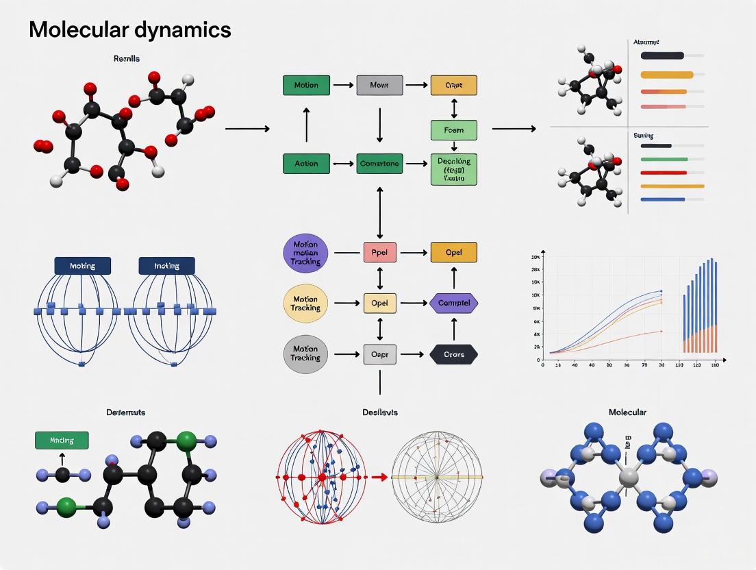
Molecular Dynamics Workflow: From Newton's Equations to Scientific Insight
The following diagram illustrates the comprehensive workflow of a molecular dynamics simulation, highlighting how Newton's laws are applied at each stage:
Molecular Dynamics Simulation Workflow
Experimental Protocols: Practical Implementation for Research Applications
Standard Molecular Dynamics Protocol for Protein-Ligand Systems
System Preparation:
- Obtain initial coordinates for the protein and ligand from experimental structures (Protein Data Bank) or homology modeling.
- Parameterize the ligand using quantum chemical calculations (GAUSSIAN, ORCA) or force field toolkits (CGenFF, ACPYPE).
- Solvate the system in a water box (TIP3P, TIP4P water models) with dimensions ensuring at least 10 Å between the protein and box edges.
- Add counterions to neutralize system charge using Monte Carlo ion placement methods.
Energy Minimization:
- Perform steepest descent minimization for 5,000 steps to remove steric clashes.
- Continue with conjugate gradient minimization until the energy convergence criterion is met (force tolerance < 1000 kJ/mol/nm).
- Verify minimized structure by analyzing potential energy and maximum force components.
System Equilibration:
- Conduct NVT equilibration for 100 ps with position restraints on heavy atoms (force constant 1000 kJ/mol/nm²) at 300 K using the Berendsen thermostat.
- Perform NPT equilibration for 100-500 ps with semi-isotropic pressure coupling at 1 bar using the Parrinello-Rahman barostat.
- Gradually release position restraints during additional NPT equilibration until all restraints are removed.
Production Simulation:
- Run unrestrained MD simulation for timescales appropriate to the biological process (typically 100 ns to 1 μs).
- Use a 2-fs time step with bonds to hydrogen atoms constrained using LINCS.
- Employ Particle Mesh Ewald method for long-range electrostatics with 10 Å real-space cutoff.
- Save atomic coordinates every 10-100 ps for subsequent analysis.
Advanced Sampling Methods for Enhanced Conformational Exploration
For processes with high energy barriers or rare events, standard MD protocols may be insufficient. Advanced sampling techniques include:
Umbrella Sampling: Apply harmonic biases along a reaction coordinate to sample high-energy regions, then reconstruct the free energy surface using the Weighted Histogram Analysis Method (WHAM).
Metadynamics: Add history-dependent Gaussian potentials along collective variables to discourage revisiting of conformational space, effectively filling energy wells to facilitate barrier crossing.
Replica Exchange MD (REMD): Run parallel simulations at different temperatures (or Hamiltonian parameters), allowing periodic exchange of configurations between replicas according to the Metropolis criterion, enhancing conformational sampling.
Research Reagent Solutions: Essential Components for Molecular Dynamics
Table 3: Essential Research Reagents and Computational Tools for Molecular Dynamics
| Reagent/Tool | Function | Application Context | Example Implementations |
|---|---|---|---|
| Force Fields | Define potential energy functions | Parameterize interatomic interactions | CHARMM, AMBER, OPLS, GROMOS |
| Water Models | Solvent representation | Biomolecular solvation environment | TIP3P, TIP4P, SPC, TIP5P |
| Integration Algorithms | Solve equations of motion | Propagate system through time | Verlet, Velocity Verlet, Leap-frog |
| Enhanced Sampling Methods | Accelerate rare events | Overcome energy barriers | Umbrella Sampling, Metadynamics, REMD |
| Analysis Tools | Extract properties from trajectories | Calculate observables and statistics | MDAnalysis, GROMACS tools, VMD |
| Quantum Chemistry Software | Parameter development | Derive force field parameters | GAUSSIAN, ORCA, NWChem |
Applications in Drug Development and Biomedical Research
Molecular dynamics simulations leveraging Newton's equations have become indispensable tools in modern drug discovery and development pipelines. Recent advances highlighted in comprehensive reviews demonstrate how MD simulations provide critical insights for polymer design in pharmaceutical formulations and drug delivery systems [3]. Key applications include:
Drug-Target Binding Mechanisms: MD simulations enable researchers to characterize the binding pathways of small molecule drugs to their protein targets, providing atomic-level details of binding kinetics and thermodynamics that inform structure-based drug design.
Membrane Permeability Prediction: Simulations of drug molecules crossing lipid bilayers yield permeability coefficients that correlate with experimental measurements, aiding in the optimization of drug candidates for improved bioavailability.
Protein Conformational Changes: Long-timescale simulations capture large-scale protein rearrangements that often play critical roles in biological function and drug action, including allosteric regulation and activation mechanisms.
Polymer-Based Drug Delivery Systems: As highlighted in recent literature, MD techniques enable the rational design of polymers for controlled drug release by modeling polymer-drug interactions, degradation kinetics, and release mechanisms at the atomic scale [3].
The integration of machine learning approaches with molecular dynamics represents a frontier in the field, with potential to dramatically accelerate sampling and improve prediction accuracy for complex biological systems relevant to drug development.
Validation and Analysis: Connecting Simulation to Experimental Data
Trajectory Analysis Methods
The validation of molecular dynamics simulations requires rigorous comparison with experimental data through comprehensive trajectory analysis:
Structural Properties:
- Root Mean Square Deviation (RMSD) to assess structural stability
- Root Mean Square Fluctuation (RMSF) to identify flexible regions
- Radius of gyration to monitor global compaction
Dynamic Properties:
- Mean Square Displacement (MSD) for diffusion coefficient calculation
- Velocity autocorrelation functions for spectral density
- Hydrogen bond lifetime analysis for interaction stability
Energetic Properties:
- Potential energy time series for stability assessment
- Free energy calculations using umbrella sampling or metadynamics
- Binding energy estimation through MM/PBSA or MM/GBSA methods
Statistical Mechanics Connection
Molecular dynamics simulations provide a bridge between Newton's classical equations and statistical mechanics through the ergodic hypothesis, which states that the time average of a property along an MD trajectory equals the ensemble average. This connection enables the calculation of thermodynamic observables:
[ \langle A \rangle = \lim{T \to \infty} \frac{1}{T} \int0^T A(t)dt ]
where ( A ) is any observable property, and the angle brackets denote the ensemble average. This fundamental relationship allows researchers to extract experimentally relevant thermodynamic quantities from atomic-level simulations based on Newtonian mechanics.
From its formulation in the 17th century to its current application in cutting-edge molecular simulations, Newton's fundamental equation of motion continues to provide the theoretical foundation for understanding and predicting physical behavior across scales. The transformation of ( \mathbf{F} = m\mathbf{a} ) into a computational algorithm for tracking atomic trajectories represents one of the most successful applications of classical physics in modern scientific research. As molecular dynamics methodologies continue to evolve, particularly through integration with machine learning approaches and advances in high-performance computing [3], Newton's laws remain firmly at the core of these developments. For researchers in drug development and biomedical sciences, mastering the principles and applications outlined in this guide provides the necessary foundation to leverage molecular dynamics simulations for investigating complex biological processes, designing novel therapeutics, and advancing scientific knowledge at the atomic scale.
In molecular dynamics (MD) simulations, the precise tracking of atomic motion relies on the fundamental laws of classical mechanics, where the forces acting on every atom determine their trajectories and velocities. At the heart of this computational methodology lies the force field, a mathematical model that describes the potential energy of a system as a function of the nuclear coordinates. Force fields serve as the critical link between atomic structure and molecular behavior, enabling the prediction of physicochemical properties from the atomistic level upward. Their accuracy is paramount, as the force field directly governs the simulated evolution of the molecular system, making it a cornerstone of research in structural biology, materials science, and computer-aided drug design [5] [6].
This technical guide details the core principles of force fields, their functional forms, parameterization strategies, and application within MD workflows. The content is framed to support a broader thesis on the basic principles of molecular dynamics atomic motion tracking research, providing researchers and drug development professionals with a comprehensive overview of the tools and methodologies essential for rigorous simulation studies.
Fundamental Concepts and Classification
A force field is a computational model composed of a functional form and a corresponding set of parameters. The functional form is a collection of mathematical functions that describe how the potential energy of a system depends on the positions of its atoms. The parameters are the numerical values that define the specific characteristics of different atom types and their interactions [5]. In MD simulations, the force on each particle is derived as the negative gradient of this potential energy with respect to the particle's coordinates [5].
Force fields can be systematically classified based on several key attributes, as illustrated in the ontology below [7].
The primary classification of force fields is based on their modeling approach and level of detail:
- Modeling Approach: Component-specific force fields are developed to describe a single, specific substance (e.g., a particular water model), offering high accuracy for that substance at the cost of transferability. In contrast, transferable force fields are constructed from building blocks (e.g., atom types or functional groups) that can be applied to a wide range of molecules, providing broad applicability [5] [7]. This guide focuses predominantly on transferable force fields, which form the basis for most biomolecular simulations.
- Level of Detail: The model resolution is a key differentiator.
- All-Atom: Explicitly represents every atom in the system, including hydrogen atoms. This provides the highest level of detail but is computationally demanding [5] [8].
- United-Atom: Treats hydrogen atoms attached to carbon (e.g., in methyl or methylene groups) as a single interaction center with the carbon atom. This reduces the number of particles and increases computational efficiency [5] [7].
- Coarse-Grained: Groups multiple atoms into a single "bead" or interaction site, sacrificing chemical details to simulate larger systems and longer timescales [5] [6].
Mathematical Formulation of Force Fields
The total potential energy ( E_{\text{total}} ) in a typical additive force field is a sum of bonded and nonbonded interaction terms [5] [8] [6]:
[ E{\text{total}} = E{\text{bonded}} + E_{\text{nonbonded}} ]
Where the bonded energy is further decomposed as:
[ E{\text{bonded}} = E{\text{bond}} + E{\text{angle}} + E{\text{dihedral}} ]
And the nonbonded energy is given by:
[ E{\text{nonbonded}} = E{\text{electrostatic}} + E_{\text{van der Waals}} ]
The following sections detail the standard functional forms for each term.
Bonded Interactions
Bonded interactions describe the energy associated with the covalent bond structure of the molecule.
Bond Stretching: The energy required to stretch or compress a chemical bond from its equilibrium length is typically modeled by a harmonic potential, analogous to a spring obeying Hooke's law [5] [6]: [ E{\text{bond}} = \sum{\text{bonds}} \frac{k{ij}}{2}(l{ij} - l{0,ij})^2 ] Here, ( k{ij} ) is the force constant, ( l{ij} ) is the instantaneous bond length, and ( l{0,ij} ) is the equilibrium bond length between atoms ( i ) and ( j ). For a more realistic description that allows for bond dissociation, the Morse potential can be used, though it is computationally more expensive [5].
Angle Bending: The energy penalty for distorting the angle between three bonded atoms from its equilibrium value is also commonly described by a harmonic function [8] [6]: [ E{\text{angle}} = \sum{\text{angles}} \frac{k{\theta{ijk}}}{2}(\theta{ijk} - \theta{0,ijk})^2 ] Here, ( k{\theta{ijk}} ) is the angle force constant, ( \theta{ijk} ) is the instantaneous angle, and ( \theta{0,ijk} ) is the equilibrium angle formed by atoms ( i ), ( j ), and ( k ).
Dihedral Torsions: This term describes the energy associated with rotation around a central bond connecting four atoms. It is typically modeled by a periodic cosine series [5] [6]: [ E{\text{dihedral}} = \sum{\text{dihedrals}} k{\chi{ijkl}} (1 + \cos(n\chi{ijkl} - \delta)) ] Here, ( k{\chi{ijkl}} ) is the dihedral force constant, ( n ) is the multiplicity (defining the number of energy minima in a 360° rotation), ( \chi{ijkl} ) is the dihedral angle, and ( \delta ) is the phase angle.
Improper Torsions: These terms are not true torsions but are used to enforce out-of-plane bending, such as maintaining the planarity of aromatic rings and other conjugated systems [5]. Their functional form can vary between force fields.
Cross-terms: Some advanced force fields include cross-terms that account for coupling between different internal coordinates, such as the influence of bond stretching on angle bending [8].
Nonbonded Interactions
Nonbonded interactions occur between atoms that are not directly bonded or are separated by more than three covalent bonds. They are computationally intensive as they must be calculated for a vast number of atom pairs.
Van der Waals Forces: These short-range forces account for attractive dispersion and repulsive Pauli exclusion interactions. The most common model is the Lennard-Jones (LJ) 12-6 potential [5] [6]: [ E{\text{van der Waals}} = \sum{i \neq j} \epsilon{ij} \left[ \left( \frac{R{\min, ij}}{r{ij}} \right)^{12} - 2 \left( \frac{R{\min, ij}}{r{ij}} \right)^6 \right] ] Here, ( \epsilon{ij} ) is the well depth, ( R{\min, ij} ) is the distance at which the potential is minimum, and ( r{ij} ) is the distance between atoms ( i ) and ( j ). The ( r^{-12} ) term describes repulsion, while the ( r^{-6} ) term describes attraction.
Electrostatic Interactions: These long-range forces arise from the attraction between partial charges on different atoms. They are modeled using Coulomb's law [5] [6]: [ E{\text{electrostatic}} = \sum{i \neq j} \frac{qi qj}{4\pi\varepsilon0 r{ij}} ] Here, ( qi ) and ( qj ) are the partial charges on atoms ( i ) and ( j ), ( r{ij} ) is their separation, and ( \varepsilon0 ) is the vacuum permittivity. The accurate assignment of atomic partial charges is critical, as this term often dominates the potential energy, especially in polar systems and ionic compounds [5].
Table 1: Summary of Standard Force Field Potential Energy Functions
| Interaction Type | Standard Functional Form | Key Parameters |
|---|---|---|
| Bond Stretching | ( E = \frac{k{ij}}{2}(l{ij} - l_{0,ij})^2 ) | Force constant ( k{ij} ), equilibrium length ( l{0,ij} ) |
| Angle Bending | ( E = \frac{k{\theta{ijk}}}{2}(\theta{ijk} - \theta{0,ijk})^2 ) | Force constant ( k{\theta{ijk}} ), equilibrium angle ( \theta_{0,ijk} ) |
| Dihedral Torsion | ( E = k{\chi{ijkl}} (1 + \cos(n\chi_{ijkl} - \delta)) ) | Force constant ( k{\chi{ijkl}} ), multiplicity ( n ), phase ( \delta ) |
| Van der Waals | ( E = \epsilon{ij} \left[ \left( \frac{R{\min, ij}}{r{ij}} \right)^{12} - 2 \left( \frac{R{\min, ij}}{r_{ij}} \right)^6 \right] ) | Well depth ( \epsilon{ij} ), minimum distance ( R{\min, ij} ) |
| Electrostatics | ( E = \frac{qi qj}{4\pi\varepsilon0 r{ij}} ) | Partial atomic charges ( qi ), ( qj ) |
Force Field Parameterization
Parameterization is the process of determining the numerical values for the force field parameters (e.g., ( l{0,ij} ), ( k{ij} ), ( q_i )), and it is crucial for the accuracy and reliability of the model [5]. This process involves several key concepts and methodologies.
The Concept of Atom Typing
A foundational principle in transferable force fields is atom typing. Instead of treating every atom of an element as identical, atoms are assigned a "type" based on their element, hybridization state, and local chemical environment [8]. For example, an oxygen atom in a carbonyl group and an oxygen atom in a hydroxyl group would be assigned different atom types with distinct parameters [5]. This allows the force field to more accurately capture the physics of different chemical functionalities.
Parameterization Strategies and Workflows
The parameters for a force field can be derived from experimental data, high-level quantum mechanical (QM) calculations, or a hybrid approach that combines both [5] [9].
- Quantum Mechanical Data: Ab initio QM calculations can provide high-quality data on molecular geometries, conformational energies, and electrostatic potentials. This data is used to fit parameters for bonded terms (bond lengths, angles, torsions) and to assign partial atomic charges, for example, by fitting to the electrostatic potential (ESP) [5] [6].
- Experimental Data: Macroscopic experimental properties, such as liquid densities, enthalpies of vaporization, and free energies of solvation, are used to refine parameters, particularly for nonbonded interactions (Lennard-Jones and electrostatic terms) [5] [9]. This ensures the force field reproduces key bulk properties.
Modern parameterization often employs systematic, automated optimization algorithms. A prominent example is the ForceBalance method, which optimizes parameters by minimizing a weighted objective function that quantifies the difference between simulated properties and reference data (both QM and experimental) [9]. This method can efficiently handle parameter interdependencies and uses thermodynamic fluctuation formulas to compute parametric derivatives of properties without running multiple expensive simulations [9].
The following diagram illustrates a generalized automated parameterization workflow.
Advanced Force Field Types
While traditional "additive" force fields use fixed partial charges, more advanced models are being developed to address their limitations.
Polarizable Force Fields: A key limitation of additive force fields is the lack of explicit electronic polarizability—the ability of a molecule's electron cloud to distort in response to its local electric field [6]. Additive force fields attempt to account for this implicitly by using enhanced, "effective" charges, but this approach fails in environments of differing polarity (e.g., when a ligand moves from aqueous solution to a protein binding pocket) [6]. Polarizable force fields explicitly model this response, leading to a more physical representation. One common approach is the Drude oscillator model (or induced dipole model), where a charged dummy particle (Drude particle) is attached to each atom by a spring, allowing the dipole moment to fluctuate [6]. These force fields show improved accuracy in simulating ion permeation, protein-ligand binding, and interactions at lipid-water interfaces [6].
Reactive Force Fields: Most standard force fields cannot model chemical reactions because their harmonic bond potentials do not allow for bond breaking and formation. Reactive force fields (such as ReaxFF) address this by using bond-order formalisms, where the strength of a bond and its associated parameters depend on the local geometry, enabling dynamic changes in connectivity during a simulation [5] [10].
Machine-Learned Force Fields (MLFFs): MLFFs represent a paradigm shift. They use machine learning models (e.g., neural networks) trained directly on high-quality QM data to predict the potential energy and forces of a system [11]. The VASP software, for instance, implements on-the-fly MLFFs where a Bayesian-learning algorithm is used during an MD simulation. If the uncertainty in the force prediction exceeds a threshold, an ab initio QM calculation is triggered, and the new data is used to update the MLFF, iteratively improving its accuracy and reliability [11]. This approach aims to achieve the accuracy of QM at a fraction of the computational cost.
Table 2: Comparison of Additive and Advanced Force Fields
| Feature | Additive (Non-polarizable) | Polarizable (e.g., Drude) | Machine-Learned (MLFF) |
|---|---|---|---|
| Electrostatics | Fixed partial atomic charges | Fluctuating dipoles (inducible) | Learned from QM electron density |
| Bond Breaking | Not possible (harmonic bonds) | Not typically possible | Possible (depending on training) |
| Computational Cost | Low | ~5x higher than additive [9] | Variable (high training, lower inference) |
| Primary Data Source | Experiment & QM | High-level QM | Ab initio QM calculations |
| Key Advantage | Speed, stability for MD | Physical accuracy in different environments | High QM-level accuracy |
| Key Challenge | Implicit treatment of polarization | Parameter complexity, cost | Transferability, computational demand |
Essential Research Reagents and Software
Table 3: Essential Resources for Force Field Application and Development
| Resource / Tool | Type | Primary Function |
|---|---|---|
| CHARMM General FF (CGenFF) [6] | Transferable Additive Force Field | Parameters for drug-like molecules compatible with CHARMM biomolecular FFs. |
| General AMBER FF (GAFF) [6] | Transferable Additive Force Field | Parameters for small organic molecules compatible with AMBER biomolecular FFs. |
| Merck Molecular FF (MMFF94) [8] | Transferable Additive Force Field | Well-parameterized for small, drug-like molecules; often used for ligand geometry optimization. |
| OPLS-AA/OPLS5 [6] [12] | Transferable Additive/Polarizable FF | Widely used force field for proteins, organic liquids, and drug discovery (e.g., in FEP+). |
| ForceBalance [9] | Parameterization Software | Automated, systematic tool for optimizing force field parameters against reference data. |
| NIST IPR [10] | Force Field Database | Repository of interatomic potentials, particularly for metals, semiconductors, and ceramics. |
| AnteChamber [6] | Parameter Assignment Tool | Automated tool for generating GAFF/AMBER topologies and parameters for new molecules. |
| ParamChem [6] | Parameter Assignment Tool | Web-based service for generating CGenFF/CHARMM topologies and parameters for new molecules. |
| OpenMM [9] [7] | MD Simulation Engine | High-performance, GPU-accelerated toolkit for running molecular dynamics simulations. |
Protocol for Force Field Parameterization
The following is a generalized protocol for parameterizing a new molecule within an existing transferable force field framework, reflecting modern best practices [9] [6].
Initial Structure and Atom Typing:
- Obtain a 3D molecular structure from a database or via quantum chemical geometry optimization.
- Use an automated tool (e.g., ParamChem for CGenFF or AnteChamber for GAFF) to assign atom types based on the chemical structure and connectivity.
Parameter Assignment:
- The automated tool will assign initial parameters for all bonds, angles, dihedrals, and partial charges. These are typically based on analogy to existing parameters in the force field's database.
- Carefully review the assigned parameters, paying special attention to dihedral terms and partial charges, as these are often the greatest sources of error and require refinement.
Target Data Selection:
- Gather high-quality target data for optimization. This should include:
- Quantum Mechanical Data: Conformational energies, torsional energy profiles, and electrostatic potential (ESP) maps derived from QM calculations (e.g., at the DFT or MP2 level of theory).
- Experimental Data: Liquid properties such as density (( \rho )) and enthalpy of vaporization (( \Delta H_{vap} )), if available.
- Gather high-quality target data for optimization. This should include:
Iterative Optimization and Validation:
- Employ an optimization system like ForceBalance. The objective function ( O(p) ) to be minimized is often of the form: [ O(p) = \sum{i} wi \left( \frac{X{i}^{\text{sim}}(p) - X{i}^{\text{ref}}}{\sigmai} \right)^2 + R(p) ] where ( p ) are the parameters, ( X{i}^{\text{sim}} ) and ( X{i}^{\text{ref}} ) are the simulated and reference properties, ( wi ) is a weight, ( \sigma_i ) is the uncertainty in the reference data, and ( R(p) ) is a regularization term to prevent overfitting [9].
- The optimization loop involves running simulations, comparing results to target data, and updating parameters until convergence is achieved.
- Crucially, validate the final parameters against a set of experimental data that was not included in the training set (e.g., free energy of solvation, viscosity, or NMR observables) to ensure the model's transferability and robustness.
Force fields are the fundamental engine that powers molecular dynamics research, translating atomic coordinates into forces and energies that drive simulated motion. The choice and quality of the force field directly determine the physical realism and predictive power of the simulation. The field is dynamic, with ongoing research focused on improving accuracy through polarizable models, extending applicability through systematic parameterization tools like ForceBalance, and pushing the boundaries of efficiency and accuracy with machine-learned potentials. For researchers engaged in atomic motion tracking, a deep understanding of force field construction, limitations, and application protocols is not merely beneficial—it is essential for producing rigorous, reliable, and interpretable scientific results.
The genesis of computational physics and the theoretical foundations of modern molecular dynamics (MD) share a common origin: a numerical experiment on a nonlinear string conducted in 1953 by Enrico Fermi, John Pasta, Stanislaw Ulam, and Mary Tsingou [13] [14]. What began as a study of thermalization in solids has evolved into a rich field of nonlinear physics that underpins our understanding of atomic-scale dynamics in complex biomolecular systems. The Fermi-Pasta-Ulam-Tsingou (FPUT) problem represents one of the earliest uses of digital computers for mathematical research and simultaneously launched the study of nonlinear systems [13]. This historical journey from the FPUT paradox to contemporary biomolecular simulation represents more than a chronological progression; it embodies the evolution of our conceptual understanding of how energy flows and distributes in complex many-body systems—a fundamental question that remains central to interpreting MD trajectories in computational biology and drug design.
The core insight connecting these seemingly disparate fields lies in their shared physical and mathematical foundations. Both involve solving Newton's equations of motion for complex, nonlinear systems—whether for a one-dimensional string or a massive biomolecule comprising hundreds of thousands of atoms [15]. The FPUT problem revealed unexpected behavior in such systems, while modern MD simulations leverage these insights to predict the behavior of biological macromolecules with remarkable accuracy. This article traces this intellectual and technical lineage, examining how a foundational paradox in statistical mechanics paved the way for computational tools that now drive innovation in drug development and materials science.
The FPUT Problem: A Paradigm Shift in Nonlinear Physics
The Original Experiment and Its Unexpected Outcome
In the summer of 1953, Fermi, Pasta, Ulam, and Tsingou conducted computer simulations on the MANIAC computer at Los Alamos National Laboratory to study how energy distributes in a nonlinear system [13]. Their model consisted of a one-dimensional chain of 64 mass points connected by springs with nonlinear forces, governed by the discrete equation of motion:
[ m\ddot{x}j = k(x{j+1} + x{j-1} - 2xj)[1 + \alpha(x{j+1} - x{j-1})] ]
where (x_j(t)) represents the displacement of the j-th particle from its equilibrium position, and α controls the strength of the nonlinear quadratic term [13]. The team also studied cubic (β) and piecewise linear approximations.
Fermi and colleagues expected that the nonlinear interactions would cause the system to thermalize rapidly, with energy eventually distributed equally among all vibrational modes according to the equipartition theorem of statistical mechanics [13] [16]. Contrary to these expectations, their simulations revealed a remarkably different behavior: instead of thermalizing, the system exhibited complicated quasi-periodic behavior with near-recurrences to the initial state—a phenomenon now known as FPUT recurrence [13] [14]. This apparent paradox challenged fundamental assumptions about statistical mechanics and ergodicity, showing that even with nonlinear interactions, the system did not necessarily behave ergodically over computationally accessible timescales.
Mathematical Formulations and Key Concepts
The FPUT problem can be formulated through several mathematical frameworks that have proven essential for understanding its behavior:
- α-FPUT Model: Features a cubic potential, (V_α(r) = \frac{r^2}{2} + \frac{α}{3}r^3), providing asymmetric nonlinearity [16].
- β-FPUT Model: Employs a quartic potential, (V_β(r) = \frac{r^2}{2} + \frac{β}{4}r^4), creating symmetric nonlinearity [16].
- Normal Mode Transformation: The transformation ( \left( \begin{array}{c} qn \ pn \end{array} \right) = \sqrt{\frac{2}{N+1}} \sum{k=1}^N \left( \begin{array}{c} Qk \ P_k \end{array} \right) \sin \left( \frac{nk\pi}{N+1} \right) ) diagonalizes the harmonic lattice but leaves coupling terms in the presence of nonlinearities [16].
- Connection to Integrable Systems: The α-FPUT model can be viewed as a truncation of the Toda lattice, which has the potential (V{\text{Toda}}(r) = V0[e^{λr} - 1 - λr]) and is completely integrable [16].
Table 1: Key Models in the FPUT Paradigm
| Model | Potential Energy | Nonlinearity Type | Key Properties |
|---|---|---|---|
| Harmonic | (\frac{r^2}{2}) | Linear | Exactly solvable, no energy sharing |
| α-FPUT | (\frac{r^2}{2} + \frac{α}{3}r^3) | Cubic, asymmetric | Truncation of Toda lattice |
| β-FPUT | (\frac{r^2}{2} + \frac{β}{4}r^4) | Quartic, symmetric | Not a truncation of integrable system |
| Toda Lattice | (V_0(e^{λr} - 1 - λr)) | Exponential | Completely integrable, soliton solutions |
The discovery of the FPUT recurrence phenomenon prompted six decades of research into nonlinear systems, leading to groundbreaking developments including soliton theory [13] [17], chaos theory [14], and wave turbulence theory [14]. The connection to the Korteweg-de Vries (KdV) equation through the continuum limit was particularly significant, as it revealed the existence of soliton solutions—special waves that maintain their shape while propagating [13].
From Nonlinear Strings to Atomic Simulations: The Emergence of Molecular Dynamics
Fundamental Principles of Molecular Dynamics
Molecular dynamics simulations track the motion of individual atoms and molecules over time by numerically solving Newton's equations of motion for complex many-body systems [18]. Often described as a "microscope with exceptional resolution," MD provides atomic-scale insights into physical and chemical processes that are difficult or impossible to observe experimentally [18]. The core principles of MD directly extend the conceptual framework established by the FPUT problem—both involve understanding how energy distributes and dynamics evolve in nonlinear systems with many degrees of freedom.
The mathematical foundation of MD relies on Newton's second law applied to each atom i in a system:
[ mi \frac{d^2\mathbf{r}i}{dt^2} = \mathbf{F}i = -\nablai U(\mathbf{r}1, \mathbf{r}2, ..., \mathbf{r}_N) ]
where (mi) is the atomic mass, (\mathbf{r}i) is the position vector, (\mathbf{F}_i) is the force acting on the atom, and (U) is the potential energy function describing interatomic interactions [18]. This system of coupled differential equations is structurally similar to the FPUT lattice equations, though dramatically more complex in dimensionality and interaction potentials.
The MD Workflow: From Initial Conditions to Analysis
The standard workflow for MD simulations comprises several well-defined stages, each with its own technical considerations:
Initial Structure Preparation: The process begins with preparing the initial atomic coordinates, often obtained from crystallographic databases (e.g., Protein Data Bank for biomolecules) or built from scratch for novel materials [18].
System Initialization: Initial velocities are assigned to all atoms, typically sampled from a Maxwell-Boltzmann distribution corresponding to the desired simulation temperature [18].
Force Calculation: This computationally intensive step determines forces between atoms based on interatomic potentials. Modern approaches include machine learning interatomic potentials (MLIPs) trained on quantum chemistry data [18] [15].
Time Integration: Using algorithms like Verlet or leap-frog, the equations of motion are solved numerically to update atomic positions and velocities. Time steps typically range from 0.5 to 1.0 femtoseconds to accurately capture the fastest atomic motions [18].
Trajectory Analysis: The resulting time-series data of atomic positions and velocities are analyzed to extract physically meaningful insights, such as structural properties, dynamics, and thermodynamic quantities [18].
Figure 1: Molecular Dynamics Simulation Workflow
Technical Parallels: FPUT Concepts in Modern Biomolecular Simulation
Metastable States and the Timescale Problem
The FPUT problem introduced the concept of metastable states—quasi-stationary states that delay the approach to thermal equilibrium [16]. In the FPUT context, this metastable state is characterized by the system remaining localized in mode space for unexpectedly long durations, exhibiting recurrences to the initial condition instead of rapid thermalization [16]. This phenomenon finds direct parallels in biomolecular simulations, where systems often become trapped in metastable conformational states for timescales that challenge computational resources.
In FPUT systems, researchers have quantified the lifetime of metastable states ((t_m)) using measures like spectral entropy (η) to track the distance from equipartition [16]. Similarly, in biomolecular simulations, analysis techniques like Markov state models are employed to identify and characterize metastable states and transitions between them. The FPUT recurrence timescale has been shown to follow power-law scaling with system parameters [16], analogous to the timescale problems encountered in simulating rare events in biomolecular dynamics, such as protein folding or ligand unbinding.
Energy Transfer and Equipartition
The original FPUT study fundamentally questioned the applicability of the equipartition theorem to nonlinear systems—a concern that remains relevant in modern MD. In FPUT systems, energy initially concentrated in low-frequency modes unexpectedly fails to distribute equally among all modes over computationally accessible timescales [13] [16]. In biomolecular simulations, similar issues manifest as difficulties in achieving proper thermalization or as non-ergodic behavior where certain regions of phase space are inadequately sampled.
Table 2: Quantitative Measures in FPUT and MD Simulations
| Property | FPUT Context | Biomolecular MD Context | Measurement Technique |
|---|---|---|---|
| Energy Distribution | Mode energies (E_k) | Potential energy terms | Normal mode analysis, energy decomposition |
| Timescales | Recurrence time (T_R) | Correlation times, folding times | Autocorrelation functions, Markov models |
| Metastability | Spectral entropy η | Free energy landscapes | Principal component analysis, MSMs |
| Thermalization | Approach to equipartition | Temperature equilibration | Thermostat coupling, kinetic temperature |
| Nonlinearity | α, β parameters | Force field anharmonic terms | Potential energy surface analysis |
Research has shown that the route to thermalization in FPUT systems occurs through specific resonance mechanisms, particularly four-wave and six-wave resonances as described by wave turbulence theory [14] [16]. These findings have implications for understanding how energy flows in biomolecules—for instance, how vibrational energy from ATP hydrolysis might propagate through specific channels in enzymes or how allosteric signals are transmitted through protein structures.
The Modern Biomolecular Simulation Toolkit
Advanced Force Fields and Nuclear Quantum Effects
Contemporary biomolecular simulations rely on sophisticated force fields that accurately represent interatomic interactions. The Arrow force field represents a significant advancement as it is parameterized entirely using quantum mechanical calculations without reliance on experimental data [15]. This ab initio approach achieves chemical accuracy (∼0.5 kcal/mol) for neutral organic compounds in the liquid phase, enabling reliable prediction of properties like solvation free energies and partition coefficients [15].
Key innovations in modern force fields include:
- Polarizability: Modeling how atomic charge distributions respond to their environment, enabling transferability from gas to liquid phase [15].
- Charge Penetration Effects: Accounting for the overlap of electron clouds between nearby atoms [15].
- Anisotropic Atomic Shapes: Representing the directional nature of atomic interactions beyond simple spheres [15].
- Nuclear Quantum Effects (NQE): Modeling quantum mechanical behavior of atomic nuclei using techniques like ring polymer MD, which is particularly important for simulating hydrogen bonding and proton transfer reactions [15].
The inclusion of NQE has proven essential for achieving accurate free energy predictions, bringing computational results into excellent agreement with experimental measurements for various molecular systems [15].
Specialized Simulation Techniques
Modern biomolecular simulation employs specialized methodologies to address specific scientific questions:
- Enhanced Sampling Methods: Techniques like metadynamics, replica exchange, and accelerated MD overcome timescale limitations by promoting transitions between metastable states [19].
- Free Energy Calculations: Methods such as thermodynamic integration and free energy perturbation provide quantitative predictions of binding affinities and partition coefficients crucial for drug design [15].
- Multiscale Simulations: QM/MM approaches combine quantum mechanical accuracy for a reaction center with molecular mechanics efficiency for the surrounding environment [15].
- Machine Learning Potentials: MLIPs trained on quantum chemistry datasets enable accurate simulations of complex material systems that were previously computationally prohibitive [18].
Figure 2: Advanced Biomolecular Simulation Techniques
Applications in Drug Discovery and Materials Science
Biomolecular Applications
Molecular dynamics simulations have become indispensable tools in pharmaceutical research and development. A recent study on uncoupling protein 1 (UCP1) illustrates the power of MD simulations to elucidate biological mechanisms at atomic resolution [20]. Researchers employed all-atom MD simulations combined with membrane conductance measurements and site-directed mutagenesis to identify novel pathways for fatty acid anion translocation at the UCP1 protein-lipid interface [20]. This work revealed how key arginine residues (R84 and R183) form stable complexes with fatty acid anions, facilitating transport essential for mitochondrial thermogenesis.
Additional biomolecular applications include:
- Protein-Ligand Binding: Predicting binding modes and affinities for drug candidates [15].
- Membrane Permeability: Studying how molecules traverse lipid bilayers [20].
- Allosteric Regulation: Identifying long-range communication pathways in proteins [18].
- Drug Solubility and Formulation: Predicting solvation free energies and partition coefficients [15].
Materials and Industrial Applications
Beyond biomedicine, MD simulations drive innovation in materials science and industrial processes:
- Ion Conductivity: Analyzing diffusion mechanisms in solid electrolytes for battery applications [18].
- Mechanical Properties: Computing stress-strain relationships and predicting yield strength in structural materials [18].
- Phase Behavior: Studying crystallization, glass formation, and phase transitions [18].
- Nanomaterial Design: Understanding self-assembly and properties of nanostructured materials [18].
Table 3: Key Research Reagents and Computational Tools
| Tool Category | Specific Examples | Function | Relevance to FPUT Concepts |
|---|---|---|---|
| Force Fields | AMBER, CHARMM, Arrow FF | Define interatomic potentials | Nonlinear interaction potentials |
| Analysis Software | MDAnalysis, VMD, GROMACS | Process MD trajectories | Mode analysis, recurrence quantification |
| Enhanced Sampling | PLUMED, MetaD | Accelerate rare events | Metastable state characterization |
| Quantum Calibration | DFT, CCSD(T) | Generate training data for ML potentials | Connection to fundamental physics |
| Structure Databases | PDB, Materials Project | Provide initial configurations | Initial condition dependence |
Future Perspectives: FAIR Principles and Next-Generation Simulations
The biomolecular simulation community is currently embracing the FAIR principles (Findability, Accessibility, Interoperability, and Reusability) to transform research practices [17]. A recent initiative signed by 128 researchers advocates for creating a comprehensive database for MD simulation data, aiming to democratize the field and enhance the impact of simulations on life science research [17]. This development represents a natural evolution from the early days of computational science exemplified by the FPUT problem—where each simulation was a unique, barely reproducible effort—toward a future of open, collaborative, and cumulative science.
Future directions in biomolecular simulation include:
- Integration with AI and Machine Learning: Leveraging deep learning for structure prediction, force field development, and analysis of high-dimensional data [18] [15].
- Exascale Computing: Harnessing next-generation supercomputers to access biologically relevant timescales and system sizes [17].
- Automated Workflows: Developing streamlined protocols for non-experts to perform reliable simulations [18].
- Experimental Validation and Integration: Strengthening connections between simulation predictions and experimental observables [20].
The intellectual journey from the Fermi-Pasta-Ulam problem to modern biomolecular simulation demonstrates how fundamental research into basic principles of nonlinear systems can yield transformative practical applications. What began as a numerical experiment on a simple nonlinear string has evolved into a sophisticated computational framework that provides unprecedented insights into biological processes at atomic resolution. The FPUT problem introduced conceptual challenges—metastable states, limited ergodicity, and the importance of nonlinear resonances—that continue to resonate in contemporary MD simulations of biomolecules.
As simulation methodologies advance, incorporating more accurate physical models, machine learning approaches, and FAIR data principles, the legacy of the FPUT problem endures in our continued fascination with understanding how energy flows and distributes in complex systems. This historical continuum, stretching from Fermi's vision to today's cutting-edge biomolecular simulations, underscores the enduring value of foundational research in shaping the scientific tools of tomorrow—tools that are increasingly essential for addressing challenges in drug discovery, materials science, and beyond.
The ergodic hypothesis is a foundational principle in statistical mechanics that links the microscopic dynamics of atoms and molecules to the macroscopic thermodynamic properties we observe. This hypothesis states that, over long periods, the time a system spends in any region of its phase space is proportional to the volume of that region [21]. In practical terms, this means that all accessible microstates with the same energy are equally probable over a sufficiently long timeframe. For researchers tracking atomic motion in molecular dynamics, this principle provides the crucial justification for using statistical ensembles to predict molecular behavior. The profound implication is that the time average of a property for a single system equates to the average of that property across an ensemble of identical systems at one instant [22]. This equivalence enables scientists to extract meaningful thermodynamic information from the chaotic motion of individual atoms, forming the theoretical bedrock for molecular dynamics simulations and atomic motion tracking research.
Within the context of molecular dynamics and atomic motion tracking, ergodicity provides the mathematical justification for why simulating a single system over time can yield the same statistical properties as studying multiple identical systems simultaneously. This is particularly vital in drug development and materials science, where researchers must predict bulk behavior from limited simulation data. The hypothesis, first proposed by Boltzmann in the late 19th century, remains intensely relevant today as advanced experimental techniques like ultrafast X-ray scattering and computational methods enable unprecedented tracking of individual atomic motions [23] [24].
Core Theoretical Framework
Time Averages vs. Ensemble Averages
In statistical mechanics, two distinct averaging methods provide the connection between microscopic dynamics and macroscopic observables:
Time Average: The average value of a property obtained from tracking a single system over an extended period. For an observable ( f ), this is mathematically expressed as ( \overline{f} = \lim{T \to \infty} \frac{1}{T} \int0^T f(x(t)) dt ) [22]. In molecular dynamics research, this corresponds to tracking atomic positions and energies throughout a simulation trajectory.
Ensemble Average: The average value of a property across a theoretical collection (ensemble) of identical systems at a specific moment, expressed as ( \langle f \rangle = \int f(x) \rho(x) dx ), where ( \rho(x) ) represents the probability density in phase space [22]. This approach is used when analyzing multiple parallel simulations or experimental replicates.
The ergodic hypothesis fundamentally asserts the equivalence of these two averages: ( \overline{f} = \langle f \rangle ) [22]. For molecular dynamics researchers, this equivalence means that sufficiently long simulations of a single molecular system can provide the same statistical information as studying multiple identical systems simultaneously—a crucial justification for the methodology underpinning computational drug design and materials science.
Mathematical Formulation
The mathematical rigor behind ergodicity is established through several key theorems:
Birkhoff's Ergodic Theorem (1931) provides the formal foundation, stating that for measure-preserving dynamical systems, the time average of a function along trajectories equals the space average for almost all starting points [22]. Mathematically, this is expressed as ( \lim{T \to \infty} \frac{1}{T} \int0^T f(T^t x) dt = \int f(x) d\mu(x) ), where ( \mu ) represents the invariant measure.
Liouville's Theorem further supports this framework by establishing that for Hamiltonian systems, the local density of microstates following a particle path through phase space remains constant [21]. This conservation property implies that if microstates are uniformly distributed initially, they remain so over time, providing a mathematical basis for the equal probability of accessible states.
The following diagram illustrates the conceptual relationship between these averaging methods in ergodic systems:
Ergodicity in Molecular Dynamics and Atomic Motion Tracking
Computational Validation Through Molecular Dynamics
Molecular Dynamics (MD) simulations serve as the primary computational tool for testing and leveraging ergodic principles in studying atomic-scale phenomena. These simulations apply Newton's laws of motion to model molecular behavior over time, with discrete time steps typically on the femtosecond scale ((10^{-15}) seconds) to capture atomic vibrations and movements [25]. The connection to ergodicity emerges through the simulation ensembles employed:
Table 1: Molecular Dynamics Ensembles and Their Relation to Ergodicity
| Ensemble Type | Conserved Quantities | Connection to Ergodicity | Primary Research Applications |
|---|---|---|---|
| NVE (Microcanonical) | Number of particles (N), Volume (V), Energy (E) | Directly corresponds to isolated systems exploring constant-energy surface; fundamental test of ergodic hypothesis | Gas-phase simulations, molecular reactions, fundamental validation studies |
| NVT (Canonical) | Number of particles (N), Volume (V), Temperature (T) | Ergodic exploration of states at fixed temperature; enables comparison with experimental conditions | Biological systems at physiological conditions, protein folding studies |
| NPT (Isothermal-Isobaric) | Number of particles (N), Pressure (P), Temperature (T) | Ergodic sampling under constant pressure and temperature; mimics common laboratory conditions | Liquid simulations, large biomolecular systems, materials design |
MD simulations validate ergodicity by demonstrating that time averages of properties like energy, pressure, and molecular conformation converge to ensemble averages over sufficiently long simulation times [25]. This convergence is particularly crucial in drug discovery, where researchers simulate drug-protein binding events and rely on ergodic sampling to ensure adequate exploration of conformational space for accurate binding affinity predictions.
Experimental Validation Through Advanced Atomic Tracking
Recent experimental breakthroughs in tracking atomic and electronic motion provide striking validation of ergodic principles:
In a landmark study at SLAC National Accelerator Laboratory, researchers used ultrafast X-ray laser pulses from the Linac Coherent Light Source (LCLS) to track the motion of a single valence electron during the dissociation of hydrogen from an ammonia molecule [23]. The experimental protocol involved:
- Sample Preparation: Creating an enclosure of high-density ammonia gas
- Photoexcitation: Using an ultraviolet laser pulse to initiate the chemical reaction
- Probing: Scattering X-rays from LCLS off electrons during the reaction
- Data Collection: Capturing the scattered X-rays to reconstruct electron positions
- Theoretical Validation: Comparing results with advanced quantum simulations
The entire process occurred within 500 femtoseconds, capturing the electron rearrangement that drives nuclear repositioning [23]. This direct observation of electron motion throughout a complete reaction pathway demonstrates the fundamental principle that molecular systems explore accessible states over time—a core requirement for ergodic behavior.
Complementary research at the Max Planck Institute has developed techniques to track atomic motion in single graphene nanoribbons using femtosecond coherent anti-Stokes Raman spectroscopy (CARS) within a scanning tunneling microscope (STM) [24]. This approach can track atomic motions on timescales as short as 70 femtoseconds, providing direct experimental observation of phase space exploration in nanoscale systems.
Ergodicity Breaking and Modern Research Applications
When Ergodicity Fails: Limitations and Broken Ergodicity
Despite its foundational status, the ergodic hypothesis has well-established limitations. Ergodicity breaking occurs when systems fail to explore their entire accessible phase space within observable timescales [21] [22]. Notable examples include:
- Glassy Systems: Structural glasses, spin glasses, and polymers exhibit extremely slow relaxation times due to complex energy landscapes with many local minima [22]. These systems remain trapped in non-equilibrium states for experimentally relevant timescales.
- Spontaneous Symmetry Breaking: Ferromagnetic materials below the Curie temperature maintain non-zero magnetization instead of exploring all possible states [21].
- Biological Systems: Large biomolecules like proteins may have folding timescales exceeding practical simulation times, leading to inadequate sampling [25].
The Fermi-Pasta-Ulam-Tsingou experiment of 1953 provided an early surprising counterexample, where a nonlinear system failed to equilibrate as expected [21]. Instead of energy equipartition, the system exhibited recurrent behavior with energy returning to initial modes—a direct violation of ergodic expectations that led to the discovery of solitons.
Implications for Drug Development and Molecular Design
For pharmaceutical researchers, understanding ergodicity and its limitations has profound practical implications:
- Drug Binding Calculations: Molecular dynamics simulations of drug-receptor interactions rely on ergodic sampling to accurately compute binding free energies. Inadequate sampling caused by ergodicity breaking can lead to inaccurate predictions of drug efficacy [25].
- Protein Folding Studies: The prediction of protein structure from sequence depends on the assumption that simulations can explore the relevant conformational space. Non-ergodic behavior in folding pathways presents significant challenges for reliable prediction [25].
- Enhanced Sampling Methods: Techniques like metadynamics and replica exchange are specifically designed to address ergodicity breaking by encouraging systems to overcome energy barriers and explore wider regions of phase space [25].
Recent computational advancements address these challenges through massive datasets like Open Molecules 2025 (OMol25), which contains over 100 million 3D molecular snapshots calculated with density functional theory [26]. This unprecedented resource, representing six billion CPU hours of computation, enables training machine learning interatomic potentials that can simulate systems with up to 350 atoms across most of the periodic table, dramatically improving ergodic sampling for complex molecular systems [26].
Experimental Protocols and Research Tools
Key Experimental Methodologies
Cutting-edge experimental techniques for tracking atomic and electronic motion employ sophisticated protocols to validate ergodic principles:
Ultrafast X-ray Scattering Protocol [23]:
- Sample Preparation: Purified molecular samples (e.g., ammonia) are prepared in high-density gas phase or thin-film environments
- Pump Laser Excitation: Ultraviolet laser pulses (typically 100-400 nm) initiate photochemical reactions with precise temporal control
- X-ray Probing: Femtosecond X-ray pulses from free-electron lasers scatter from electron clouds
- Detection: Area detectors capture diffraction patterns with single-shot capability
- Data Reconstruction: Computational algorithms transform time-resolved diffraction into real-space electron density maps
- Theoretical Comparison: Advanced quantum simulations validate interpretations of electron dynamics
STM-CARS Protocol for Atomic Tracking [24]:
- Sample Fabrication: Nanoscale materials (e.g., graphene nanoribbons) are synthesized on atomically flat substrates
- Laser Excitation: Two time-delayed broadband laser pulses generate vibrational wave packets
- STM Detection: Scanning tunneling microscope tips detect local electronic changes induced by atomic motion
- Signal Processing: Coherent anti-Stokes Raman spectroscopy extracts specific vibrational frequencies
- Atomic Reconstruction: Position-dependent signals are reconstructed into time-resolved atomic trajectories
The Scientist's Toolkit: Essential Research Reagents and Materials
Table 2: Key Research Reagents and Materials for Atomic Motion Studies
| Reagent/Material | Function in Research | Application Examples |
|---|---|---|
| Ultrafast Laser Systems | Generate femtosecond optical pulses for initiating and probing reactions | Pump-probe spectroscopy, coherent control experiments |
| X-ray Free Electron Lasers | Provide intense, femtosecond X-ray pulses for scattering experiments | Linac Coherent Light Source (LCLS), European XFEL facilities |
| Scanning Tunneling Microscopes | Atomic-scale imaging and local spectroscopy | Single-molecule studies, surface science, nanomaterial characterization |
| High-Purity Molecular Samples | Ensure reproducible experimental conditions with minimal impurities | Ammonia for dissociation studies, custom-synthesized organic molecules |
| Cryogenic Cooling Systems | Reduce thermal noise and stabilize fragile quantum states | Helium flow cryostats, closed-cycle refrigerators |
| Ultrahigh Vacuum Chambers | Create pristine environments free from contamination | Surface science experiments, single-molecule tracking |
| Quantum Chemistry Software | Perform advanced simulations for experimental interpretation | Density functional theory, ab initio molecular dynamics |
Future Directions and Research Frontiers
The integration of ergodic principles with emerging technologies is creating new research frontiers:
Machine Learning-Enhanced Molecular Dynamics leverages neural network potentials trained on massive datasets like OMol25 to achieve accurate simulations at dramatically reduced computational cost, effectively improving ergodic sampling for complex systems [26] [25]. These approaches can predict molecular properties with density functional theory accuracy while being thousands of times faster, enabling previously impossible simulations of biologically relevant systems.
Quantum-Classical Hybrid Methods combine quantum mechanical accuracy with classical simulation efficiency, particularly important for studying processes like electron transfer and catalytic reactions where electronic structure changes drive atomic motion [25]. The recent experimental tracking of single electrons during chemical reactions provides crucial validation for these approaches [23].
Single-Molecule Experimental Techniques are increasingly capable of directly testing ergodic assumptions by tracking individual molecules over extended periods, revealing molecular heterogeneity that ensemble measurements average out [22]. These techniques are particularly valuable for studying biological systems where ergodicity breaking may have functional significance.
The following diagram illustrates the integrated workflow combining computational and experimental approaches in modern ergodicity research:
As these technologies mature, the ergodic hypothesis continues to provide the conceptual framework connecting nanoscale atomic motion to macroscopic observables, enabling researchers to design more effective drugs, novel materials, and advanced biomolecules through a fundamental understanding of molecular behavior across timescales.
The fidelity of a molecular dynamics (MD) simulation is fundamentally contingent upon the integrity of its initial setup. The processes of defining accurate initial structures, applying appropriate boundary conditions, and meticulously preparing the system constitute the foundational steps that determine the physical relevance and numerical stability of the entire simulation. Within the broader context of basic principles of molecular dynamics atomic motion tracking research, a rigorous setup is paramount for distinguishing physical phenomena from computational artifacts and for generating trajectories that can be meaningfully compared with experimental data. This guide details the core methodologies and current best practices for preparing MD simulations, with a focus on protocols relevant to researchers, scientists, and drug development professionals.
The initial atomic coordinates, or the starting structure, define the system's initial conformational state. The choice and preparation of this structure directly influence the simulation's equilibration time and the biological relevance of the sampled dynamics.
- Experimental Structures: The most reliable starting points are experimentally determined structures from databases such as the Protein Data Bank (RCSB PDB) [27]. These provide atomistic details of macromolecules but often require preprocessing to add missing residues, protons, or correct for unresolved loops.
- Previous Simulations: Structures obtained from previous MD simulations can be used as starting points for new simulations, for instance, to extend simulation time or to study a different thermodynamic state. A critical consideration in such cases is the handling of molecules that may cross the periodic box boundaries in the saved structure. It is often permissible to proceed, as periodic boundary conditions will correctly map atoms that exit the box to the opposite side, but verification of the system's integrity is necessary [28].
- De Novo and Homology Models: For targets without an experimental structure, computational models built through homology modeling (e.g., with MODELLER [27]) or other de novo methods are used. These require careful validation and often extensive equilibration.
Structure Preprocessing and Energy Minimization
Raw initial structures frequently contain steric clashes or geometrically unfavorable conformations. Energy minimization (EM) is a critical preprocessing step that adjusts atomic coordinates to find a local minimum on the potential energy surface, relieving these strains before dynamics begin [29].
The energy of a molecule is a function of its nuclear coordinates, and EM algorithms iteratively adjust these coordinates to reduce the net forces on the atoms. The general update formula is: xnew = xold + correction
Common minimization algorithms include [29]:
- Steepest Descent: A robust method for initial steps that moves atoms opposite the direction of the largest gradient. It is computationally inexpensive per step but can be slow to converge.
- Conjugate Gradient: A more efficient method that incorporates information from previous steps to avoid oscillation, leading to faster convergence towards the minimum.
Table 1: Key Energy Minimization Algorithms
| Method | Principle | Computational Cost | Typical Use Case |
|---|---|---|---|
| Steepest Descent | Moves atoms against the largest force gradient | Low per step, but requires many steps | Initial minimization of poorly structured systems with steric clashes |
| Conjugate Gradient | Combines current gradient with previous search direction | Moderate per step, fewer steps than Steepest Descent | Refined minimization after initial Steepest Descent steps |
| Newton-Raphson | Uses first and second derivatives (Hessian) for high accuracy | Very high per step | Final precision minimization for small systems |
The following workflow outlines a standard procedure for obtaining and preparing an initial structure for simulation.
Boundary Conditions and System Definition
Boundary conditions (BCs) are a critical computational tool for simulating a small part of a much larger system, such as a solute in a continuous solvent, while avoiding unphysical surface effects.
Types of Boundary Conditions
- Periodic Boundary Conditions (PBC): PBCs are the most common approach in biomolecular simulations. The simulation box containing the solute and solvent is treated as a repeating unit cell in all three spatial dimensions. As a molecule moves and an atom exits the box through one face, it simultaneously re-enters the box through the opposite face [28]. This maintains a constant number of atoms and effectively models a bulk environment. Forces between atoms are calculated using minimum image conventions, where each atom interacts with the closest periodic image of every other atom.
- Isolated Boundary Conditions (IBC): Also known as vacuum or gas-phase BCs, this approach places the molecule in a vacuum without periodic images. It is less computationally intensive but ignores solvation effects, which can be critical for modeling biological processes. Its use is typically limited to gas-phase calculations or very initial testing [29].
Implementing Periodic Boundary Conditions
The following diagram illustrates the logic and consequences of applying PBCs in a molecular dynamics simulation, addressing a common challenge when using structures from previous simulations.
Integrated System Preparation Workflow
Preparing a solvated, neutralized, and energetically stable system for production MD is a multi-step process. Integrated web-based platforms like CHARMM-GUI have streamlined this workflow, providing a reproducible interface for building complex systems and generating input files for numerous simulation packages like GROMACS, AMBER, NAMD, and OpenMM [30]. The general protocol involves:
- Solvation: The preprocessed biomolecule is placed in a box of explicit solvent molecules, most commonly water models like TIP3P or SPC/E.
- Neutralization and Ion Concentration: Counterions (e.g., Na⁺, Cl⁻) are added to replace water molecules at positions of favorable electrostatic potential, first to neutralize the system's net charge, and then to achieve a physiologically relevant salt concentration.
- Final Energy Minimization and Equilibration: The fully assembled system undergoes a final round of energy minimization to relieve any steric strain introduced during solvation and ion placement. This is followed by a phased equilibration process, where positional restraints on the solute are gradually released while the solvent and ions relax around it.
Table 2: Key System Preparation Steps and Their Functions
| Preparation Step | Primary Function | Key Considerations |
|---|---|---|
| Solvation | Embeds the solute in a realistic chemical environment (e.g., aqueous solution) | Choice of water model; Box size and shape (e.g., cubic, rhombic dodecahedron) |
| Neutralization | Adds counterions to achieve a net zero charge for the system | Ion placement is based on solving the Poisson-Boltzmann equation to find favorable electrostatic locations |
| Ion Concentration | Adds salt ions to match experimental or physiological conditions (e.g., 150 mM NaCl) | Ionic strength affects biomolecular electrostatics and stability |
| Final Energy Minimization | Relieves atomic clashes introduced during the building process, especially between solute and solvent | A combination of Steepest Descent and Conjugate Gradient methods is typically used |
| Equilibration | Relaxes the system to the target temperature and density while preserving the solute's structure | Performed in stages with strong, then weak, positional restraints on the solute heavy atoms |
The Scientist's Toolkit: Essential Research Reagents and Software
The following table details key software tools and computational "reagents" essential for modern molecular dynamics research, particularly in drug development where understanding membrane permeability and ligand binding is crucial [31] [32] [27].
Table 3: Essential Research Reagent Solutions for MD Simulation Setup
| Tool / Reagent | Category | Primary Function in Setup |
|---|---|---|
| RCSB PDB [27] | Data Repository | Source for experimentally determined initial atomic coordinates of proteins, nucleic acids, and complexes. |
| CHARMM-GUI [30] | Web-Based Platform | Streamlines system building: solvation, ion placement, lipid bilayer assembly, and input file generation for major MD engines. |
| AMBER [31] | MD Software Suite | Provides the LEaP module for system preparation and the PMEMD engine for GPU-accelerated dynamics and free energy calculations. |
| GROMACS [31] | MD Software Suite | A highly optimized, open-source package for MD simulation and analysis, known for exceptional performance on GPU hardware. |
| LAMMPS [33] | MD Simulator | A versatile, open-source code for modeling materials (metals, semiconductors) and soft matter, supporting a wide range of potentials. |
| Packmol [28] | Packing Tool | Used to pre-assemble complex molecular systems, such as lipid bilayers, by packing molecules into defined regions. |
| Force Fields (e.g., AMBER, CHARMM) [31] | Parameter Set | Defines the functional form and parameters for bonded and non-bonded interactions (the "law of physics") for the simulation. |
| Water & Ion Models (e.g., TIP3P, SPC/E) | Solvent Model | Provides the parameters for explicitly representing water molecules and ions in the simulated solvent environment. |
From Theory to Therapy: MD Workflows and Applications in Drug Discovery
Molecular dynamics (MD) simulation has become an indispensable computational method for understanding the physical basis of structures, functions, and dynamics in materials science, drug discovery, and biosciences [18] [34]. This technique functions as a "computational microscope," providing atomic-level insights into dynamic processes that are often difficult or impossible to observe experimentally [18]. The core principle involves numerically solving Newton's equations of motion for a system of atoms over time, generating trajectories that reveal fundamental mechanisms underlying complex physical and chemical phenomena [35] [18]. This technical guide delineates the standard MD workflow from initialization to analysis, framed within the broader context of atomic motion tracking research, to provide researchers and drug development professionals with a comprehensive reference for conducting rigorous simulations.
The complete MD workflow encompasses multiple stages, from initial structure preparation to final trajectory analysis. Each stage must be meticulously executed to ensure physically meaningful results. The following diagram provides a high-level overview of this iterative process, illustrating the logical sequence and dependencies between key stages:
Step-by-Step Workflow Methodology
Step 1: Initial Structure Preparation
Every MD simulation begins with preparing the initial atomic configuration of the target system. For biomolecular simulations, such as protein-ligand complexes relevant to drug discovery, experimental structures are typically sourced from the Protein Data Bank (PDB) [36] [18]. Small molecule structures can be obtained from databases like PubChem or ChEMBL [18]. For materials science applications, crystal structures are often retrieved from repositories such as the Materials Project or AFLOW [18].
Critical Considerations: Database structures frequently require preprocessing before simulation. This may involve adding missing atoms, correcting protonation states, or removing crystallographic artifacts. Tools like PDBFixer are commonly employed for these tasks [36]. For novel compounds without experimental structures, initial models must be constructed from scratch using molecular modeling techniques or emerging AI-based structure prediction tools like AlphaFold2 [18]. The accuracy of this initial model directly impacts simulation reliability, making validation through geometric checks and comparison with known structural motifs essential [18].
Step 2: System Initialization and Equilibration
Once the initial structure is prepared, the simulation system must be initialized with appropriate physical conditions. This involves assigning initial atomic velocities sampled from a Maxwell-Boltzmann distribution corresponding to the desired simulation temperature [37] [18]. The system may also be solvated in explicit water models and ions added to achieve physiological or specific ionic concentrations.
Ensemble Selection: The choice of thermodynamic ensemble determines how the system interacts with its environment during simulation:
- NVE Ensemble: Maintains constant Number of particles, Volume, and Energy. This microcanonical ensemble is used for testing energy conservation but is less common for production runs simulating experimental conditions [37].
- NVT Ensemble: Maintains constant Number of particles, Volume, and Temperature. This canonical ensemble uses thermostats (e.g., Nose-Hoover) to couple the system to a heat bath, ensuring temperature fluctuations around the target value [37].
- NPT Ensemble: Maintains constant Number of particles, Pressure, and Temperature. This isothermal-isobaric ensemble uses both thermostats and barostats (e.g., Martyna-Tobias-Klein) to maintain constant pressure, allowing simulation cell dimensions to fluctuate [37].
Practical Implementation: In QuantumATK, for example, NVT simulations utilize the Nose-Hoover thermostat with parameters such as reservoir temperature (e.g., 300 K or 1000 K for high-temperature studies) [37]. For NPT simulations, the barostat timescale parameter controls the amplitude and frequency of volume oscillations around the target pressure [37].
Steps 3 & 4: Force Calculation and Time Integration
The computational core of MD simulation involves iteratively calculating interatomic forces and integrating Newton's equations of motion to update atomic positions and velocities.
Force Calculation: Interatomic forces are derived from potential energy functions (force fields), which mathematically describe the energy landscape of the atomic system. These typically include terms for bond stretching, angle bending, torsional rotations, and non-bonded van der Waals and electrostatic interactions [35]. This step is the most computationally intensive part of MD simulations, often employing cutoff methods to ignore interactions beyond a certain distance and spatial decomposition algorithms to distribute workload across multiple processors [18]. Machine Learning Interatomic Potentials (MLIPs) represent a recent advancement, enabling accurate and efficient force calculations trained on quantum chemistry data [18].
Time Integration: The velocity Verlet algorithm is commonly used for numerical integration due to its favorable energy conservation properties and stability over long simulations [19] [18]. This symplectic integrator conserves a shadow Hamiltonian, contributing to its numerical stability [18]. The integration time step is critical for balancing accuracy and computational efficiency, typically set between 0.5-1.0 femtoseconds to capture the fastest atomic motions (e.g., hydrogen atom vibrations) while maintaining reasonable simulation throughput [18].
Step 5: Trajectory Analysis
The raw output of MD simulations consists of time-series atomic coordinates and velocities (trajectories). Transforming this data into scientifically meaningful insights requires careful analysis. Multiple analysis techniques can be applied to the same trajectory to extract different physical properties, as summarized in the following table:
Table 1: Essential Analysis Methods for MD Trajectories
| Analysis Method | Physical Property | Application Context | Key Output |
|---|---|---|---|
| Radial Distribution Function (RDF) [18] | Structural order | Liquid/amorphous materials, solvation | Coordination numbers, local structure |
| Mean Square Displacement (MSD) [18] | Particle mobility | Diffusion, ion conductivity | Diffusion coefficient (D) |
| Principal Component Analysis (PCA) [18] | Collective motions | Protein dynamics, phase transitions | Essential dynamics, free energy landscapes |
| Stress-Strain Calculation [18] | Mechanical properties | Material strength, deformation | Young's modulus, yield stress |
Implementation Considerations: For robust diffusion coefficient calculation, the MSD should exhibit a linear regime where particles demonstrate random-walk behavior. The diffusion coefficient D is then calculated using the Einstein relation for a three-dimensional system: ( D = \frac{1}{6N} \lim{t \to \infty} \frac{d}{dt} \sum{i=1}^{N} \langle [ri(t) - ri(0)]^2 \rangle ), where N is the number of particles, r_i(t) is the position of particle i at time t, and the angle brackets represent ensemble averaging [18]. For RDF analysis, the function g(r) describes how atomic density varies as a function of distance from a reference atom, with sharp periodic peaks indicating crystalline order and broader peaks suggesting liquid or amorphous structure [18].
Successful MD simulations require both specialized software tools and carefully parameterized molecular components. The following table catalogs essential "research reagents" in the computational domain:
Table 2: Essential Research Reagent Solutions for Molecular Dynamics
| Tool/Component | Type | Function/Purpose | Implementation Examples |
|---|---|---|---|
| Force Fields [35] | Parameter set | Defines interatomic potentials | CHARMM, AMBER, Tersoff, ReaxFF |
| Solvation Models [36] | Water/membrane | Provides biological environment | Explicit water, lipid bilayers |
| Thermostats [19] [37] | Algorithm | Regulates simulation temperature | Berendsen, Nose-Hoover, NHC |
| Barostats [19] [37] | Algorithm | Maintains constant pressure | MTK, Berendsen barostat |
| Integrators [19] [18] | Algorithm | Solves equations of motion | Velocity Verlet, Leap-frog |
| Analysis Suites [38] [36] | Software | Processes trajectory data | MDTraj, MDSuite, MDAnalysis |
| Visualization Tools [34] | Software | Visualizes structures & dynamics | VMD, NGLView, Molrender |
Selection Guidelines: Force field choice depends on the system composition, with biomolecular force fields like CHARMM and AMBER optimized for proteins and nucleic acids, while reactive force fields like ReaxFF are suited for chemical reactions [35]. Modern analysis suites like MDSuite utilize database structures (MDS-DB) to efficiently manage large trajectory datasets and enable memory-safe, parallelized, and GPU-accelerated analysis [38]. Visualization tools have evolved from basic frame-by-frame viewers to advanced systems incorporating GPU acceleration, virtual reality, and web-based technologies for enhanced perception of complex dynamics [34].
Advanced Considerations and Emerging Trends
As MD simulations continue to evolve, several advanced considerations are shaping current research practices. In situ workflow approaches are gaining traction, where analysis is tightly coupled with simulation execution rather than performed as a separate post-processing step [39]. This paradigm reduces I/O pressure and data footprint by processing data as it is generated, with implementations following either helper-core (analysis co-located with simulation) or in-transit (analysis on dedicated nodes) placement strategies [39].
Automation and AI integration represent another frontier, with tools like MDCrow demonstrating how large language model (LLM) agents can automate complex MD workflows through expert-designed tools for simulation setup, analysis, and information retrieval [36]. Simultaneously, machine learning interatomic potentials are dramatically expanding the accuracy and scope of systems accessible to MD simulation [18]. These advancements are particularly valuable for drug development professionals investigating protein-ligand interactions, binding mechanisms, and allosteric regulation at atomic resolution, providing critical insights that complement experimental structural biology approaches.
For researchers engaged in atomic motion tracking, the standard MD workflow provides a rigorous framework for connecting atomic-scale dynamics to macroscopic observables. Through careful implementation of each workflow stage—from structure preparation through advanced analysis—MD simulations serve as a powerful instrument for scientific discovery across materials science, chemistry, and pharmaceutical development.
Molecular dynamics (MD) simulations provide an atomic-resolution "computational microscope" for observing biomolecular motion, complementing experimental structural biology. The value of these simulations is unlocked not by the raw trajectories of atomic coordinates, but through sophisticated analysis of the resulting data. This whitepaper serves as a technical guide to four cornerstone analysis techniques: Root Mean Square Deviation (RMSD) and Root Mean Square Fluctuation (RMSF), which quantify structural stability and flexibility; Principal Component Analysis (PCA), which identifies essential collective motions; and Free Energy Landscapes (FEL), which reveal the thermodynamic states and barriers governing molecular function. Framed within the broader principles of MD and atomic motion tracking research, this review provides researchers and drug development professionals with the foundational knowledge to interpret and leverage these powerful methods, complete with detailed protocols and practical insights.
Molecular dynamics simulations predict the time evolution of a molecular system by numerically solving Newton's equations of motion for every atom, generating trajectories that capture atomic positions and velocities over time [40] [41]. These trajectories, often encompassing millions of data points, contain a complete record of the simulated system's behavior. However, this raw data is overwhelmingly complex, making analysis a critical step for extracting meaningful biological and physical insights [18]. The core challenge lies in reducing this high-dimensional data into intelligible patterns that describe functionally relevant motions, stability, and thermodynamics.
The four techniques covered in this guide form the essential toolkit for this task. RMSD and RMSF provide fundamental, single-value metrics for assessing global structural stability and local flexibility, respectively. Going beyond these basic measures, PCA reduces the dimensionality of the complex atomic motions to reveal the large-scale, collective movements that are often functionally important. Finally, Free Energy Landscapes provide a thermodynamic framework for understanding the probabilities of different conformational states and the barriers between them. Together, these methods transform atomic trajectories into a quantitative understanding of molecular behavior, with profound implications for interpreting function, allostery, and ligand binding in drug discovery [42] [43] [44].
Quantifying Structural Stability and Flexibility
Root Mean Square Deviation (RMSD)
Theoretical Foundation: RMSD is a fundamental metric for quantifying the global structural change of a biomolecule (e.g., a protein) relative to a reference structure over the course of a simulation. It measures the average distance between the atoms of two structures after they have been optimally superimposed [45]. A stable RMSD profile indicates that the protein has equilibrated and is sampling conformations around a stable average structure, while significant drifts suggest large-scale conformational changes or potential instability.
Calculation Methodology: The RMSD is calculated using the formula: $$RMSD(t) = \sqrt{\frac{1}{N} \sum{i=1}^{N} |\vec{r}i(t) - \vec{r}_i^{ref}|^2}$$ where:
- ( N ) is the number of atoms selected for the fit (typically Cα atoms for protein backbone analysis).
- ( \vec{r}_i(t) ) is the position of atom ( i ) at time ( t ).
- ( \vec{r}_i^{ref} ) is the position of atom ( i ) in the reference structure (often the initial energy-minimized structure or an average structure) [42] [45].
The structures are first aligned to the reference frame to eliminate the effects of overall rotation and translation, ensuring the RMSD reflects internal conformational changes.
Interpretation and Application: In practice, researchers calculate the RMSD for the protein backbone or Cα atoms throughout the trajectory. A time-series plot of RMSD reveals the system's equilibration phase and its subsequent stability. For instance, in a study targeting the influenza polymerase PB2 cap-binding domain, a stable RMSD for the protein-ligand complex over 500 ns indicated a stable binding interaction, crucial for validating the potential of a discovered inhibitor [42]. A steady RMSD implies the simulation is sampling a consistent conformational state, while large, sustained changes can indicate conformational transitions or unfolding.
Root Mean Square Fluctuation (RMSF)
Theoretical Foundation: While RMSD provides a global measure, RMSF quantifies the local flexibility of individual residues or atoms. It is particularly useful for identifying flexible loops, terminal, and hinge regions that undergo large fluctuations, which are often key to biological function like ligand binding and allostery [45].
Calculation Methodology: RMSF is calculated as the square root of the time-averaged squared deviation of an atom's position from its average position: $$RMSF(i) = \sqrt{\frac{1}{T} \sum{t=1}^{T} |\vec{r}i(t) - \langle\vec{r}_i\rangle|^2}$$ where:
- ( T ) is the total number of time frames in the trajectory.
- ( \vec{r}_i(t) ) is the position of atom ( i ) at time ( t ).
- ( \langle\vec{r}_i\rangle ) is the time-averaged position of atom ( i ) [45].
Interpretation and Application: RMSF is typically calculated per residue and plotted as a bar graph. Peaks in the RMSF plot correspond to regions of high flexibility. In drug discovery, comparing the RMSF of a protein in its apo (unbound) and holo (ligand-bound) states can reveal which regions are stabilized upon inhibitor binding. This was demonstrated in a study on MTHFD2 enzyme inhibitors, where analysis confirmed the stability of the binding site in complex with natural compounds [43]. This residue-level insight is critical for understanding functional dynamics and for targeting specific regions in rational drug design.
Table 1: Key Differences and Applications of RMSD and RMSF
| Feature | Root Mean Square Deviation (RMSD) | Root Mean Square Fluctuation (RMSF) |
|---|---|---|
| Scope | Global, whole-molecule stability | Local, per-residue/atom flexibility |
| What it Measures | Conformational change from a reference | Amplitude of atomic fluctuations around an average |
| Primary Application | Assessing system equilibration and overall stability | Identifying flexible loops, binding sites, and hinge regions |
| Typical Plot | Time series (RMSD vs. Time) | Bar graph (RMSF vs. Residue Number) |
Dimensionality Reduction and Free Energy Analysis
Principal Component Analysis (PCA)
Theoretical Foundation: The positional data from an MD trajectory is extremely high-dimensional. PCA is a dimensionality reduction technique that identifies the collective motions in a biomolecule that account for the largest proportion of the total structural variance [18] [45]. It separates the essential, large-amplitude motions (often functionally relevant) from the irrelevant, low-amplitude local fluctuations.
Calculation Methodology:
- Covariance Matrix Construction: The analysis begins by building the covariance matrix of atomic fluctuations. For a system of ( N ) atoms, the 3N×3N matrix ( C ) is constructed from the deviation of atomic coordinates from their mean positions. The elements are: ( C{ij} = \langle (ri - \langle ri\rangle) (rj - \langle r_j\rangle) \rangle ), where ( i, j ) run over all Cartesian coordinates [45].
- Diagonalization: This covariance matrix is then diagonalized to obtain a set of eigenvectors and eigenvalues.
- Interpretation of Output:
- Eigenvectors (Principal Components, PCs): These represent the directions of collective motion in the configurational space. The first PC (PC1) is the direction of largest variance, PC2 the second largest, and so on.
- Eigenvalues: The magnitude of each eigenvalue corresponds to the mean-square fluctuation along its corresponding eigenvector. The sum of all eigenvalues equals the total mean-square fluctuation of the system [45].
Interpretation and Application: By projecting the MD trajectory onto the first two or three principal components, one can visualize the dominant motions as a low-dimensional "essential dynamics" plot. This reveals the conformational subspace sampled by the protein and can help identify metastable states and transitions between them. PCA is invaluable for understanding large-scale functional movements, such as domain motions in enzymes or allosteric transitions [18] [45].
Free Energy Landscapes (FEL)
Theoretical Foundation: Free energy landscapes provide a thermodynamic perspective on molecular conformations. They describe the probability of a system adopting a particular configuration, with low-energy basins corresponding to stable or metastable states and high-energy barriers representing the resistance to transitioning between these states [44]. Understanding this landscape is key to elucidating mechanisms of folding, binding, and conformational change.
Construction Methodology: Free energy landscapes are typically constructed as a function of one or two collective variables (CVs) or reaction coordinates, which are chosen to describe the process of interest. The free energy ( G ) is related to the probability ( P(s) ) of finding the system at a point ( s ) in the CV space: $$G(s) = -kB T \ln P(s)$$ where ( kB ) is Boltzmann's constant and ( T ) is the temperature [42].
Common choices for CVs include:
- Root Mean Square Deviation (RMSD) of specific domains.
- Radius of Gyration (Rg), which measures compactness.
- Principal Components (PCs) from PCA.
- Distances, angles, or dihedrals defining a specific conformational change.
In the RG-RMSD-based free energy landscape approach, the free energy is calculated as a function of the radius of gyration and the RMSD, providing a 2D map that correlates structural compactness with global conformational change [42].
Interpretation and Application: On a 2D free energy landscape plot, the x and y axes are the chosen CVs (e.g., PC1 and PC2, or Rg and RMSD), and the free energy is represented by a color gradient (e.g., blue for low energy, red for high energy). The deep blue basins represent the most stable conformational states, while the saddles and peaks between them are transition states. For example, in the study of influenza PB2 inhibitors, the free energy profile was used to highlight Compound 4 as the most promising candidate due to its single, deep free energy minimum, indicating high stability and strong binding affinity [42]. This analysis directly links structural dynamics to thermodynamic stability and binding efficacy.
Table 2: Comparison of Advanced Analysis Techniques
| Feature | Principal Component Analysis (PCA) | Free Energy Landscape (FEL) |
|---|---|---|
| Primary Goal | Identify dominant collective motions | Map thermodynamic stability of states |
| Basis of Calculation | Variance in atomic coordinates (dynamics) | Probability distribution along collective variables (thermodynamics) |
| Key Output | Eigenvectors (directions of motion) and Projections | Free Energy (G) as a function of 1 or 2 variables |
| Visualization | Scatter plot of trajectory projections (PC1 vs PC2) | 2D contour or 3D surface plot (Energy vs CVs) |
| Main Application | Understanding functional large-scale motions; Dimensionality reduction | Identifying stable states, metastable intermediates, and transition barriers |
Experimental Protocols and Methodologies
Detailed Protocol: MD Simulation and Analysis of a Protein-Ligand Complex
This protocol is adapted from recent studies on influenza PB2 and MTHFD2 enzyme inhibitors [42] [43].
1. System Setup and Simulation
- Initial Structure Preparation: Obtain the protein structure from the PDB (e.g., 4CB5). Prepare the structure by removing water molecules and heteroatoms, adding hydrogen atoms, and assigning protonation states using tools like Chimera [42].
- Ligand Parameterization: Generate topology and parameter files for the small molecule ligand using the antechamber tool and the General Amber Force Field (GAFF) [42].
- Solvation and Neutralization: Place the protein-ligand complex in a simulation box (e.g., a TIP3P water box) with a buffer distance (e.g., 10-12 Å). Add counterions (e.g., Na⁺ or Cl⁻) to neutralize the system's net charge [42] [43].
- Energy Minimization: Perform energy minimization in stages to remove steric clashes, first with restraints on heavy atoms, then without restraints.
- Equilibration: Gradually heat the system to the target temperature (e.g., 300 K) over 100 ps in the NVT ensemble. Then, equilibrate for 1 ns in the NPT ensemble (1 atm) to achieve correct density [42].
- Production MD: Run a long, unbiased production simulation (e.g., 300-500 ns). Save atomic coordinates at regular intervals (e.g., every 10-100 ps) for analysis [42] [43].
2. Trajectory Analysis
- RMSD and RMSF: After aligning the trajectory to a reference frame (e.g., the initial protein backbone), calculate the backbone RMSD to assess stability and the per-residue RMSF to identify flexible regions using tools like GROMACS or CPPTRAJ [43] [45].
- Principal Component Analysis (PCA):
- Preparation: Create a stable, well-fitted trajectory by removing global translation and rotation.
- Covariance Matrix: Build and diagonalize the mass-weighted covariance matrix of the Cα atomic positions.
- Projection: Project the trajectory onto the first few principal components to visualize the essential dynamics [45].
- Free Energy Landscape (FEL) Construction:
- Collective Variables: Select appropriate CVs, such as PC1 and PC2 from the PCA, or Rg and RMSD.
- Probability Calculation: Compute the 2D probability distribution, ( P(\text{CV1}, \text{CV2}) ), by binning the data from the production trajectory.
- Energy Calculation: Apply the relation ( G(\text{CV1}, \text{CV2}) = -k_B T \ln P(\text{CV1}, \text{CV2}) ) to obtain the free energy landscape [42].
3. Binding Energetics (MM/PBSA)
- To quantify binding affinity, use the Molecular Mechanics/Poisson-Boltzmann Surface Area method on snapshots from the trajectory. Calculate the free energy of binding, ( \Delta G{bind} ), as: ( \Delta G{bind} = \langle E{MM} \rangle + \langle G{solv} \rangle - T\langle S \rangle ) where ( E{MM} ) is the gas-phase interaction energy, ( G{solv} ) is the solvation free energy, and ( T\langle S \rangle ) is the entropic contribution (often omitted for relative comparisons) [42] [43].
Research Reagent Solutions
Table 3: Essential Software and Tools for MD Analysis
| Tool Name | Type/Function | Key Use in Analysis |
|---|---|---|
| GROMACS [46] | MD Simulation Software | High-performance engine for running simulations; contains built-in tools for RMSD, RMSF, PCA, and more. |
| AMBER [42] | MD Simulation Suite | Includes modules for running simulations (PMEMD) and analysis (CPPTRAJ, MMPBSA.py). |
| CPPTRAJ [45] | Trajectory Analysis Tool | Part of AMBER tools; a powerful and versatile program for processing and analyzing MD trajectories. |
| MDAnalysis [45] | Python Library | Provides a Python API for analyzing trajectories from various formats (GROMACS, NAMD, AMBER). |
| VMD [45] | Visualization Software | Used for visualizing trajectories, structures, and analysis results such as RMSF mappings onto structures. |
| CHARMM/AMBER/GROMOS [41] | Force Fields | Empirical potential functions defining interatomic interactions; critical for simulation accuracy. |
Proteins are inherently dynamic macromolecules that interconvert between multiple conformational states to perform their biological functions. The therapeutic effect of drug molecules arises from their specific binding to only some conformations of target proteins, thereby modulating essential biological activities by altering the conformational landscape of these proteins [47]. Understanding these conformational changes and ligand binding events is crucial for deciphering biomolecular function and facilitating rational drug design. Molecular dynamics (MD) simulations have emerged as a powerful tool for studying biomolecular systems, offering in-depth insights into the dynamic behaviors of proteins and their interactions with ligands [48]. This technical guide explores the core principles, methodologies, and applications of MD simulations and related computational approaches for tracking atomic motion and predicting protein-ligand interactions, framed within the broader context of basic principles of molecular dynamics atomic motion tracking research.
The inherent flexibility of proteins presents a significant challenge for computational methods. Traditional experimental techniques for determining three-dimensional structures of protein-ligand complexes, such as X-ray crystallography, cryo-electron microscopy, and NMR spectroscopy, all have limitations regarding the quality of crystallization, resolution, and the ability to solve large protein structures [49]. These limitations have spurred the development of computational methods that can complement experimental approaches and provide atomic-level insights into dynamic processes that are difficult to capture experimentally [49].
Theoretical Foundations of Biomolecular Simulations
Physical Principles of Protein-Ligand Interactions
Protein-ligand interactions are central to the in-depth understanding of protein functions in biology because proteins accomplish molecular recognition through binding with various molecules [49]. These interactions are governed by several types of non-covalent forces that collectively determine the binding affinity and specificity:
- Hydrogen bonds: These are polar electrostatic interactions that can be described in the form of D—H … A, where D and A stand for an electron donor and acceptor atom, respectively. Hydrogen bonds have a strength of about 5 kcal/mol and contribute a stabilizing force to macromolecules that is of great importance to their structure [49].
- Ionic interactions: These are electronic attractions between oppositely charged ionic pairs. In aqueous solutions, ions are surrounded by a shell of water molecules, which influences their interaction energetics [49].
- Van der Waals interactions: When atoms in different molecules are close enough, transient dipoles in the electron clouds lead to an intermolecular force. The strength of Van der Waals interaction is approximately 1 kcal/mol, weaker than typical hydrogen bonds [49].
- Hydrophobic interactions: These result from the tendency of nonpolar molecules to exclude water and aggregate in adequate surroundings. Hydrophobic interactions are often considered as driven by entropy gain [49].
The contributions of these non-covalent interactions are intimately linked to two fundamental thermodynamic quantities: entropy and enthalpy. The enthalpic and entropic changes occurring before and after complex formation determine how tightly the protein and ligand bind together, quantified by the Gibbs binding free energy: ΔGbind = ΔH - TΔS [49]. The net driving force for binding is balanced between entropy (the tendency to achieve the highest degree of randomness) and enthalpy (the tendency to achieve the most stable bonding state) [49].
Molecular Recognition Models
Three conceptual models of ligand-protein binding have been proposed to explain the mechanism for molecular recognition:
- Lock-and-key model: This model, theorized by Fisher, proposes that the binding interface should be matched complementarily. The protein and ligand are rigid, meaning their conformations are identical before and after binding. This is considered an entropy-dominated binding process [49].
- Induced-fit model: Koshland's model suggests that conformational change occurs in the protein during binding to accommodate the ligand with the best amino acid configuration. In most cases, only minor conformational changes arise in proteins after ligand binding [49].
- Conformational selection model: In this model, ligands bind selectively to the most suitable conformational state among an ensemble substates. In an extended conformational selection recognition mechanism, ligands first bind to a protein in the initial favorable protein conformational state, which may be followed by further conformational rearrangement [49].
Force Fields in Molecular Dynamics
In the context of chemistry and molecular modeling, a force field is a computational model that describes the forces between atoms within molecules or between molecules [5]. Force fields refer to the functional form and parameter sets used to calculate the potential energy of a system on the atomistic level and are typically used in molecular dynamics or Monte Carlo simulations [5].
The basic functional form of potential energy for modeling molecular systems includes intramolecular interaction terms for interactions of atoms linked by covalent bonds, and intermolecular terms that describe long-range electrostatic and van der Waals forces [5]. This can be expressed as:
Etotal = Ebonded + Enonbonded
Where: Ebonded = Ebond + Eangle + Edihedral Enonbonded = Eelectrostatic + Evan der Waals
Table 1: Key Components of Molecular Force Fields
| Component | Mathematical Form | Physical Basis | Common Potentials |
|---|---|---|---|
| Bond Stretching | Ebond = kij/2(lij - l0,ij)² | Vibrational energy of covalent bonds | Harmonic, Morse |
| Angle Bending | Eangle = kijk/2(θijk - θ0,ijk)² | Energy of bond angle deformation | Harmonic |
| Dihedral Torsion | Edihedral = kijkl/2[1 + cos(nφ - δ)] | Energy barrier of bond rotation | Periodic cosine |
| Van der Waals | ELJ = 4ε[(σ/r)¹² - (σ/r)⁶] | Dispersion and repulsion forces | Lennard-Jones |
| Electrostatic | ECoulomb = (1/4πε₀)qiqj/rij | Charge-charge interactions | Coulomb's Law |
Parameters for force fields are derived from classical laboratory experiment data, calculations in quantum mechanics, or both [5]. Force fields can be categorized as all-atom (providing parameters for every type of atom), united-atom (treating hydrogen and carbon atoms in methyl groups as one interaction center), or coarse-grained (sacrificing chemical details for higher computing efficiency, often used in long-time simulations of macromolecules) [5].
Methodological Approaches and Computational Frameworks
Molecular Dynamics Simulations
Molecular dynamics (MD) simulations are a powerful computational technique for studying the dynamic behavior of biomolecular systems at atomic resolution. MD simulations contribute to our understanding of protein structures, conformational changes, and the mechanisms underlying protein-ligand interactions by numerically solving Newton's equations of motion for all atoms in the system [48]. The methodology relies on force fields to describe the potential energy surface and forces between atoms, enabling the simulation of thermodynamic and kinetic properties of biological macromolecules.
MD simulations employ various algorithms to integrate the equations of motion, with the Langevin dynamics and Verlet integrator being commonly used approaches. These simulations can capture events ranging from femtoseconds to milliseconds, depending on system size and computational resources. Enhanced sampling techniques, such as metadynamics, replica-exchange MD, and accelerated MD, have been developed to overcome the timescale limitations of conventional MD and facilitate the observation of rare events like large-scale conformational changes and ligand binding/unbinding processes.
Molecular Docking Methodologies
Molecular docking stands as a pivotal element in computer-aided drug design (CADD), consistently contributing to advancements in pharmaceutical research [49]. In essence, molecular docking employs computer algorithms to identify the best match between two molecules, akin to solving intricate three-dimensional jigsaw puzzles [49]. More specifically, the molecular docking challenge entails predicting the accurate bound association state based on the atomic coordinates of two molecules [49].
Traditional docking methods frequently treat proteins as rigid entities or permit only selected side chains to move to manage computational costs [47]. However, this approach often fails to account for the protein flexibility that is crucial for accurate prediction of binding modes, particularly when the apo (unbound) and holo (bound) protein conformations differ significantly.
Advanced Sampling and Deep Learning Approaches
Recent advances have integrated machine learning with physical principles to enhance the efficiency of sampling biomolecular conformations. DynamicBind is one such deep learning method that employs equivariant geometric diffusion networks to construct a smooth energy landscape, promoting efficient transitions between different equilibrium states [47]. Unlike traditional docking methods, DynamicBind efficiently adjusts the protein conformation from its initial AlphaFold prediction to a holo-like state, capable of handling a wide range of large conformational changes during prediction [47].
This approach shares similarities with Boltzmann generators, as it allows for direct and efficient sampling of low-energy states from the learned model [47]. However, unlike traditional Boltzmann generators that are typically constrained to the systems they were trained on, DynamicBind is a generalizable model that can handle new proteins and ligands [47].
Diagram 1: DynamicBind dynamic docking workflow (76 characters)
Quantitative Analysis of Method Performance
Performance Benchmarking
Comprehensive evaluation of computational methods for predicting protein-ligand interactions is essential for assessing their practical utility. Performance benchmarks typically measure accuracy using metrics such as ligand root-mean-square deviation (RMSD) from experimental structures, success rates in pose prediction, and performance in virtual screening.
Table 2: Performance Comparison of Docking Methods on Benchmark Datasets
| Method | Type | PDBbind Test Set\nLigand RMSD < 2Å / < 5Å | MDT Test Set\nLigand RMSD < 2Å / < 5Å | Clash Score | Large Conformational Changes |
|---|---|---|---|---|---|
| DynamicBind | Deep Learning | 33% / 65% | 39% / 68% | Low | Excellent |
| DiffDock | Deep Learning | ~19% / ~55%* | ~25% / ~60%* | Moderate | Good |
| GNINA | Force Field-Based | ~15% / ~50%* | ~20% / ~55%* | High | Limited |
| GLIDE | Force Field-Based | ~18% / ~52%* | ~22% / ~57%* | High | Limited |
| VINA | Force Field-Based | ~12% / ~45%* | ~18% / ~50%* | High | Limited |
*Estimated values based on comparative performance data [47]
DynamicBind demonstrates state-of-the-art performance in docking and virtual screening benchmarks, achieving a fraction of ligand RMSD below 2Å (5Å) of 33% (65%) on the PDBbind test set and 39% (68%) on the Major Drug Target (MDT) test set [47]. Under stringent criteria (ligand RMSD < 2Å, clash score < 0.35), the success rate of DynamicBind (0.33) is 1.7 times higher than the best baseline DiffDock (0.19) [47].
Analysis Tools for Molecular Dynamics
Specialized software tools have been developed to analyze MD simulation data efficiently. The mdapy library is designed for pre- and postprocessing molecular dynamics simulation data, benefiting from just-in-time compile technology to achieve similar speed to those written in C++ while maintaining the accessibility of Python [50].
Table 3: Key Features of MD Analysis Software (mdapy)
| Feature | Description | Applications | Performance |
|---|---|---|---|
| Neighbor Finding | Fast parallel implementation in periodic and free boundary systems | Environment analysis, clustering | Outperforms OVITO and freud in benchmarks [50] |
| Atomic Entropy Fingerprint | Newer method for quantifying atomic environments | Characterizing disorder, phase transitions | Highly parallelized on CPU/GPU [50] |
| Radial Distribution Function | Standard structural correlation analysis | Liquid structure, solvation shells | Optimized for large systems [50] |
| Centrosymmetry Parameter | Identifying crystal defects and local disorder | Material deformation, plasticity | Efficient C++ core with Python API [50] |
| Lindemann Index | Atomic-level analysis of melting processes | Nanoparticles, phase transitions | Useful for atomic-scale melting characterization [50] |
Mdapy implements a fast module to find the neighbors of particles in both free and periodic boundaries, based on which it offers a wide variety of methods to analyze atomic environments [50]. The package can directly read the DUMP and DATA format defined in LAMMPS code, and in practice, it accepts any other format by converting it into NumPy ndarray format [50]. This design philosophy enables seamless integration with abundant scientific ecosystems in the Python community and easy cooperation with other analysis codes like OVITO or freud [50].
Experimental Protocols and Methodologies
Molecular Dynamics Simulation Protocol
A typical MD simulation protocol for studying protein conformational changes and ligand binding includes the following steps:
System Preparation:
- Obtain initial protein coordinates from experimental structures or prediction tools like AlphaFold
- Parameterize ligands using tools such as CGenFF or GAFF
- Solvate the system in an appropriate water model (TIP3P, TIP4P)
- Add ions to neutralize system charge and achieve physiological concentration
Energy Minimization:
- Perform steepest descent followed by conjugate gradient minimization
- Remove bad contacts and relieve steric clashes
- Convergence criterion typically 100-1000 kJ/mol/nm
System Equilibration:
- Position-restrained MD in NVT ensemble (constant Number, Volume, Temperature)
- Gradually heat system from 0K to target temperature (310K for physiological)
- Position-restrained MD in NPT ensemble (constant Number, Pressure, Temperature)
- Achieve stable density and potential energy
Production Simulation:
- Unrestrained MD in NPT ensemble
- Sufficient length to observe phenomena of interest (ns to μs timescales)
- Save trajectories at appropriate intervals (1-100 ps)
Analysis:
- Calculate RMSD, RMSF, radius of gyration
- Analyze hydrogen bonds, salt bridges, and hydrophobic contacts
- Perform principal component analysis to identify essential dynamics
- Compute binding free energies using MM/PBSA or related methods
DynamicBind Implementation Protocol
The DynamicBind method executes "dynamic docking," performing prediction of protein-ligand complex structure while accommodating substantial protein conformational changes [47]. The implementation protocol consists of:
Input Preparation:
- Provide apo-like structures (AlphaFold-predicted conformations) in PDB format
- Provide small-molecule ligands in SMILES or SDF format
- Generate seed ligand conformation using RDKit [47]
Initialization:
- Randomly place the ligand around the protein
- Initialize model parameters based on pre-trained weights
Iterative Refinement:
- Execute 20 iterations with progressively smaller time steps
- For the first 5 steps: only change ligand conformation
- For remaining steps: simultaneously translate and rotate protein residues while modifying side-chain chi angles and optimizing ligand pose [47]
Feature Processing:
- At each step, feed features and coordinates into SE(3)-equivariant interaction module
- Generate predicted translation, rotation, and dihedral updates for current state [47]
Structure Selection:
- Generate multiple candidate complex structures
- Apply contact-LDDT (cLDDT) scoring module to select most suitable complex
- Use predicted cLDDT correlation with actual ligand RMSD for quality assessment [47]
Diagram 2: Energy landscape comparison (52 characters)
Research Reagents and Computational Tools
Table 4: Essential Research Reagents and Computational Tools
| Tool/Reagent | Type | Function | Application Context |
|---|---|---|---|
| LAMMPS | Software | Molecular dynamics simulator | Large-scale atomic/molecular simulations [50] |
| GROMACS | Software | Molecular dynamics package | Biomolecular simulations with high performance [50] |
| HOOMD-blue | Software | Particle dynamics package | GPU-accelerated MD simulations [50] |
| mdapy | Python Library | Analysis toolkit for MD data | Pre- and postprocessing simulation data [50] |
| OVITO | Software | Visualization and analysis | Scientific visualization of atomic systems [50] |
| DynamicBind | Deep Learning Model | Dynamic docking | Predicting ligand-specific protein conformations [47] |
| AlphaFold | AI System | Protein structure prediction | Generating initial apo structures for docking [47] |
| RDKit | Cheminformatics | Chemical informatics | Ligand conformation generation [47] |
| Force Fields | Parameters | Interatomic potentials | Defining energy landscape in simulations [5] |
Applications in Drug Discovery and Challenges
Applications in Structure-Based Drug Design
Molecular docking and MD simulations have wide-ranging applications in structure-based drug design, given the prevalent use of small compounds as drug candidates [49]. Key applications include:
- Virtual Screening: Rapid evaluation of large compound libraries to identify potential hits [49] [47]
- Lead Optimization: Guiding chemical modifications to improve binding affinity and selectivity [49]
- Structure-Activity Relationships: Elucidating the structural basis of biological activity [49]
- Cryptic Pocket Identification: Discovering hidden binding sites that emerge due to protein flexibility [47]
- Mechanistic Studies: Understanding the molecular basis of protein function and inhibition [47]
DynamicBind has demonstrated particular utility in identifying cryptic pockets in unseen protein targets, showing potential in accelerating the development of small molecules for previously undruggable targets [47]. The method can accommodate a wide range of large protein conformational changes, such as the well-known DFG-in to DFG-out transition in kinase proteins, a challenge that has been formidable for other methods [47].
Current Challenges and Limitations
Despite significant advances, several challenges remain in simulating conformational changes and predicting ligand binding:
- Timescale Limitations: Many biologically relevant conformational changes occur on timescales that are computationally demanding to simulate using conventional MD [47]
- Force Field Accuracy: Inaccuracies in force fields can lead to deviations from true energetic landscapes [5]
- Sampling Efficiency: Rugged energy landscapes with high barriers lead to rare transitions between biologically relevant states [47]
- Ligand Parameterization: Accurate representation of novel chemical entities remains challenging [5]
- Solvation Effects: Explicit treatment of solvent molecules significantly increases computational cost
- Validation: Experimental data for validating predicted conformational changes is often limited
The application of docking, especially in the context of protein-small molecule interactions, holds wide-ranging implications for structure-based drug design [49]. However, traditional docking methods often treat proteins as rigid or only partially flexible to manage computational costs [47]. This limitation becomes particularly problematic when using AlphaFold-predicted structures of apoproteins as inputs for docking, as they often yield ligand pose predictions that do not align well with ligand-bound co-crystallized holo-structures [47].
The simulation of conformational changes and ligand binding represents a crucial frontier in computational structural biology and drug discovery. Molecular dynamics simulations provide powerful tools for studying biomolecular systems at atomic resolution, offering insights into dynamic behaviors of proteins and their interactions with ligands [48]. Molecular docking methods complement these approaches by enabling rapid prediction of protein-ligand complex structures [49].
Recent advances in deep learning, exemplified by methods like DynamicBind, have demonstrated remarkable capabilities in predicting ligand-specific protein conformations and handling large conformational changes that challenge traditional approaches [47]. The integration of physical principles with machine learning offers promising avenues for overcoming the sampling limitations of conventional MD simulations and the flexibility limitations of traditional docking methods.
As force field parameters continue to improve [5] and computational methods advance, the ability to accurately simulate biomolecular function and predict ligand binding will play an increasingly important role in understanding biological mechanisms and accelerating drug discovery. These computational approaches provide powerful tools for deciphering biomolecular function by simulating the dynamic atomic motions that underlie biological processes.
The integration of advanced computational techniques has catalyzed a paradigm shift in modern drug discovery. This whitepaper examines the synergistic application of pharmacophore development and lead optimization within the framework of molecular dynamics (MD) and atomic motion tracking research. By leveraging unprecedented datasets and artificial intelligence (AI), researchers can now accelerate the identification and optimization of therapeutic candidates with enhanced precision. We provide a comprehensive technical guide detailing methodologies, benchmarking data, and practical protocols that empower researchers to harness these technologies effectively, ultimately reducing development timelines and improving success rates in preclinical pipelines.
Traditional drug discovery paradigms face formidable challenges characterized by lengthy development cycles, prohibitive costs, and high preclinical attrition rates. The process from lead compound identification to regulatory approval typically spans over 12 years with cumulative expenditures exceeding $2.5 billion, with clinical trial success probabilities declining precipitously from Phase I (52%) to Phase II (28.9%), culminating in an overall success rate of merely 8.1% [51]. Computational molecular modeling has catalyzed a fundamental shift, enabling precise simulation of receptor-ligand interactions and optimization of lead compounds while leveraging atomic-level insights often unattainable through experimental methods alone [51] [52].
Molecular dynamics simulations provide the foundational framework for understanding atomic motion in biomolecular systems. By applying Newton's laws of motion, MD simulations model and analyze the behavior of atoms and molecules over time, calculating forces between particles using potential energy functions (force fields) that describe atomic interactions including bond stretching, angle bending, van der Waals forces, and electrostatic interactions [52]. This atomic motion tracking capability is particularly valuable for studying dynamic processes such as ligand binding, protein folding, and conformational changes that occur over time, providing crucial information about how molecules interact and evolve [52].
Theoretical Foundations: Pharmacophores and Lead Optimization
Pharmacophore Modeling Fundamentals
A pharmacophore represents an abstract description of molecular features necessary for molecular recognition by a biological target. It encompasses steric and electrochemical factors that govern binding interactions between proteins and ligands [53]. Pharmacophores map these features in 3D space, providing a simple means to screen compound libraries virtually, characterize the binding of existing leads, and optimize their performance [53]. This approach supports de novo drug design, multi-target drug design, and activity profiling to drive small molecule R&D, with or without target-structured data [53].
Modern pharmacophore modeling techniques include:
- Structure-based pharmacophores: Generated automatically from receptor binding sites or receptor-ligand complexes
- Ligand-based pharmacophores: Built from sets of active ligands when structural data is unavailable
- Ensemble pharmacophores: Overcoming limitations of classical algorithms for very large/diverse compound sets with potential multiple modes of action [53]
Lead Optimization Principles
Lead optimization is the critical stage where hit compounds are purposely reshaped into drug-like candidates through iterative chemical modifications [54]. A lead molecule may be biologically active but is almost always flawed – it may be unstable in biological systems, bind to off-targets, or exhibit poor pharmacokinetic properties. Rather than discarding such compounds, researchers systematically address these issues through structural modifications [54].
The lead optimization process acts as both a filter and a builder – it filters out unstable or unsafe options while building up a smaller set of stronger drug candidates deserving to move forward in the pipeline [54]. This stage decides whether a compound has a future or should be discarded, typically requiring 5-10 iterations or more to achieve satisfactory drug-like properties [54].
Methodologies and Experimental Protocols
Pharmacophore Development Workflow
The standard workflow for pharmacophore development and application involves three key phases: Build, Apply, and Design [53].
Figure 1: Comprehensive Pharmacophore Development and Application Workflow
Building Pharmacophore Models:
- Data Source Selection: Choose appropriate structural data sources based on availability:
- Feature Identification: Automatically generate pharmacophores identifying critical features including hydrogen bond donors/acceptors, hydrophobic regions, aromatic rings, and charged centers [53]
- Hypothesis Generation: Create geometric, feature-based queries that may include shape-similarity constraints and "forbidden" spaces [53]
- Validation: Perform rigorous pharmacophore validation using control compounds with known activity to assess model predictive power [53]
Applying Pharmacophore Models:
- Conformational Analysis: Build and search databases of 3D conformations, considering the full conformational space of ligands [53]
- Virtual Screening: Conduct robust pharmacophore screening studies against compound libraries [53]
- Off-Target Profiling: Explore off-target activity and drug repurposing using extensive databases like PharmaDB, which contains approximately 240,000 receptor-ligand pharmacophore models built from and validated using the scPDB [53]
Design and Characterization:
- Library Enumeration: Enumerate reaction- or core-based libraries, including ionization states, tautomers, and isomers [53]
- Property Filtering: Filter poor candidates with undesirable functional groups using Lipinski and Veber rules or custom criteria [53]
- Multi-parameter Optimization: Calculate numerous physicochemical and fingerprint properties, optimizing combinatorial libraries using Pareto optimization, diversity, and similarity analysis [53]
Lead Optimization Protocols
The lead optimization workflow follows an iterative design-make-test-analyze cycle that integrates computational and experimental approaches [54].
Figure 2: Iterative Lead Optimization Workflow
Preliminary SAR Exploration:
- Hit Validation: Confirm biological activity, reproducibility, and selectivity of initial hit compounds [54]
- Analog Design: Design a focused library of lead analogues based on initial structure-activity relationship (SAR) data [54]
- Synthesis: Synthesize and characterize lead compounds and their analogues [54]
Comprehensive Profiling:
- Biological Screening: Test compounds for target potency and selectivity against related targets [54]
- ADMET Screening: Conduct absorption, distribution, metabolism, excretion, and toxicity profiling using in vitro and in silico methods [54]
- Structural Analysis: Employ NMR spectrometry, X-ray crystallography, or cryo-EM to determine compound structures and target interactions [54]
Iterative Optimization:
- Data Integration: Combine biological, ADMET, and structural data to inform design decisions [54]
- Compound Prioritization: Use multi-parameter optimization and predictive tools to select promising candidates for further optimization [54]
- Loop Iteration: Repeat the design-make-test-analyze cycle until compounds meet predefined criteria for advancement [54]
Molecular Dynamics Simulation Protocols
MD simulations provide critical insights into atomic-level interactions and dynamics that inform both pharmacophore development and lead optimization.
System Setup:
- Force Field Selection: Choose appropriate force fields (e.g., CHARMM, AMBER, including RNA-specific variants like χOL3) based on the system being studied [52] [55]
- Solvation and Ionization: Solvate the system in appropriate water models and add ions to achieve physiological concentration and neutrality [52]
- Energy Minimization: Perform energy minimization to remove steric clashes and unfavorable interactions [52]
Simulation Execution:
- Equilibration: Run simulations under NVT (constant Number of particles, Volume, and Temperature) and NPT (constant Number of particles, Pressure, and Temperature) ensembles to equilibrate the system [52]
- Production Run: Execute production simulations with appropriate time steps (typically 1-2 femtoseconds) [52]
- Enhanced Sampling: Apply enhanced sampling techniques (e.g., metadynamics, replica exchange) for rare events or improved conformational sampling [52]
Analysis Methods:
- Trajectory Analysis: Calculate root-mean-square deviation (RMSD), radius of gyration, and other structural metrics [55] [56]
- Binding Free Energy: Employ molecular mechanics-generalized Born surface area (MM/GBSA) or similar methods to calculate binding free energies [56]
- Interaction Analysis: Identify key protein-ligand interactions, hydrogen bonds, and hydrophobic contacts [52] [56]
Computational Tools and Datasets
Key Software Solutions
The drug discovery software landscape has evolved significantly, with multiple platforms offering specialized capabilities for pharmacophore modeling and lead optimization.
Table 1: Key Software Solutions for Pharmacophore Development and Lead Optimization
| Software | Primary Application | Key Features | Licensing Model |
|---|---|---|---|
| BIOVIA Discovery Studio [53] | Pharmacophore-based Design | CATALYST Pharmacophore Modeling, PharmaDB with ~240,000 models, virtual screening, de novo design | Commercial |
| MOE (Molecular Operating Environment) [57] | Comprehensive Molecular Modeling | Molecular docking, QSAR modeling, pharmacophore modeling, structure-based design | Modular licensing |
| Schrödinger [57] | Quantum Mechanics & Free Energy Calculations | Live Design platform, GlideScore, DeepAutoQSAR, free energy perturbation (FEP) | Modular licensing, higher cost |
| deepmirror [57] | AI-driven Hit-to-Lead Optimization | Generative AI engine, property prediction, protein-drug binding prediction, up to 6x speed acceleration | Single package |
| Cresset Flare V8 [57] | Protein-Ligand Modeling | Free Energy Perturbation (FEP), MM/GBSA, molecular dynamics, Torx platform for hypothesis-driven design | Modular pricing |
| Optibrium StarDrop [57] | AI-guided Lead Optimization | Patented rule induction, sensitivity analysis, QSAR models for ADME, Cerella AI platform integration | Modular pricing |
| DataWarrior [57] | Cheminformatics & Machine Learning | Open-source, chemical intelligence, QSAR model development, multiple visualization options | Open source |
Revolutionary Datasets and AI Models
The recent release of Open Molecules 2025 (OMol25) represents a quantum leap in computational chemistry resources. This unprecedented dataset contains more than 100 million 3D molecular snapshots whose properties have been calculated with density functional theory (DFT) [26].
Table 2: OMol25 Dataset Specifications and Applications
| Parameter | Specification | Significance |
|---|---|---|
| Dataset Size | 100+ million molecular snapshots [26] | Most chemically diverse molecular dataset for training MLIPs ever built |
| Computational Cost | 6 billion CPU hours [26] | 10x greater than any previous dataset, equivalent to 50+ years on 1,000 laptops |
| System Size | Up to 350 atoms [26] | 10x larger than previous datasets (typically 20-30 atoms) |
| Element Coverage | Most of the periodic table including heavy elements and metals [26] | Substantially more complex than previous limited-element datasets |
| Chemical Diversity | Biomolecules, electrolytes, metal complexes [26] | Covers scientifically relevant molecular systems of real-world complexity |
| Primary Application | Training Machine Learned Interatomic Potentials (MLIPs) [26] | Provides DFT-level accuracy 10,000x faster, enabling simulation of large atomic systems |
This dataset enables the training of Machine Learned Interatomic Potentials (MLIPs) that can provide predictions of DFT caliber but run 10,000 times faster, unlocking the ability to simulate large atomic systems that were previously computationally prohibitive [26]. The Meta FAIR lab has released an open-access universal model trained on OMol25 and other open-source datasets, designed to work "out of the box" for many applications [26].
The Scientist's Toolkit: Essential Research Reagents and Materials
Table 3: Essential Research Reagents and Computational Tools
| Reagent/Tool | Function | Application Context |
|---|---|---|
| BIOVIA CATALYST [53] | Pharmacophore modeling and analysis | Build, validate, and apply pharmacophore models for virtual screening |
| OMol25 Dataset [26] | Training AI/ML models for molecular property prediction | Provides unprecedented chemical diversity for training accurate MLIPs |
| Amber with χOL3 [55] | RNA-specific force field for MD simulations | Specialized for RNA structure refinement and stability assessment |
| PharmaDB [53] | Database of ~240,000 receptor-ligand pharmacophore models | Off-target profiling and drug repurposing studies |
| MM/GBSA [56] | Molecular Mechanics/Generalized Born Surface Area method | Binding free energy calculations for protein-ligand complexes |
| AutoDock/Vina/Glide [58] | Molecular docking algorithms | Predicting drug-target interactions and binding modes |
| Free Energy Perturbation (FEP) [57] | Calculate relative binding free energies | Lead optimization through precise prediction of binding affinity changes |
| DeepAutoQSAR [57] | Machine learning for molecular properties | Predicting ADMET and other key compound characteristics |
Practical Applications and Case Studies
MD-Guided Antimicrobial Development
A recent study demonstrates the integrated application of these methodologies in developing broad-spectrum antimicrobial cellulose derivatives [56]. Researchers functionalized cellulose tricarboxylate (CTC) with ethyl-3-(4-chlorophenyl)-2-cyanoacrylate (W) and 2-chloro-3-hydrozinoquinoxaline (R) to create nanocomposites CTC/W and CTC/R [56]. Following synthesis and characterization using FTIR, SEM, and TEM, the compounds underwent antimicrobial testing and computational validation.
The research team employed molecular docking followed by molecular dynamics simulations to understand the interaction mechanisms with biological targets. For the most active compounds, they performed MD simulations to calculate binding free energies using MM/GBSA methods [56]. Computational studies revealed that CTC/W showed high affinity toward the active site of E. coli beta-Ketoacyl-acyl carrier protein synthase III (Ec FabH), providing a strong platform for new structure-based design efforts [56]. This integrated approach demonstrates how computational methods guide the development of biologically active compounds with validated mechanisms of action.
RNA Model Refinement Using MD
A systematic benchmark of MD simulations applied to RNA models from the CASP15 community experiment provides practical guidelines for biomolecular refinement [55]. Using Amber with the RNA-specific χOL3 force field across 61 models representing diverse targets, researchers found that short simulations (10-50 ns) can provide modest improvements for high-quality starting models, particularly by stabilizing stacking and non-canonical base pairs [55].
In contrast, poorly predicted models rarely benefit and often deteriorate, regardless of their difficulty class. Longer simulations (>50 ns) typically induced structural drift and reduced fidelity [55]. These findings yield actionable guidelines for selective, resource-conscious use of MD in RNA modeling, positioning MD as most effective for fine-tuning reliable models and quickly testing their stability rather than as a universal corrective method [55].
The integration of pharmacophore modeling, lead optimization strategies, and molecular dynamics simulations has transformed the drug discovery landscape. The availability of unprecedented datasets like OMol25, coupled with advanced AI-driven platforms, enables researchers to accelerate the identification and optimization of therapeutic candidates with enhanced precision [26] [51]. Practical benchmarks provide actionable insights for optimal application of these resources, such as using short MD simulations for refining already-reliable models rather than attempting to correct poor-quality structures [55].
Future developments will likely focus on enhanced integration of machine learning approaches with physical simulation methods. AI-driven platforms can already speed up the drug discovery process by up to six times in real-world scenarios and help reduce ADMET liabilities in optimization programs [57]. As these technologies continue to evolve, their implementation within standardized workflows will further reduce development timelines and improve success rates, ultimately delivering better therapeutics to patients through more efficient and targeted discovery processes.
Molecular dynamics (MD) simulations have emerged as a transformative tool in the design and optimization of advanced drug delivery systems, enabling researchers to probe atomic-level interactions that dictate the performance of nanocarriers and pharmaceutical formulations. By providing a computational framework to model the physical movements of atoms and molecules over time, MD simulations illuminate complex dynamical processes that are often inaccessible to experimental observation alone [41]. This technical guide frames the application of MD within the broader context of basic principles of molecular dynamics atomic motion tracking research, detailing how this methodology accelerates the rational design of nanocarriers and solid dosage forms. The integration of MD with emerging technologies such as machine learning and high-performance computing is revolutionizing pharmaceutical development, offering unprecedented capabilities to predict material properties, optimize formulation parameters, and elucidate biological interactions before committing to costly and time-consuming experimental workflows [59] [60] [61].
Fundamental Principles of Molecular Dynamics in Drug Formulation
At its core, molecular dynamics simulation numerically solves Newton's equations of motion for a system of interacting atoms, generating a trajectory that describes how the positions and velocities of atoms evolve over time [41]. In classical "all-atom" MD, the model system consists of atoms representing both solute and solvent, contained within a simulation box with periodic boundary conditions typically employed to emulate a larger molecular system [41]. The interactions between atoms are described by empirical potential energy functions known as force fields, which include terms for bonded interactions (bonds, angles, dihedrals) and non-bonded interactions (electrostatic and van der Waals forces) [41]. Specialized force fields have been developed for proteins, biological lipids, and small molecules, enabling realistic simulation of complex pharmaceutical systems [41].
The analytical power of MD simulations stems from their ability to capture thermodynamic and kinetic properties relevant to drug formulation. Through statistical mechanics, trajectories obtained from MD simulations can be used to calculate essential formulation properties including solubility, stability, miscibility, and release profiles [62]. Modern MD implementations leverage enhanced sampling techniques and free energy perturbation (FEP) methods to overcome timescale limitations and quantitatively predict binding affinities, solubility parameters, and partition coefficients critical to rational formulation design [62] [41]. When applied to nanocarrier systems, MD provides unique insights into drug-carrier interactions, encapsulation efficiency, and stability under physiological conditions [59] [63].
Quantitative Analysis of Nanocarrier Properties via MD Simulations
Table 1: Key Properties of Nanocarriers Accessible Through MD Simulations
| Property Category | Specific Parameters | MD Methodologies | Formulation Impact |
|---|---|---|---|
| Structural Properties | Size distribution, Shape anisotropy, Surface area-to-volume ratio | All-atom MD, Coarse-grained MD [64] | Determines circulation time, biodistribution, and targeting efficiency |
| Mechanical Properties | Young's modulus, Shear modulus, Deformation behavior | Steered MD, Stress-strain calculations [65] | Affects cellular uptake, tissue penetration, and drug release kinetics |
| Interaction Parameters | Drug-carrier binding affinity, Polymer-drug miscibility, Membrane adhesion energy | Free energy perturbation (FEP), Umbrella sampling [62] [41] | Controls loading capacity, stability, and targeted delivery |
| Dynamic Behavior | Diffusion coefficients, Release profiles, Aggregation propensity | Mean squared displacement (MSD) analysis, Radial distribution function [65] | Influences drug solubility, release rates, and stability in biological fluids |
Table 2: Experimentally Validated MD Predictions for Anticancer Nanocarriers
| Nanocarrier System | Key MD-Derived Insights | Experimental Validation | Reference |
|---|---|---|---|
| α-lactalbumin nanotubes | Short, tubular, low-rigidity carriers optimize multistep delivery | Demonstrated superior blood circulation, tumor penetration, and cancer cell uptake | [64] |
| DWCNT-shielded drugs | Enhanced structural integrity with 10.85-fold increase in elastic modulus | Reduced MSD (0.872 Ų vs. 2.39 Ų) confirming improved stability | [65] |
| Amorphous solid dispersions | Molecular interactions governing release mechanisms and loss of release | Correlation between polymer-drug interactions and dissolution profiles | [62] |
| Lipid-based nanoparticles | Prediction of solubilization, aggregation, and release behavior | Confirmed through in vitro release testing and stability studies | [62] |
MD Simulation Methodologies for Nanocarrier Design
System Setup and Force Field Selection
The initial setup of an MD simulation for nanocarrier design requires careful consideration of system composition and simulation parameters. Beginning with atomic coordinates from experimental structures or computational modeling, the nanocarrier and drug molecules are solvated in water or physiological solution within a simulation box that maintains appropriate periodic boundary conditions [41]. Force field selection is critical, with specialized force fields such as CHARMM, AMBER, GROMOS, and OPLS-AA providing parameters for proteins, lipids, polymers, and small molecules [41]. For larger systems and longer timescales, coarse-grained (CG) models map several atoms to single interaction sites, significantly reducing computational cost while retaining essential physicochemical properties [62] [64]. The Martini force field has proven particularly effective for simulating lipid-based nanocarriers and their interactions with biological membranes [41].
Simulation Workflow and Analysis Protocols
A typical MD workflow encompasses energy minimization to remove steric clashes, equilibration in canonical (NVT) and isothermal-isobaric (NPT) ensembles to stabilize temperature and density, followed by production simulation for data collection [41]. Analysis of the resulting trajectory focuses on both structural and dynamic properties. Radial distribution functions (RDF) reveal spatial organization between drug and carrier components, while mean squared displacement (MSD) calculations quantify molecular mobility and diffusion coefficients [65]. Interaction energies between drug and excipient molecules can be calculated to predict compatibility and stability, with binding free energies providing quantitative measures of affinity [41]. For nanocarrier-membrane interactions, specialized analyses track penetration depth, orientation, and disruption of the lipid bilayer, all critical predictors of cellular uptake efficiency and potential cytotoxicity [59] [64].
MD Simulation Workflow
Research Reagent Solutions: Computational Tools for MD Simulations
Table 3: Essential Software Tools for MD Simulations in Drug Delivery
| Tool Category | Specific Software | Key Functionality | Application Examples |
|---|---|---|---|
| MD Engines | GROMACS [41], AMBER [41], NAMD [41], CHARMM [41], Desmond [62] | High-performance MD simulation execution | Biomolecular simulations, free energy calculations, enhanced sampling |
| Analysis Suites | MDAnalysis [66], VMD, PyMOL | Trajectory analysis, visualization, and measurement | Calculation of RDF, MSD, interaction energies, and structural properties |
| Force Fields | CHARMM36 [41], AMBER ff19SB [41], GROMOS [41], OPLS-AA [41], Martini (CG) [41] | Parameterization of molecular interactions | Atomistic and coarse-grained simulations of proteins, lipids, and polymers |
| Specialized Modules | FEP+ [62], Jaguar [62], MS CG [62] | Free energy calculations, quantum mechanics, coarse-grained simulations | Solubility prediction, binding affinity estimation, large system modeling |
Advanced Integration: AI-Driven Formulation Optimization
The convergence of MD simulations with artificial intelligence represents a paradigm shift in pharmaceutical formulation design. Generative AI methods can now synthesize digital versions of drug products from images of exemplar formulations, creating structural variations that can be optimized in silico before physical manufacturing [60]. This approach employs conditional generative adversarial networks (GANs) guided by critical quality attributes such as particle size, drug loading, and porosity to generate realistic digital product variants [60]. The resulting structures can be analyzed using virtual MD simulations to predict performance metrics including dissolution profiles and release kinetics, enabling high-throughput screening of formulation candidates without resource-intensive experimentation [60] [67].
This AI-driven framework effectively addresses the qualitative (Q1), quantitative (Q2), and structural (Q3) aspects of pharmaceutical dosage form design [60]. For instance, generative AI has successfully predicted percolation thresholds of excipients in oral tablets and optimized drug distribution in long-acting implants, with generated structures exhibiting comparable properties to physical samples [60]. When combined with MD simulations that elucidate molecular-level interactions, this integrated approach enables comprehensive formulation optimization across multiple length and time scales, significantly accelerating the development of nanocarriers and solid dosage forms with precisely tailored performance characteristics.
AI-MD Integration Framework
Molecular dynamics simulations have established themselves as indispensable tools in the rational design and optimization of drug delivery systems, providing atomic-level insights that bridge the gap between molecular structure and formulation performance. The integration of MD with artificial intelligence and high-performance computing represents the future of pharmaceutical development, enabling predictive design of nanocarriers and dosage forms with precisely tailored properties. As force fields continue to improve in accuracy and computational resources expand accessibility to longer timescales and larger systems, the role of MD in formulation development will undoubtedly grow. By embracing these computational methodologies alongside targeted experimental validation, researchers can accelerate the development of advanced drug delivery systems with optimized efficacy, stability, and safety profiles, ultimately bringing better medicines to patients in a more efficient and cost-effective manner.
Navigating Computational Challenges: Accuracy, Speed, and Scalability in MD
Molecular dynamics (MD) simulation is a cornerstone technique for studying the physical movements of atoms and molecules over time, with critical applications in drug development, materials science, and biophysics. The core challenge in MD research lies in balancing three interdependent parameters: the size of the molecular system being simulated, the integration time step used to propagate the simulation, and the total temporal duration that can be practically achieved. This tripartite relationship directly determines the computational cost, feasibility, and scientific value of MD studies. In the context of atomic motion tracking research, investigators must navigate fundamental trade-offs between spatial resolution (system size), temporal resolution (time step), and observable timescale (simulation duration) within finite computational resources. Recent advances in machine learning potentials and specialized hardware are transforming these traditional constraints, offering new pathways for scientific discovery.
Fundamental Parameters and Their Interdependence
System Size (Number of Atoms)
The number of atoms in an MD simulation directly dictates the computational memory requirements and the number of force calculations needed per time step. Traditional all-atom simulations range from small systems of a few thousand atoms (e.g., individual proteins) to massive systems exceeding one million atoms (e.g., viral capsids or complex molecular assemblies). The STMV (Satellite Tobacco Mosaic Virus) system, containing 1,067,095 atoms, represents a typical large-scale benchmark in MD studies [68]. System size affects not only the computational workload but also the biological relevance—larger systems can capture more complex molecular interactions but require substantially more resources.
Time Step (Integration Interval)
The integration time step represents the temporal resolution of the simulation and is constrained by the fastest atomic vibrations in the system, typically bond stretching involving hydrogen atoms. Conventional MD simulations employ time steps of 1-2 femtoseconds (fs) to maintain stability and energy conservation [69] [68]. Exceeding this limit without corrective measures leads to simulation instability and inaccurate physics. Multiple time step (MTS) algorithms and machine learning approaches now enable effective time steps an order of magnitude larger while preserving accuracy [70] [71].
Total Simulation Duration
The total simulated time determines what physical phenomena can be observed. While MD trajectories can extend to milliseconds or longer using enhanced sampling techniques, standard production simulations often target microsecond to millisecond timescales for studying biologically relevant processes like protein folding or ligand binding. The relationship between wall-clock time and simulated time is expressed as simulation throughput (ns/day), which varies dramatically with hardware selection and system size [69] [68].
Table 1: Representative MD System Sizes and Performance Characteristics
| System Description | Number of Atoms | Typical Performance Range (ns/day)* | Primary Applications |
|---|---|---|---|
| Myoglobin (implicit solvent) | 2,492 | 500-1,150 [68] | Method validation, quick tests |
| DHFR (JAC Production) | 23,558 | 1,300-1,700 [68] | Protein-ligand interactions |
| FactorIX | 90,906 | 350-530 [68] | Blood coagulation studies |
| Cellulose | 408,609 | 150-170 [68] | Materials science, polymers |
| STMV (explicit solvent) | 1,067,095 | 50-110 [68] | Viral capsid assembly |
*Performance measured on high-end GPUs (NVIDIA RTX 5090, H200, etc.)
Quantifying the Computational Trade-offs: Hardware Considerations
GPU Performance and Selection Guidelines
Graphics Processing Units (GPUs) have become essential for accelerating MD simulations, offering order-of-magnitude speedups over CPUs [72]. The choice of GPU significantly impacts both simulation speed and cost efficiency:
Table 2: GPU Performance Benchmarks for MD Simulations (T4 Lysozyme, ~44,000 atoms)
| GPU Model | VRAM | Simulation Speed (ns/day) | Relative Cost Efficiency* | Best Use Cases |
|---|---|---|---|---|
| NVIDIA H200 | 80-141GB | 555 [69] | High (13% better than T4) | Large systems, ML-enhanced MD |
| NVIDIA L40S | 48GB | 536 [69] | Best value | Cost-effective production runs |
| NVIDIA RTX 5090 | 32GB | 109.75-1655† [68] | Excellent | Single-GPU workstations |
| NVIDIA A100 | 40-80GB | 250 [69] | Good | Balanced speed and affordability |
| NVIDIA H100 | 80GB | 450 [69] | Moderate | AI/ML workflows |
| NVIDIA V100 | 32GB | 237 [69] | Poor | Legacy systems |
| NVIDIA T4 | 16GB | 103 [69] | Baseline | Budget option, development |
*Cost efficiency normalized against T4 baseline as reported in benchmarks [69] †Range represents performance across different system sizes from STMV (large) to DHFR (small) [68]
Impact of System Size on Hardware Selection
The optimal hardware configuration depends heavily on the target system size. For simulations under 100,000 atoms, the NVIDIA RTX PRO 4500 Blackwell provides excellent price-to-performance, matching more expensive cards for small systems [68]. For large systems exceeding 500,000 atoms, high-memory cards like the NVIDIA RTX 6000 Ada (48GB) or H200 deliver significantly better performance despite higher costs [72] [68]. Mid-range options like the L40S and RTX 5090 offer the best balance for typical research systems containing 50,000-300,000 atoms.
Methodological Advances for Computational Efficiency
Machine Learning-Enhanced Time Step Optimization
Recent breakthroughs in machine learning are addressing the fundamental time step limitations of traditional MD. The Machine Learning-enhanced Multiple Time Step (ML-MTS) method demonstrates two innovative approaches [70]:
Scheme 1: ML force estimates replace expensive high-level quantum calculations, achieving speedups of two orders of magnitude over standard velocity Verlet integration while maintaining hybrid-DFT accuracy.
Scheme 2: ML corrections applied to the fast component of force calculations enable a fourfold increase in time step compared to modern ab initio MTS algorithms without stability loss, yielding nearly 10× speedup over conventional approaches.
Concurrently, structure-preserving ML integrators based on learning the mechanical action of the system enable significantly extended time steps while conserving energy and maintaining physical fidelity [71] [73]. These approaches learn symplectic maps equivalent to learning the generating function of Hamiltonian mechanics, preserving the geometric structure of phase space evolution even with time steps two orders of magnitude beyond conventional stability limits [73].
Neural Network Potentials for Accuracy and Efficiency
Neural network potentials (NNPs) have emerged as a transformative technology bridging the accuracy of quantum mechanics and the efficiency of classical force fields. The EMFF-2025 potential provides DFT-level accuracy for C, H, N, O-based energetic materials while being computationally efficient enough for large-scale molecular dynamics simulations [74]. Through transfer learning strategies, EMFF-2025 achieves high accuracy with minimal additional DFT calculations, demonstrating the potential for rapid adaptation to new chemical systems.
Meta's Open Molecules 2025 (OMol25) dataset and associated Universal Model for Atoms (UMA) represent another leap forward, providing pre-trained NNPs on over 100 million quantum chemical calculations [75]. These models demonstrate essentially perfect performance on molecular energy benchmarks while enabling simulations of systems previously computationally prohibitive. Users report that these models provide "much better energies than the DFT level of theory I can afford" and "allow for computations on huge systems that I previously never even attempted to compute" [75].
Table 3: Comparison of Advanced Sampling and Integration Methods
| Method | Time Step | Accuracy Preservation | Speedup Factor | Best Applications |
|---|---|---|---|---|
| Conventional Verlet | 1-2 fs | Reference | 1× | Standard production MD |
| Multiple Time Step (MTS) | 4-8 fs | High (with appropriate force splitting) | 2-4× | Systems with clear timescale separation |
| Machine Learning MTS [70] | 8-32 fs | DFT-level | 10-100× | Ab initio MD, reactive systems |
| Structure-Preserving ML [71] [73] | 20-200 fs | Excellent energy conservation | 10-100× | Long-timescale dynamics |
| Neural Network Potentials [74] [75] | 1-2 fs (but faster force evaluation) | Near-DFT accuracy | 10-1000× (vs QM) | Large systems requiring quantum accuracy |
Experimental Protocols and Best Practices
Protocol for Validating ML-Accelerated Simulations
The EMFF-2025 development team established a rigorous validation protocol that serves as a template for implementing ML-accelerated MD [74]:
Training Database Construction: Curate diverse structural configurations representing the chemical space of interest, using active learning approaches like the Deep Potential generator (DP-GEN) framework to ensure comprehensive coverage.
Transfer Learning Implementation: Leverage pre-trained models (e.g., DP-CHNO-2024) and fine-tune with system-specific data, significantly reducing the required training data and computational cost.
Energy and Force Validation: Compare ML-predicted energies and forces against DFT reference calculations, targeting mean absolute errors (MAE) within ±0.1 eV/atom for energy and ±2 eV/Å for forces [74].
Property Prediction Benchmarking: Validate against experimental crystal structures, mechanical properties, and thermal decomposition behaviors across multiple compounds.
Stability Assessment: Run extended simulations (nanoseconds to microseconds) to verify numerical stability and physical consistency, using principal component analysis (PCA) and correlation heatmaps to analyze sampling quality [74].
Workflow for Optimizing Simulation Throughput
I/O Optimization Strategies
Disk I/O represents a frequently overlooked bottleneck in MD simulations. Benchmark studies demonstrate that optimizing trajectory output frequency can improve simulation throughput by up to 4× [69]:
- Problem: Frequent trajectory saving (e.g., every 10 steps) causes significant GPU idle time due to data transfer between GPU and CPU memory.
- Solution: Increase saving intervals to 1,000-10,000 steps (2-20ps), reducing I/O interruptions while maintaining sufficient spatial resolution for analysis.
- Validation: Monitor GPU utilization to ensure computational resources remain efficiently occupied; target ≥90% utilization for production runs.
Table 4: Key Research Reagents and Computational Resources for Modern MD
| Resource Category | Specific Tools | Function/Purpose | Application Context |
|---|---|---|---|
| Neural Network Potentials | EMFF-2025 [74], Meta UMA/eSEN [75], ANI-nr | Bridge quantum accuracy with classical MD speed | Energetic materials, drug discovery, reactive systems |
| MD Simulation Engines | AMBER [68], GROMACS [72], NAMD [72], OpenMM [69] | Core molecular dynamics algorithms | Biomolecular simulations, materials science |
| Enhanced Sampling Methods | ML-MTS [70], Structure-preserving integrators [71] | Extend accessible timescales | Rare events, slow conformational changes |
| Quantum Chemistry Data | OMol25 dataset [75], ANI-2x, SPICE | Training data for NNPs | Model development, transfer learning |
| Specialized Hardware | NVIDIA H200/L40S/RTX 5090 [69] [68] | Accelerate MD calculations | Production simulations, large systems |
| Validation Benchmarks | Wiggle150 [75], GMTKN55 [75] | Assess model accuracy | Method development, force field validation |
| Cloud Platforms | Nebius, Scaleway, Hyperstack [69] | Provide access to specialized hardware | Resource-flexible research programs |
The balancing of system size, time steps, and simulation duration in molecular dynamics represents a fundamental computational trilemma that continues to evolve with methodological and hardware advances. The integration of machine learning approaches—particularly neural network potentials and intelligent integrators—is fundamentally reshaping the trade space, enabling researchers to access larger systems, longer timescales, and higher accuracy than previously possible. For the drug development professional, these advances translate to more reliable predictions of binding affinities, more accurate characterization of allosteric mechanisms, and ultimately, reduced development timelines for therapeutic interventions. As the field progresses toward increasingly automated and intelligent simulation frameworks, researchers must maintain rigorous validation practices while embracing the unprecedented computational efficiencies offered by these transformative technologies.
In the realm of molecular dynamics (MD), the force field serves as the fundamental computational model that defines the potential energy surface governing atomic motions and interactions. [5] By providing the functional forms and parameters used to calculate the potential energy of an atomistic system, force fields enable the simulation of molecular behavior through numerical integration of Newton's equations of motion. [18] The accuracy of these simulations hinges entirely on the force field's ability to faithfully represent underlying physical laws. However, conventional force fields incorporate significant simplifications that limit their predictive power for complex biological systems and processes relevant to drug discovery. [76]
The basic functional form of most molecular mechanics force fields decomposes the total energy into bonded and non-bonded components: E_total = E_bonded + E_nonbonded, where E_bonded = E_bond + E_angle + E_dihedral and E_nonbonded = E_electrostatic + E_van der Waals. [5] This additive approximation, while computationally efficient, fails to capture the complex, coupled nature of molecular interactions. As science is "a game of successive approximations," our current force fields represent transient understanding that requires continual refinement, particularly for simulating biological systems where oversimplification becomes especially dangerous. [76]
This technical guide examines three fundamental limitations in conventional force fields—electrostatics, solvation, and van der Waals interactions—within the broader context of molecular dynamics research. We explore the physical origins of these limitations, quantitative assessments of their impact, and emerging computational strategies that promise to overcome these challenges for more accurate biomolecular simulations and drug development.
Fundamental Limitations in Electrostatic Treatments
The Monopole Approximation and Its Consequences
The representation of electrostatics presents one of the most significant challenges in force field development. Conventional force fields—including AMBER, CHARMM, and OPLS—utilize a monopole approximation wherein atomic charges are placed at atom centers and interactions are computed using Coulomb's law. [76] [5] This approach assumes spherical symmetry of electron density around atoms, a simplification that fails to capture the anisotropic nature of molecular electron clouds.
The limitations of this approximation become quantitatively evident when comparing electrostatic potentials derived from force fields against quantum mechanical calculations. For a water molecule, the best possible monopole fit to the quantum electrostatic potential yields a root mean square (RMS) error exceeding 8% at 363 grid points surrounding the molecule. [76] The incorporation of dipole moments reduces this error to approximately 1%, while further addition of quadrupole moments achieves remarkable accuracy with less than 0.1% error. [76] These errors propagate significantly in molecular interactions; when two water molecules, each with 8% error in their electrostatic fields, interact, the resulting geometries deviate substantially from experimentally determined structures. [76]
Table 1: Quantitative Errors in Electrostatic Potential Representations for a Water Molecule
| Electrostatic Representation | RMS Error (%) | Mathematical Complexity | Computational Cost |
|---|---|---|---|
| Monopole (Point Charges) | >8% | Low | Low |
| + Dipole Moments | ~1% | Medium | Medium |
| + Quadrupole Moments | <0.1% | High | High |
The biological implications of inadequate electrostatic treatment extend to fundamental structural elements. For instance, monopole electrostatics inherently favor linear hydrogen bonding arrangements because "the minimum energy orientation of two interacting dipoles is linear." [76] However, high-resolution crystallographic data analyzed without constraints from monopole-based force fields reveals a smooth, single-minimum distribution of backbone torsional angles in protein helices rather than the bifurcated distribution between α- and 3₁₀-helical conformations traditionally assumed. [76] This suggests that previous structural interpretations may have been biased by force field limitations.
Beyond Monopoles: Multipole Electrostatics and Polarizability
Second-generation force fields address these limitations through more sophisticated electrostatic treatments. The AMOEBA (Atomic Multipole Optimized Energetics for Biomolecular Applications) force field incorporates atomic multipole moments through quadrupoles and includes explicit polarizability to describe mutually induced charge perturbations. [76] This approach distinguishes between electrostatic anisotropy (requiring multipoles) and polarizability, which are distinct physical phenomena with different computational implications.
The performance advantage of advanced electrostatic treatments emerges clearly in comparative studies. In molecular dynamics simulations of β-hairpin peptides stabilized by cation-π and aromatic-aromatic interactions, AMOEBA successfully predicted over 80% of observed nuclear Overhauser effect (NOE) patterns across a series of peptides with varying degrees of lysine methylation. [76] By contrast, three monopole force fields (AMBER, CHARMM, OPLS) failed to consistently predict the same NOEs for any of the four peptides, highlighting the divergent biases introduced by their respective parameterizations. [76]
Table 2: Comparison of Force Field Performance in β-Hairpin Peptide Simulations
| Force Field | Electrostatic Treatment | NOE Prediction Accuracy | Remarks |
|---|---|---|---|
| AMOEBA | Multipole + Polarizable | >80% | Consistent across all four peptides |
| AMBER | Monopole | Inconsistent | Variable performance across peptides |
| CHARMM | Monopole | Inconsistent | Variable performance across peptides |
| OPLS | Monopole | Inconsistent | Variable performance across peptides |
Solvation and Implicit Solvent Models
Limitations of Explicit Solvent Models
Solvation profoundly influences molecular structure, dynamics, and function, making accurate solvation treatment essential for meaningful MD simulations. Explicit solvent models, which represent individual solvent molecules, incur substantial computational costs that limit simulation timescales and system sizes. [77] The calculation of non-bonded forces, particularly electrostatic interactions, scales quadratically with atom count, creating fundamental bottlenecks. [77] Additionally, accurately resolving high-frequency atomic vibrations necessitates femtosecond-scale integration steps, further restricting accessible simulation timescales for biologically relevant processes. [77]
Implicit solvent models, which represent solvation effects through a continuous dielectric medium, offer computational efficiency but introduce their own limitations. Most implicit solvent models have been "calibrated primarily with results from explicit calculations with monopole force fields," [76] thereby inheriting and potentially amplifying the electrostatic inaccuracies of their parent models. This circular dependency limits the predictive accuracy of implicit solvation for novel molecular systems or non-standard environments.
Machine Learning Approaches to Solvation
Recent advances in machine learning interatomic potentials (MLIPs) offer promising alternatives for addressing solvation challenges. MLIPs trained on large datasets of quantum mechanical calculations can predict atomic energies and forces with remarkable precision and efficiency, enabling accurate simulations of solvated systems that were previously computationally prohibitive. [18]
The Open Molecules 2025 (OMol25) dataset represents a landmark resource in this domain, containing over 100 million 3D molecular snapshots with properties calculated using density functional theory (DFT). [26] This unprecedented dataset, incorporating biomolecules, electrolytes, and metal complexes with up to 350 atoms, captures a vast range of molecular interactions and dynamics. [26] MLIPs trained on OMol25 can provide DFT-level predictions approximately 10,000 times faster than direct DFT calculations, dramatically expanding the accessible timescales for studying solvation dynamics. [26]
Van der Waals Interactions and Dispersion Forces
Functional Forms and Parameterization Challenges
Van der Waals interactions, encompassing both attractive dispersion forces and repulsive exchange interactions, are typically modeled using the Lennard-Jones potential or related functions like the Mie potential. [5] The Lennard-Jones potential follows the form E_LJ = 4ε[(σ/r)¹² - (σ/r)⁶], where the r¹² term represents repulsive interactions and the r⁶ term captures attractive dispersion forces. [5]
The parameterization of van der Waals interactions presents significant challenges. Parameters (ε and σ) are often derived through heuristic approaches using experimental data such as enthalpy of vaporization, enthalpy of sublimation, and liquid densities, combined with quantum mechanical calculations. [5] This parameterization process has been criticized for its subjectivity and limited reproducibility, [5] particularly as parameters transferred from small molecules may not accurately represent interactions in complex macromolecular environments.
Transferability and System-Specific Limitations
Force fields face fundamental trade-offs between transferability and accuracy. "Component-specific" parametrization optimizes parameters for a single substance (e.g., water), while "transferable" force fields design parameters as building blocks applicable to different substances (e.g., methyl groups in alkanes). [5] Unfortunately, transferable parameters often fail to capture context-dependent interactions in heterogeneous biomolecular systems.
The representation of aromatic and charge-aromatic interactions exemplifies these limitations. Conventional force fields with monopole electrostatics struggle to accurately model these interactions, which are "dominated by polarization" and require more sophisticated treatments. [76] While some force fields approximate these effects through internal continuum models, physically rigorous approaches necessitate explicit treatment of polarization and electron density anisotropy. [76]
Experimental Protocols for Force Field Validation
Comparative MD Simulations of β-Hairpin Peptides
Objective: To evaluate force field performance in simulating peptide conformational dynamics and comparing against experimental NMR data.
System Preparation:
- Obtain initial peptide structures (Ac-R-W-V-W-V-N-G-Orn-K(Me)n-I-L-Q-NH2, where n = 0, 1, 2, 3) from molecular modeling or database sources
- Solvate peptides in explicit water boxes using tools like TLEAP or CHARMM-GUI
- Apply appropriate ion concentrations to achieve physiological salinity
Simulation Protocol:
- Energy minimization using steepest descent algorithm (5,000 steps)
- System equilibration in NVT ensemble (100 ps, position restraints on heavy atoms)
- System equilibration in NPT ensemble (100 ps, position restraints on heavy atoms)
- Production MD simulation (100 ns per system) using Langevin dynamics at 300 K with collision frequency of 1 ps⁻¹
- Pressure regulation using Monte Carlo barostat at 1 atm pressure
- Bond constraints applied to bonds involving hydrogen atoms (SHAKE or LINCS algorithms)
- Non-bonded cutoffs set to 8-10 Å with particle mesh Ewald for long-range electrostatics
Analysis Methods:
- Calculate interproton distances from MD trajectories
- Convert distances to predicted NOE intensities using the r⁻⁶ approximation
- Compare predicted NOE patterns with experimental NMR data
- Quantify agreement using correlation coefficients and root mean square deviations
This protocol revealed that only the AMOEBA force field consistently reproduced >80% of experimental NOEs across all peptide variants, while monopole force fields showed inconsistent performance. [76]
Machine Learning Interatomic Potential Training
Objective: To develop accurate and efficient MLIPs using quantum mechanical data.
Dataset Curation:
- Collect diverse molecular configurations from existing databases and simulations
- Perform additional DFT calculations to fill chemical space gaps, focusing on underrepresented systems
- For OMol25, compute properties using density functional theory with advanced functionals and basis sets
- Include biomolecules, electrolytes, and metal complexes with up to 350 atoms [26]
Training Protocol:
- Represent molecular structures using invariant or equivariant descriptors
- Partition data into training (80%), validation (10%), and test (10%) sets
- Train neural networks to predict atomic energies and forces using DFT data as labels
- Employ early stopping based on validation set performance
- Implement data augmentation techniques to enhance model robustness
Validation Methods:
- Evaluate energy and force predictions against quantum mechanical calculations
- Assess conservation of energy and momentum in MD simulations
- Compare simulated properties (RDFs, diffusion coefficients) with experimental data
- Test extrapolation capability to unseen molecular systems
MLIPs trained on datasets like OMol25 can achieve DFT-level accuracy while running approximately 10,000 times faster, enabling previously inaccessible simulations. [26]
Visualization of Force Field Concepts and Relationships
Emerging Solutions and Research Directions
Machine Learning Interatomic Potentials
The integration of machine learning with molecular modeling represents a paradigm shift in addressing force field limitations. MLIPs learn the relationship between molecular structure and potential energy from quantum mechanical data, effectively capturing complex physical interactions without explicit functional forms. [18] The recently released OMol25 dataset, with over 100 million DFT-calculated molecular snapshots, provides an unprecedented resource for training such models. [26] With a computational cost of six billion CPU hours (equivalent to over 50 years on 1,000 typical laptops), this dataset offers comprehensive coverage of diverse chemical spaces, including challenging elements like heavy metals and complex biomolecular interactions. [26]
Frameworks like BioMD demonstrate the capability of ML approaches to simulate long-timescale biomolecular processes, such as protein-ligand binding and unbinding, that remain inaccessible to conventional MD. [77] BioMD employs a hierarchical framework combining forecasting and interpolation to generate all-atom trajectories, successfully producing ligand unbinding paths for 97.1% of protein-ligand systems within ten attempts. [77] This performance highlights the potential of ML-based approaches to overcome timescale limitations in studying pharmacologically relevant processes.
Advanced Sampling and Enhanced Dynamics
While force field improvements enhance the accuracy of energy surfaces, capturing rare events still requires advanced sampling techniques. Methods like metadynamics extend MD capabilities by introducing history-dependent bias potentials that discourage revisiting previously explored states, effectively enhancing sampling of rare events and transition pathways. [77] This approach systematically fills free-energy wells, enabling exploration of biologically relevant processes beyond the reach of standard MD.
Generative models like Distributional Graphormer (DiG) offer complementary approaches by directly predicting equilibrium distributions of molecular systems. [78] Inspired by annealing processes in thermodynamics, DiG uses deep neural networks to transform simple distributions toward equilibrium distributions conditioned on molecular descriptors. [78] This framework enables efficient generation of diverse conformations and state density estimations orders of magnitude faster than conventional methods, demonstrating particular utility for proteins with multiple functional states like adenylate kinase. [78]
Table 3: Key Computational Resources for Addressing Force Field Limitations
| Resource | Type | Primary Function | Access/Reference |
|---|---|---|---|
| OMol25 Dataset | Training Dataset | Provides 100M+ DFT molecular snapshots for MLIP training | [26] |
| AMOEBA Force Field | Polarizable Force Field | Multipole electrostatics with explicit polarization for accurate molecular dynamics | TINKER Package [76] |
| BioMD | Generative Model | Simulates long-timescale protein-ligand dynamics via hierarchical framework | [77] |
| Distributional Graphormer (DiG) | Deep Learning Framework | Predicts equilibrium distributions for molecular systems | [78] |
| Metadynamics | Enhanced Sampling Method | Accelerates rare event sampling through history-dependent bias potentials | [77] |
| TensorMol | ML Interatomic Potential | Comb neural networks with physical constraints for chemical accuracy | Emerging Technology |
| OpenMM | MD Simulation Platform | High-performance toolkit for molecular simulation with GPU acceleration | Open Source Platform |
| MISATO Dataset | Quantum Chemical Dataset | Protein-ligand complexes with QM/MM calculations for ML training | [77] |
| Density Functional Theory | Quantum Mechanical Method | Provides reference data for force field parametrization and validation | Standard Quantum Chemistry Package |
| Markov State Models | Analysis Framework | Extracts kinetic and thermodynamic information from MD trajectories | [78] |
Force field limitations in electrostatics, solvation, and van der Waals interactions represent fundamental challenges in molecular dynamics simulations, particularly for biological systems and drug discovery applications. The monopole approximation in conventional force fields introduces significant errors in electrostatic potential representation, while simplified solvation models and transferable parameters struggle to capture the complexity of molecular environments. These limitations manifest as inaccurate predictions of molecular structure, dynamics, and interactions, potentially misleading scientific interpretations and drug design efforts.
Emerging approaches based on machine learning interatomic potentials, multipole electrostatics with explicit polarization, and enhanced sampling methods offer promising paths forward. Resources like the OMol25 dataset provide the comprehensive quantum mechanical data necessary to train accurate MLIPs, while frameworks like BioMD and DiG demonstrate the potential to overcome timescale limitations that have traditionally constrained molecular simulations. As these methodologies mature and integrate, they will progressively expand the frontiers of molecular simulation, enabling more reliable predictions of biomolecular behavior and accelerating the discovery of novel therapeutic agents.
Within molecular dynamics (MD) simulations, the accurate tracking of atomic motion over time is governed by the numerical integration of Newton's equations of motion. The choice of integrator is paramount, as it directly influences the simulation's numerical stability, energy conservation, and ability to produce physically meaningful trajectories. The Verlet family of algorithms, including the original Verlet, Velocity Verlet, and Leap-Frog methods, has become the cornerstone of modern MD research due to its favorable properties [79] [80]. These integrators are classified as symplectic, meaning they preserve the phase space volume of Hamiltonian systems, which leads to superior long-term energy conservation compared to non-symplectic methods [79] [81]. For researchers in fields like drug development, where simulating the dynamics of biomolecules is essential, understanding the nuances of these algorithms is critical for designing reliable and efficient simulations. This guide provides an in-depth technical analysis of the Verlet and Leap-Frog algorithms, framing them as essential tools for atomic motion tracking within a broader MD research context.
The Core Algorithms: Mathematical Formulations
The Verlet Algorithm
The original Verlet algorithm is a direct result of using a central difference approximation to the second derivative in Newton's equations of motion [79]. The core formula calculates the new position ( \mathbf{r}(t + \Delta t) ) using the current and previous positions, along with the current acceleration:
[ \mathbf{r}(t + \Delta t) = 2\mathbf{r}(t) - \mathbf{r}(t - \Delta t) + \mathbf{a}(t)\Delta t^2 ]
where ( \mathbf{a}(t) = \mathbf{F}(t)/m ) is the acceleration computed from the force ( \mathbf{F} ) at time ( t ) [79] [80]. A key characteristic of this formulation is that it does not explicitly involve velocity. While this contributes to its stability, velocities must be inferred retrospectively for properties like kinetic energy:
[ \mathbf{v}(t) \approx \frac{\mathbf{r}(t + \Delta t) - \mathbf{r}(t - \Delta t)}{2\Delta t} ]
The Verlet algorithm is time-reversible and achieves second-order accuracy with a local discretization error on the order of ( \Delta t^4 ) [79].
The Velocity Verlet Algorithm
The Velocity Verlet algorithm is mathematically equivalent to the original Verlet method but explicitly calculates velocities at the same time points as the positions, which is more convenient for MD codes [80] [82]. It updates the system's state in two stages. First, it calculates the new position and a half-step velocity:
[ \mathbf{r}(t + \Delta t) = \mathbf{r}(t) + \mathbf{v}(t)\Delta t + \frac{1}{2}\mathbf{a}(t)\Delta t^2 ] [ \mathbf{v}(t + \frac{\Delta t}{2}) = \mathbf{v}(t) + \frac{1}{2}\mathbf{a}(t)\Delta t ]
After computing the new acceleration ( \mathbf{a}(t + \Delta t) ) from the new positions, the velocity update is completed:
[ \mathbf{v}(t + \Delta t) = \mathbf{v}(t + \frac{\Delta t}{2}) + \frac{1}{2}\mathbf{a}(t + \Delta t)\Delta t ]
This algorithm provides direct access to velocities and is widely used in production MD due to its stability and clarity [80].
The Leap-Frog Algorithm
The Leap-Frog method is a variant where positions and velocities are updated at staggered time points [80] [81]. The algorithm "leapfrogs" over the positions, with velocities defined at half-integer time steps:
[ \mathbf{v}(t + \frac{\Delta t}{2}) = \mathbf{v}(t - \frac{\Delta t}{2}) + \mathbf{a}(t)\Delta t ] [ \mathbf{r}(t + \Delta t) = \mathbf{r}(t) + \mathbf{v}(t + \frac{\Delta t}{2})\Delta t ]
While this method offers high stability and is computationally efficient, it introduces a minor complication: velocities are not synchronized with positions. The velocity at time ( t ) can be approximated for analysis as ( \mathbf{v}(t) \approx \frac{1}{2} \left[ \mathbf{v}(t - \frac{\Delta t}{2}) + \mathbf{v}(t + \frac{\Delta t}{2}) \right] ) [80] [81]. Research indicates that using the average of the kinetic energies from the previous and following half-steps provides a more accurate calculation of the instantaneous temperature than using the square of this averaged velocity [83].
Comparative Analysis of Integrator Performance
Property Comparison
Table 1: Comparative properties of common molecular dynamics integrators.
| Property | Verlet (Original) | Velocity Verlet | Leap-Frog | Euler Method |
|---|---|---|---|---|
| Numerical Stability | High [79] | High [82] | Very High [81] | Low [80] |
| Time-Reversibility | Yes [79] | Yes [82] | Yes [81] | No [80] |
| Symplectic | Yes [79] | Yes [79] | Yes [81] | No |
| Velocity Calculation | Indirect [79] | Direct, Synchronized [80] | Direct, Staggered [80] | Direct, Synchronized |
| Computational Cost | Low | Medium | Low | Very Low |
| Best For | General MD, Constraint dynamics [82] | Standard MD, Damping forces [82] | Game physics, Particle systems [82] | Prototypes, Brownian dynamics [80] |
Stability and Error Analysis
The stability of the Verlet family integrators is a key advantage. They are stable for oscillatory motion as long as the time step ( \Delta t ) is less than ( 2/\omega ), where ( \omega ) is the highest frequency vibration in the system [80] [81]. For instance, the fast C-H bond stretch has a period of about 10 fs, theoretically allowing a time step just under 3.2 fs; in practice, a 1 fs step is often used for reliability [80].
The Störmer-Verlet method's local error is on the order of ( \Delta t^4 ), as the odd-order terms in the Taylor expansion cancel out due to the time symmetry of the method [79]. This cancellation contributes significantly to the method's stability and energy conservation over long simulation times.
Practical Implementation and Protocols
Workflow for a Molecular Dynamics Simulation
The following diagram illustrates the standard workflow in an MD simulation, highlighting the role of the integration algorithm within the broader context.
Implementation Protocols
Protocol for Velocity Verlet Integration
This protocol details the steps for implementing the Velocity Verlet algorithm in a production MD environment, such as within a code like GROMACS.
Objective: To numerically integrate Newton's equations of motion for a system of N particles over a specified number of time steps, conserving energy and producing physically valid trajectories.
Procedure:
- Initialization: Read initial particle positions ( \mathbf{r}(0) ) and velocities ( \mathbf{v}(0) ). Calculate the initial forces ( \mathbf{F}(0) ) and acceleration ( \mathbf{a}(0) ) from the interaction potential.
- Main Integration Loop: For each time step ( t = 1 ) to ( N{steps} ):
- Update Positions and Half-step Velocity: [ \mathbf{r}(t + \Delta t) = \mathbf{r}(t) + \mathbf{v}(t)\Delta t + \frac{1}{2}\mathbf{a}(t)\Delta t^2 ] [ \mathbf{v}{temp} = \mathbf{v}(t) + \frac{1}{2}\mathbf{a}(t)\Delta t ]
- Force Calculation: Using the new positions ( \mathbf{r}(t + \Delta t) ), compute the new forces ( \mathbf{F}(t + \Delta t) ) and thus the new acceleration ( \mathbf{a}(t + \Delta t) ). This is the most computationally intensive step.
- Complete Velocity Update: [ \mathbf{v}(t + \Delta t) = \mathbf{v}_{temp} + \frac{1}{2}\mathbf{a}(t + \Delta t)\Delta t ]
- Data Output: At specified intervals, write trajectory data (positions, velocities) and observables (energies, temperature, pressure) to output files.
In GROMACS, this algorithm is selected with integrator = md-vv in the molecular dynamics parameter (mdp) file [80].
Protocol for Leap-Frog Integration
This protocol outlines the steps for the Leap-Frog method, the default integrator in packages like GROMACS (integrator = md) [84] [80].
Objective: To achieve stable numerical integration with staggered position and velocity updates, often yielding performance benefits.
Procedure:
- Initialization: Read initial particle positions ( \mathbf{r}(0) ). The Leap-Frog algorithm requires velocities at ( t = -\frac{\Delta t}{2} ). If starting from velocities at ( t = 0 ), an initial half-step back-calculation is needed: [ \mathbf{v}(-\frac{\Delta t}{2}) = \mathbf{v}(0) - \frac{1}{2}\mathbf{a}(0)\Delta t ]
- Main Integration Loop: For each time step:
- "Kick": Update Velocities (Half-step): Use the current acceleration to update velocities to the next half-step. [ \mathbf{v}(t + \frac{\Delta t}{2}) = \mathbf{v}(t - \frac{\Delta t}{2}) + \mathbf{a}(t)\Delta t ]
- "Drift": Update Positions (Full-step): Use the new half-step velocities to update the positions. [ \mathbf{r}(t + \Delta t) = \mathbf{r}(t) + \mathbf{v}(t + \frac{\Delta t}{2})\Delta t ]
- Force Calculation: Calculate new forces ( \mathbf{F}(t + \Delta t) ) and acceleration ( \mathbf{a}(t + \Delta t) ) from the new positions ( \mathbf{r}(t + \Delta t) ).
- Data Output and Analysis: Output positions. To output synchronized velocities, calculate ( \mathbf{v}(t) \approx \frac{1}{2}[\mathbf{v}(t - \frac{\Delta t}{2}) + \mathbf{v}(t + \frac{\Delta t}{2})] ). For accurate kinetic energy, use the average of the squared half-step velocities [83].
The Scientist's Toolkit: Essential Research Reagents
Table 2: Key software and algorithmic "reagents" in molecular dynamics simulations.
| Item | Function | Example Use Case |
|---|---|---|
| Verlet Neighbor List | Optimizes non-bonded force calculations by maintaining a list of particles within a buffered cut-off radius, reducing computation from O(N²) to O(N) [84]. | Essential for simulating large systems (e.g., solvated proteins) to make force calculations tractable. |
| Constraint Algorithms (SHAKE, LINCS, SETTLE) | Fix bond lengths (and optionally angles) to their equilibrium values, allowing for larger integration time steps by eliminating the fastest vibrational frequencies [80]. | Constraining bonds with hydrogen atoms allows increasing the time step from 1 fs to 2 fs. |
| Thermostat & Barostat | Algorithms that control temperature and pressure, respectively, by modifying particle velocities or box dimensions to simulate specific thermodynamic ensembles (NVT, NPT). | Coupling a protein-ligand system to a thermostat maintains a constant physiological temperature. |
| Force Fields | Mathematical expressions and parameters defining the potential energy surface of a system (bonded and non-bonded interactions) [84]. | A force field like CHARMM or AMBER provides parameters for simulating biomolecules. |
| Machine-Learned Interatomic Potentials (MLIPs) | Neural network potentials trained on quantum mechanical data (e.g., from Density Functional Theory) that offer near-quantum accuracy at a fraction of the computational cost [26]. | Modeling chemical reactions or systems with complex electronic properties in materials science and drug discovery. |
Advanced Considerations and Future Directions
Enhancing Performance: Beyond Basic Integration
To maximize simulation efficiency, several advanced techniques are employed in conjunction with integration algorithms:
- Multiple Time Stepping (MTS): Forces are calculated at different frequencies based on how rapidly they vary. Fast, bonded interactions are computed every time step, while slower, long-range non-bonded interactions are computed less frequently [80]. This is specified in GROMACS via
nstlistor in NAMD withnonbondedFreqandfullElectFrequency. - Hydrogen Mass Repartitioning (HMR): The mass of hydrogen atoms is increased while decreasing the mass of the attached heavy atom, keeping the total mass constant. This slows down the fastest bond vibrations, permitting time steps as large as 4 fs [80].
- Higher-Order Symplectic Integrators: Techniques like the Yoshida algorithm can generate higher-order integrators by applying the Leap-Frog method multiple times with specific, interleaved time steps, leading to even lower energy drift for a given time step [81].
The Evolving Landscape of Atomic Motion Tracking
The field of atomic-level simulation is being transformed by new data and experimental capabilities. The recent release of the Open Molecules 2025 (OMol25) dataset, containing over 100 million molecular snapshots calculated with Density Functional Theory (DFT), is poised to revolutionize the development of machine-learned interatomic potentials (MLIPs) [26]. These MLIPs can predict forces with DFT-level accuracy but thousands of times faster, enabling accurate simulations of chemically complex systems that were previously infeasible [26].
Furthermore, breakthroughs in experimental validation are emerging. The first-ever microscopy images of atomic thermal vibrations, captured using a technique called electron ptychography, allow for direct visualization of phenomena like "moiré phasons" in twisted 2D materials [85]. This provides a new, atomic-precision method to validate the predictions of MD simulations and refine our understanding of energy dissipation in nanomaterials, directly feeding back into the design of more accurate simulation methodologies for quantum and electronic devices [85].
The selection of an integration algorithm is a foundational decision in molecular dynamics research, with the Verlet and Leap-Frog methods representing the gold standard for numerical stability. Their symplectic, time-reversible nature ensures robust energy conservation, which is indispensable for obtaining reliable thermodynamic and kinetic data in applications ranging from drug design to materials science. While the core principles of these algorithms are well-established, the field is dynamic. The integration of advanced techniques like multiple time stepping and hydrogen mass repartitioning, combined with the emerging power of machine-learned potentials and novel experimental validation methods, continues to push the boundaries of what is possible in atomic motion tracking. A deep understanding of these integrators empowers researchers to design simulations that are not only computationally efficient but also physically rigorous, thereby accelerating scientific discovery.
In molecular dynamics (MD) simulations, accurately representing the solvent environment is not a mere computational detail but a fundamental aspect of determining biomolecular structure, dynamics, and function. Solvent effects profoundly influence processes ranging from protein folding and ligand binding to electron transfer reactions [86] [87]. The central challenge for researchers tracking atomic motion lies in selecting a solvation approach that balances physical realism with computational feasibility. Traditionally, this selection has been framed as a choice between two predominant paradigms: explicit solvent models, which treat each solvent molecule as a discrete entity, and implicit solvent models, which represent the solvent as a continuous dielectric medium [88] [89]. This technical guide examines the core principles, methodological trade-offs, and practical applications of these approaches within the context of molecular dynamics research, providing researchers with a framework for selecting appropriate solvation models for specific scientific inquiries.
Theoretical Foundations and Core Methodologies
Explicit Solvent Models: Atomistic Detail at a Cost
Explicit solvent modeling employs all-atom representations where each solvent molecule (typically water) is treated individually according to the same force field principles as the solute. Common water models include TIP3P, TIP4P, and SPC, each with specific parameterizations for different simulation scenarios [90] [91].
Computational Methodology:
- System Setup: The solute molecule is embedded in a box of pre-equilibrated solvent molecules, with box dimensions ensuring a sufficient cutoff distance from the solute (typically ≥10 Å) [46].
- Periodic Boundary Conditions: Applied to simulate a bulk environment, with particle mesh Ewald (PME) summation typically handling long-range electrostatic interactions [92].
- Equilibration Protocol: The system undergoes stepwise equilibration, often beginning with energy minimization followed by gradual heating to the target temperature (e.g., 300 K) and density stabilization under NPT (constant Number of particles, Pressure, and Temperature) conditions [46].
The primary advantage of this approach lies in its physical completeness; explicit models naturally capture specific solute-solvent interactions such as hydrogen bonding, water bridging, and solvent structure effects that are critical for many biological processes [86] [87].
Implicit Solvent Models: The Continuum Approximation
Implicit solvent models replace discrete solvent molecules with a continuous medium characterized by a dielectric constant (ε), significantly reducing computational complexity. These models compute solvation free energy (ΔG_solv) through two primary components [88] [86]:
- Polar Component (ΔG_ele): Accounts for electrostatic interactions between the solute charge distribution and the dielectric continuum.
- Nonpolar Component (ΔG_np): Describes cavity formation energy and van der Waals interactions, often modeled as proportional to solvent-accessible surface area (SASA).
Primary Methodological Approaches:
Poisson-Boltzmann (PB) Model: Solves the PB equation numerically to determine electrostatic potentials, providing a rigorous treatment of dielectric effects but at substantial computational cost [86].
Generalized Born (GB) Model: Approximates the PB formalism through pairwise additive calculations, offering significantly faster computation times suitable for molecular dynamics simulations [88].
Polarizable Continuum Model (PCM): Widely used in quantum chemical calculations, defining a solute cavity via overlapping spheres and solving for the solvent reaction field [86].
Table 1: Key Characteristics of Major Implicit Solvent Models
| Model Type | Theoretical Basis | Computational Cost | Key Applications |
|---|---|---|---|
| Poisson-Boltzmann (PB) | Numerical solution of PB equation | High | Detailed electrostatic analysis, fixed conformations |
| Generalized Born (GB) | Pairwise additive approximation to PB | Moderate | Molecular dynamics, conformational sampling |
| Polarizable Continuum (PCM) | Integral equation formalism | Varies (QM calculations) | Quantum chemistry, reaction modeling |
Quantitative Comparison of Performance and Trade-offs
Computational Efficiency and Sampling Speed
The computational cost differential between explicit and implicit solvent models represents one of the most significant practical considerations for researchers. This difference stems from two distinct factors: (1) the reduced number of particles in implicit solvent simulations, and (2) the elimination of solvent viscosity effects that dampen conformational transitions [88] [92].
Quantitative Speed Comparisons: Research directly comparing explicit solvent PME with TIP3P water against GB implicit solvent demonstrates that speedup factors are highly system-dependent [92]:
- Small conformational changes: Approximately 2-fold combined speedup
- Large conformational changes: ~1-100 fold speedup in sampling, with ~1-60 fold combined speedup
- Mixed changes (e.g., miniprotein folding): ~7-fold sampling speedup, ~50-fold combined speedup
These speed advantages make implicit solvent models particularly valuable for rapid conformational searching, free energy calculations, and simulations of large biomolecular systems where explicit solvent treatment would become computationally prohibitive [88].
Accuracy and Physical Realism Limitations
While offering significant computational advantages, implicit solvent models introduce specific physical approximations that can limit their accuracy for certain applications:
Key Limitations of Implicit Models:
- Lack of specific solvent interactions: Cannot capture explicit hydrogen bonding, water-bridged interactions, or ion-specific effects [86] [87]
- Simplified treatment of nonpolar interactions: Often use surface-area proportional terms that overlook atomic-level dispersion interactions [90]
- Dielectric boundary definition: Sensitivity to atomic radii parameters and solute geometry [86]
- Neglect of solvent entropy: Oversimplification of solvent reorganization effects [89]
Experimental evidence demonstrates contexts where implicit models fundamentally fail to capture essential physics. For instance, in calculating the aqueous reduction potential of carbonate radical anion, implicit solvation methods predicted only one-third of the measured value, while explicit solvation with 9-18 water molecules achieved accurate results by capturing essential hydrogen bonding networks [87].
Table 2: Comprehensive Trade-off Analysis Between Solvation Approaches
| Parameter | Explicit Solvent | Implicit Solvent |
|---|---|---|
| Computational Cost | High (thousands of solvent atoms) | Low (solvent as continuum) |
| Sampling Speed | Limited by solvent viscosity | Significantly faster (1-100×) [92] |
| Specific Solvent Effects | Naturally captured (H-bonding, bridging) | Absent or empirically approximated |
| System Size Limitations | Limited by solvent box size | Enables larger system simulation |
| Free Energy Calculations | Theoretically complete but slow sampling | Efficient but may lack specificity |
| Electrostatics Treatment | Atomistically detailed with PME | Continuum dielectric approximation |
| Parameter Sensitivity | Force field dependent | Highly sensitive to atomic radii |
Emerging Hybrid and Machine Learning Approaches
Hybrid Solvation Methodologies
To leverage the advantages of both approaches, researchers have developed several hybrid strategies:
Semi-Explicit Assembly (SEA): This innovative approach precomputes solvation properties of water around simple spheres using explicit-solvent simulations, then assembles a solute's solvation shell by combining these precomputed components. SEA captures much of the explicit solvent physics while approaching computational speeds of implicit models, demonstrating approximately 100-fold speedup compared to Poisson-Boltzmann calculations [90].
Quantum Mechanics/Molecular Mechanics (QM/MM) with Implicit Solvent: Combines QM treatment of a reactive core with MM treatment of the immediate environment, embedded in an implicit solvent continuum. This approach balances computational cost with physical accuracy for chemical reactions [93].
Machine Learning-Enhanced Solvation Models
Recent advances integrate machine learning (ML) to address limitations of traditional implicit solvent models:
ML-Augmented Continuum Models: Machine learning correctors applied to GB or PB baselines can improve accuracy while maintaining efficiency, with some implementations providing uncertainty quantification to guide model refinement [86].
Machine-Learned Potentials (MLPs): These surrogates for quantum mechanical methods enable accurate modeling of chemical processes in explicit solvent at significantly reduced computational cost. Active learning strategies combined with descriptor-based selectors efficiently build training sets that span relevant chemical and conformational space [89] [93].
Solvation Property Prediction: ML algorithms can directly predict solvation-influenced properties like aqueous solubility from MD-derived descriptors. Gradient Boosting algorithms using features such as SASA, Coulombic interactions, and estimated solvation free energies have achieved predictive R² values of 0.87 for drug solubility [46] [94].
Experimental Protocols and Research Applications
Protocol for Comparative Solvation Model Assessment
Objective: Systematically evaluate implicit vs. explicit solvent performance for a target biomolecular system.
Methodology:
- System Preparation:
- Obtain/generate initial solute structure
- For explicit solvent: embed in appropriate solvent box with ions for neutralization
- For implicit solvent: define dielectric boundary parameters (e.g., GB model with appropriate radii set)
Simulation Parameters:
- Use identical force fields for solute parameters
- Employ consistent temperature and pressure control algorithms
- Implement equivalent electrostatic treatment (cutoff vs. continuum)
Performance Metrics:
- Convergence Rate: Measure time to stable RMSD for backbone atoms
- Sampling Efficiency: Quantify transition rates between conformational states
- Energetic Consistency: Compare solvation free energies with experimental values
- Structural Accuracy: Assess deviation from experimental structures (NMR, XRD)
Validation:
- Compare with experimental observables (spectroscopic data, thermodynamic measurements)
- Benchmark against higher-level theoretical calculations where available
Application-Specific Recommendations
Protein-Ligand Binding Studies: Implicit solvent models enable rapid binding free energy estimates through MM/PBSA or MM/GBSA approaches, though they may overlook specific water-mediated interactions critical in some binding pockets [86].
Intrinsically Disordered Proteins (IDPs): The efficient conformational sampling of implicit solvents facilitates exploration of vast conformational landscapes, enabling comparison with ensemble-averaged experimental data such as SAXS or FRET [86].
Electron Transfer Reactions: Explicit solvation is essential for modeling systems with extensive solvent reorganization, such as carbonate radical anions, where implicit models capture only one-third of the measured reduction potential [87].
Drug Solubility Prediction: MD simulations with explicit solvent generate descriptors (SASA, solvation free energies, Coulombic interactions) that enable machine learning models to predict aqueous solubility with high accuracy (R² = 0.87) [46].
The Scientist's Toolkit: Essential Research Reagents and Computational Solutions
Table 3: Key Computational Tools for Solvation Modeling Research
| Tool/Resource | Type | Primary Function | Application Context |
|---|---|---|---|
| GROMACS | Software Package | High-performance MD simulation | Explicit solvent MD with PME electrostatics |
| AMBER | Software Package | Biomolecular simulation | GB implicit solvent and explicit solvent MD |
| APBS | Software Tool | Poisson-Boltzmann equation solver | Implicit solvent electrostatics calculations |
| GAFF | Force Field | Generalizable small molecule parameters | Solvent model parameterization for drug-like molecules |
| RESP Charges | Parameterization | Restrained electrostatic potential charges | Improved electrostatic description in polarizable models [91] |
| Drude Polarization Model | Force Field Extension | Explicit electronic polarization | Enhanced accuracy for ion-solvent interactions [91] |
| Semi-Explicit Assembly (SEA) | Methodology | Fast solvation free energy calculations | Rapid estimates with explicit solvent physical basis [90] |
Decision Framework and Future Directions
The choice between explicit and implicit solvent models involves balancing competing demands of computational efficiency and physical accuracy. The following decision workflow provides a systematic approach for researchers:
Future methodological developments will likely focus on several key areas:
- Integration of machine learning to create accurate, transferable solvation models with built-in uncertainty quantification [86] [89]
- Improved polarizable force fields that better capture electronic response without prohibitive computational cost [91]
- Multi-scale modeling approaches that adaptively adjust solvent resolution based on regional importance [93]
- Quantum-continuum hybrids that combine quantum mechanical treatment of reactive regions with efficient continuum solvation [86]
As these methodologies mature, the traditional explicit-implicit dichotomy will likely give way to context-adaptive solvation models that automatically optimize the trade-off between computational efficiency and physical accuracy for specific research applications.
Molecular Dynamics (MD) simulations provide atomistic-resolution insights into the behavior of biological systems, from small drug molecules to massive protein complexes. However, a significant challenge limits their application: the rough energy landscapes of biomolecules. These landscapes are characterized by many local minima separated by high-energy barriers, which govern biomolecular motion. This topography makes it easy for simulations to become trapped in non-functional conformational states, failing to sample all relevant configurations, particularly those connected to biological function such as catalysis or transport [95]. Concurrently, the presence of fast vibrational motions (e.g., bond stretching) forces the use of exceedingly small integration time steps (typically 1 femtosecond, fs), leading to a high computational cost for evaluating forces, especially with expensive potential energy models [96]. This article details two pivotal classes of strategies to overcome these limitations: enhanced sampling algorithms to traverse energy barriers and multiple time step methods to accelerate the integration of equations of motion.
Enhanced Sampling Techniques
Enhanced sampling methods are designed to facilitate the escape from local energy minima and promote a more thorough exploration of a system's conformational space. They are particularly crucial for studying large conformational changes relevant to biological function [95].
Replica-Exchange Molecular Dynamics (REMD)
Replica-Exchange Molecular Dynamics (REMD), also known as parallel tempering, addresses the sampling problem by running multiple parallel MD simulations (replicas) of the same system at different temperatures. Periodically, an attempt is made to exchange the configurations of two adjacent replicas based on a Metropolis criterion that considers their energies and temperatures. This allows a configuration trapped in a local minimum at a low temperature to be "heated up," where it can escape the minimum more easily, and then "cooled down" back to a lower temperature, thereby promoting a random walk in temperature space and enhancing conformational sampling [95].
A key advantage of REMD is its ability to efficiently sample the conformational space of biomolecules, making it widely applicable to peptide and protein folding studies. Its effectiveness is sensitive to the choice of the maximum temperature in the replica ladder; if set too high, the method can become less efficient than conventional MD [95]. Several variants have been developed to improve efficiency and applicability:
- H-REMD (Hamiltonian REMD): Exchanges are performed between replicas using different Hamiltonians (e.g., different force field parameters), enhancing sampling in dimensions other than temperature [95].
- λ-REMD: A form of H-REMD where the thermodynamic coupling parameter λ is exchanged, which is useful for probing alternative conformations and calculating binding free energies [95].
- M-REMD (Multiplexed REMD): Employs multiple replicas per temperature level, which can improve sampling convergence at a shorter simulation time, albeit at a higher total computational cost [95].
Metadynamics
Metadynamics is an enhanced sampling technique that introduces a history-dependent bias potential to discourage the system from revisiting previously explored states. This bias is constructed as a sum of Gaussian functions deposited along a small set of user-defined collective variables (CVs), which are descriptors of the system's configuration (e.g., a distance, angle, or coordination number). By "filling the free energy wells with computational sand," metadynamics forces the system to explore new regions of the CV space, enabling the reconstruction of the underlying free-energy landscape [95].
The method's success hinges on the careful selection of a few relevant CVs that describe the transition of interest. A significant advantage is that it does not require an extremely accurate initial potential energy surface, as errors tend to average out. Metadynamics has been successfully applied to a wide range of problems, including protein folding, molecular docking, and conformational changes [95]. Related methods based on similar principles include the Adaptive Biasing Force (ABF) method and hyperdynamics [95].
Simulated Annealing
Inspired by the metallurgical process of annealing, simulated annealing methods use an artificially high temperature to overcome energy barriers, which is gradually reduced during the simulation. This gradual cooling allows the system to settle into a low-energy, potentially global minimum configuration. While classical simulated annealing was long restricted to small proteins, a variant known as Generalized Simulated Annealing (GSA) has been developed to be employed at a relatively low computational cost for large macromolecular complexes [95].
Multiple Time Step Integration Methods
While enhanced sampling techniques address the challenge of conformational space, Multiple Time Step (MTS) methods tackle the problem of computational efficiency in integrating the equations of motion.
The RESPA Algorithm
The standard MTS integrator is the Reversible Reference System Propagator Algorithm (RESPA). It exploits the natural separation of timescales in molecular systems by categorizing forces into fast-varying and slow-varying components. Fast-varying forces (e.g., from bond stretching and angle bending) are integrated with a small, inner time step (δt), while slow-varying forces (e.g., from non-bonded van der Waals and electrostatic interactions) are integrated with a larger, outer time step (Δt). This strategy significantly reduces the number of expensive slow-force evaluations per simulation time unit, leading to substantial speedups [96].
Advanced MTS with Machine Learning Potentials
A recent and powerful advancement combines MTS with machine learning interatomic potentials (ML-IAPs). ML-IAPs, such as neural network potentials (NNPs), offer near-quantum mechanical accuracy at a fraction of the cost of ab initio methods but are still substantially more expensive to evaluate than classical force fields [96] [97].
The Distilled Multi-Time-Step (DMTS) strategy accelerates simulations using a foundation NNP by employing a dual-level neural network architecture [96]. A key innovation is the use of knowledge distillation to create a smaller, faster neural network model that mimics the behavior of the larger, accurate model for the fast-varying "bonded" interactions. Within the RESPA framework, this distilled model (e.g., with a 3.5 Å cutoff) is evaluated frequently with the small inner time step. The difference between the accurate foundation model's force and the distilled model's force—which primarily corresponds to the slow-varying interactions—is then applied less frequently, correcting the trajectory back to the accurate model's dynamics [96].
This DMTS approach has demonstrated the ability to preserve both static and dynamical properties while achieving large simulation speedups: nearly 4-fold in homogeneous systems and 3-fold in large solvated proteins compared to standard 1 fs integration with the foundation model [96].
Comparative Analysis of Methods
Table 1: Summary of Enhanced Sampling and Acceleration Methods
| Method | Core Principle | Primary Advantage | Key Consideration / Challenge |
|---|---|---|---|
| REMD [95] | Parallel simulations at different temperatures exchange configurations. | Efficiently samples conformational space; good for folding. | Requires many replicas; efficiency sensitive to temperature range. |
| Metadynamics [95] | History-dependent bias potential fills visited free energy minima. | Directly reconstructs free-energy surfaces; intuitive. | Choice of collective variables (CVs) is critical and non-trivial. |
| Simulated Annealing [95] | Temperature is gradually lowered from a high initial value. | Well-suited for finding global energy minima in flexible systems. | Less common for dynamics; used for structure optimization. |
| RESPA (Classical) | Forces split into fast/slow components, integrated with different time steps. | Significant speedup for classical force fields. | Risk of resonance instabilities if time steps are poorly chosen. |
| DMTS (ML Potentials) [96] | RESPA + a distilled fast model for fast forces, corrected by an accurate model. | Large speedups (3-4x) for accurate NNPs; preserves dynamics. | Requires training a distilled model; active learning can enhance stability. |
Table 2: Essential Research Reagent Solutions for Advanced MD Simulations
| Item / Resource | Function / Purpose | Example Use Case |
|---|---|---|
| MD Software Packages (Amber, GROMACS, NAMD) [95] | Provide implemented algorithms for production MD and enhanced sampling (REMD, Metadynamics). | Running standard MD and REMD simulations of solvated proteins. |
| Neural Network Potential Frameworks (DeePMD-kit, FeNNol) [96] [97] | Train and run ML-IAPs for high-accuracy simulations. | Performing DMTS simulations with the FeNNix-Bio1(M) foundation model [96]. |
| Reference Quantum Datasets (QM9, MD17, MD22) [97] | Provide high-fidelity data for training and validating ML-IAPs. | Training a general-purpose ML-IAP on diverse molecular structures. |
| Collective Variables (CVs) [95] | Low-dimensional descriptors of complex transitions for metadynamics. | Defining a distance and dihedral angle to study a ligand unbinding process. |
Experimental and Implementation Protocols
Protocol: Setting up a REMD Simulation
- System Preparation: Prepare the initial structure of the biomolecular system (e.g., a protein or peptide) and solvate it in a water box with appropriate ions.
- Replica Generation: Determine the number of replicas and the range of temperatures (typically from 300 K to a maximum of ~500 K, depending on the system). The temperatures should be spaced to ensure a sufficient exchange acceptance rate (target: 20-30%).
- Equilibration: Minimize and equilibrate each replica independently at its assigned temperature.
- Production Run: Run parallel MD simulations for each replica. Periodically (e.g., every 1-2 ps), attempt to swap configurations between neighboring replicas ( i ) and ( j ) based on the Metropolis probability: ( min(1, exp[(\betai - \betaj)(Ui - Uj)]) ), where ( \beta = 1/k_B T ) and ( U ) is the potential energy.
- Analysis: Re-assign trajectory data to their respective temperatures to analyze the conformational ensemble at the temperature of interest (e.g., 300 K) [95].
Protocol: Implementing the DMTS Strategy
- Model Selection: Choose a large, accurate foundation neural network potential, such as FeNNix-Bio1(M), as the reference model [96].
- Distillation:
- System-Specific: Run a short (<1 ns) MD with the reference model on the target system. Train a smaller, faster NNP (with a shorter cutoff, e.g., 3.5 Å) on the energies and forces from this trajectory.
- Generic: Alternatively, use a pre-trained generic fast model distilled from a diverse dataset like SPICE2.
- Integration Setup: Configure the BAOAB-RESPA integrator. The distilled model (( FENNIX{small} )) computes forces for the inner loop (1 fs step). The force difference ( \Delta F = FENNIX{large}(x) - FENNIX{small}(x) ) is applied every ( n{slow} ) steps (outer step, e.g., 3-6 fs).
- Simulation & Monitoring: Run the production simulation. For large systems like solvated proteins, employ active learning to ensure stability and maintain accuracy [96].
Workflow Diagrams
Diagram 1: REMD method workflow.
Diagram 2: DMTS integration cycle.
Benchmarking and Future Trends: Validating MD and the Rise of AI
Molecular dynamics (MD) simulations have emerged as a powerful computational microscope, enabling researchers to track atomic motion and visualize biomolecular processes with unprecedented temporal and spatial resolution. By applying Newton's laws of motion, MD simulations calculate the trajectories of atoms and molecules over time, providing dynamic insights that complement static structural snapshots [98]. However, the true power of MD is realized only when rigorously validated against experimental data. This whitepaper examines how integration with three key experimental techniques—Nuclear Magnetic Resonance (NMR), Cryo-Electron Microscopy (Cryo-EM), and Förster Resonance Energy Transfer (FRET)—establishes a robust framework for validating MD simulations, ultimately enhancing their predictive power in structural biology and drug discovery.
The fundamental challenge in MD simulations lies in their computational nature, which introduces potential limitations in force field accuracy, sampling efficiency, and timescale accessibility [98]. Without experimental validation, MD results remain theoretical predictions. The convergence of simulation and experiment represents a paradigm shift in structural biology, moving from static structural models to dynamic ensemble representations that more accurately reflect the functional reality of biomolecules in their native environments [99]. This integration is particularly crucial for understanding complex biological processes such as allosteric regulation, enzyme catalysis, and conformational changes in intrinsically disordered proteins [99].
Basic Principles of Molecular Dynamics and Experimental Validation
Fundamentals of Molecular Dynamics Simulations
Molecular dynamics simulations operate on the principles of classical mechanics, numerically solving Newton's equations of motion for all atoms in a system. The basic workflow involves several key steps: First, researchers prepare the initial structure of the target atoms or molecules, often obtained from databases such as the Protein Data Bank (PDB) for biomolecules or the Materials Project for materials science [18]. Next, the system is initialized by assigning initial velocities to all atoms, typically sampled from a Maxwell-Boltzmann distribution corresponding to the desired simulation temperature [18].
At the core of MD simulations lies the calculation of interatomic forces derived from potential energy functions known as force fields [98]. These forces are used to update atomic positions and velocities through time integration algorithms, with the Verlet algorithm being one of the most common due to its efficiency and favorable numerical properties [18]. The simulation progresses through discrete time steps, typically on the order of femtoseconds (10^-15 seconds), allowing the tracking of atomic motion over time [98]. The resulting trajectory data is then analyzed to extract meaningful physical and chemical insights, transforming raw numerical data into interpretable biological information [18].
The Critical Need for Experimental Validation
While MD simulations provide atomic-level detail and temporal resolution, their accuracy depends heavily on several factors. The choice of force field represents a significant consideration, as these mathematical functions approximate quantum mechanical interactions using classical physics [98]. Additionally, the limited timescales accessible to conventional MD simulations (typically nanoseconds to microseconds) may fail to capture biologically relevant rare events or slow conformational changes [98]. Sampling efficiency remains another challenge, as complex biomolecular systems often exhibit rugged energy landscapes with multiple metastable states.
Experimental validation addresses these limitations by providing ground truth data against which simulation results can be benchmarked. Each experimental technique offers complementary information: NMR spectroscopy probes local atomic environments and dynamics across multiple timescales [99]; Cryo-EM visualizes large macromolecular complexes in near-native states [99]; and FRET measures distances between specific sites within biomolecules [100]. Together, these techniques form a multi-faceted validation framework that tests different aspects of MD simulations, ensuring their biological relevance and predictive accuracy.
Table 1: Key Experimental Techniques for MD Validation
| Technique | Spatial Resolution | Temporal Resolution | Key Measurable Parameters | Complementary MD Data |
|---|---|---|---|---|
| NMR | Atomic-level | Picoseconds to seconds | Chemical shifts, relaxation rates, residual dipolar couplings, order parameters | Atomic trajectories, conformational ensembles, dynamics metrics |
| Cryo-EM | Near-atomic to atomic (1.5-4 Å) | Static snapshots (multiple states) | 3D density maps, conformational states, domain arrangements | Structural models, conformational pathways, large-scale motions |
| FRET | 20-100 Å range | Nanoseconds to milliseconds | Inter-dye distances, distance distributions, dynamics | Distance trajectories, conformational heterogeneity, state populations |
Nuclear Magnetic Resonance (NMR) Spectroscopy
NMR Fundamentals and Measurable Parameters
Nuclear Magnetic Resonance spectroscopy provides atomic-resolution insights into protein structure and dynamics by exploiting the magnetic properties of atomic nuclei. NMR measures several key parameters that are directly comparable with MD simulations. Chemical shifts report on the local electronic environment of nuclei, providing information about secondary structure and backbone conformation [99]. Relaxation parameters, including T1, T2, and nuclear Overhauser effects, probe motions across timescales from picoseconds to nanoseconds [99]. Residual dipolar couplings (RDCs) offer orientational constraints that reveal global structure and dynamics, while order parameters (S²) quantify the amplitude of bond vector motions [99].
NMR relaxation experiments specifically enable the characterization of protein motions across a wide range of timescales, from fast ps-ns fluctuations to slower μs-ms conformational exchanges [99]. This broad temporal coverage makes NMR particularly valuable for validating the dynamic properties predicted by MD simulations. The technique can capture everything from side-chain rotations and loop motions to larger-scale domain movements, providing a comprehensive picture of protein dynamics that complements the structural information available from other methods.
Validation Methodologies: NMR Data vs. MD Simulations
Validating MD simulations against NMR data involves multiple approaches that test different aspects of the simulation. Direct comparison of experimental and back-calculated NMR observables represents one of the most rigorous validation methods. Researchers can calculate NMR parameters such as chemical shifts, relaxation rates, and residual dipolar couplings directly from MD trajectories and compare them with experimental measurements [99]. Agreement between calculated and experimental data indicates that the simulation accurately captures both the structure and dynamics of the biomolecule.
Another powerful approach involves using NMR data as restraints in MD simulations. Techniques like NMR-restrained MD incorporate experimental measurements as energetic constraints, guiding the simulation toward conformations that satisfy both the experimental data and the force field physics [99]. This integration helps overcome sampling limitations while ensuring the resulting structures remain physically plausible. Additionally, researchers can analyze the dynamic cross-correlations and entropy estimates from both NMR and MD to compare the collective motions and flexibility predicted by each method.
Table 2: Key NMR Parameters for MD Validation
| NMR Parameter | Dynamic Timescale | Structural/Dynamic Information | MD Equivalent |
|---|---|---|---|
| Chemical Shifts | Fast (ps-ns) | Local structure, secondary structure | Chemical shift calculation from structure |
| Relaxation Rates (R1, R2) | Ps-ns | Local backbone and side-chain dynamics | Order parameters (S²) from trajectory analysis |
| Heteronuclear NOE | Ps-ns | Fast internal motions | Time correlation functions of bond vectors |
| Residual Dipolar Couplings (RDCs) | Ms-s | Global structure and orientation | RDC calculation from ensemble structures |
| Paramagnetic Relaxation Enhancement (PRE) | Nanoseconds | Long-range distances, transient states | Distance analysis from MD trajectories |
| Order Parameters (S²) | Ps-ns | Amplitude of bond vector motions | S² calculation from MD trajectories |
Experimental Protocol: NMR-Assisted MD Validation
A robust protocol for validating MD simulations with NMR data involves multiple stages. First, acquire NMR data for the target system under physiological conditions, ensuring proper sample preparation and data quality assessment. Key experiments include:
- Chemical shift assignment experiments to resolve backbone and side-chain resonances
- Relaxation measurements to probe ps-ns dynamics
- Residual dipolar coupling measurements for structural and dynamic information
- Paramagnetic relaxation enhancement for long-range distance constraints
Process the NMR data to extract experimental parameters, then perform MD simulations using appropriate force fields and simulation conditions. From the MD trajectories, calculate the corresponding NMR parameters using specialized software tools. Finally, perform quantitative comparison between experimental and back-calculated NMR data, using statistical metrics to evaluate agreement.
This protocol enables researchers to identify potential force field inaccuracies, assess sampling adequacy, and refine simulation parameters to better match experimental observations. The iterative nature of this process—where simulation results inform further experimental design and vice versa—creates a virtuous cycle of improvement in both methods.
Cryo-Electron Microscopy (Cryo-EM)
Cryo-EM Fundamentals and Resolution Advancements
Cryo-Electron Microscopy has revolutionized structural biology by enabling the visualization of proteins in near-native states without the need for crystallization [99]. This technique involves rapidly freezing biomolecular samples in vitreous ice, preserving their native conformation, and then imaging them using electron microscopy. Recent technological advances, particularly in direct electron detectors and image processing software, have pushed Cryo-EM resolution to near-atomic levels for many previously intractable targets [99].
Unlike X-ray crystallography, which typically produces a single static structure, Cryo-EM can capture multiple conformational states within a single sample. This capability stems from the fact that individual particles in a Cryo-EM dataset may be trapped in different states, allowing researchers to classify and reconstruct distinct structures from a heterogeneous population [99]. This inherent ability to sample structural heterogeneity makes Cryo-EM particularly valuable for validating the conformational ensembles generated by MD simulations.
Time-resolved Cryo-EM represents another significant advancement, enabling researchers to capture transient intermediate states by freezing samples at different time points after initiating a biochemical reaction [99]. This approach has been instrumental in elucidating the mechanics of molecular machines such as ribosomes and polymerases, which undergo complex, multistep processes during their functional cycles [99]. These time-resolved techniques provide dynamic information that can be directly compared with MD simulations of the same processes.
Validation Methodologies: Cryo-EM Maps and MD Structures
Validating MD simulations against Cryo-EM data involves several sophisticated computational approaches. The most direct method is comparing simulated structures with experimental density maps by calculating the cross-correlation between the Cryo-EM map and the electron density simulated from MD structures [99]. This quantitative metric evaluates how well the simulated conformations fit the experimental data.
Flexible fitting techniques represent another powerful approach, where MD simulations are guided by Cryo-EM density maps. Methods like molecular dynamics flexible fitting use the experimental density as an additional potential energy term, steering the simulation toward conformations that better match the experimental data while maintaining physical realism [99]. This approach is particularly valuable for interpreting medium-resolution maps where traditional modeling approaches struggle.
Ensemble refinement methods represent a more advanced validation strategy, where multiple MD structures are simultaneously refined against a single Cryo-EM map. This approach acknowledges that the experimental map may represent a weighted average of multiple conformations, similar to an MD ensemble [99]. By optimizing the ensemble weights and structures to best fit the experimental data, researchers can determine whether the simulation samples the relevant conformational space observed experimentally.
Experimental Protocol: Time-Resolved Cryo-EM for Dynamics Validation
Implementing time-resolved Cryo-EM to validate MD-predicted dynamics involves a multi-step process. First, prepare the biomolecular sample under conditions compatible with both the biological function and Cryo-EM grid preparation. Initiate the reaction of interest using a rapid mixing apparatus or photoactivation system, then freeze the sample at precisely controlled time points (typically in the millisecond range) after reaction initiation [99].
Collect Cryo-EM data for each time point using modern electron microscopes equipped with direct electron detectors. Process the images through standard single-particle analysis workflows: motion correction, contrast transfer function estimation, particle picking, 2D classification, and 3D reconstruction. For each time point, perform 3D variability analysis or similar approaches to identify distinct conformational states and their relative populations [99].
Compare the time-resolved Cryo-EM results with MD simulations by quantifying the similarity between simulated structures and experimental density maps across the time series. Analyze the progression of conformational states observed in both methods to validate the pathways and kinetics predicted by the simulation. This approach provides unprecedented insight into dynamic processes that were previously inaccessible to structural biology.
Förster Resonance Energy Transfer (FRET)
FRET Fundamentals and Distance Measurements
Förster Resonance Energy Transfer is a powerful spectroscopic technique that measures non-radiative energy transfer between a donor fluorophore and an acceptor fluorophore. The efficiency of this transfer depends strongly on the distance between the fluorophores, following an inverse sixth-power relationship, making FRET exceptionally sensitive in the 20-100 Å range [100]. This distance dependence establishes FRET as a "molecular ruler" for studying biomolecular structure and dynamics [101].
FRET experiments can be performed at both ensemble and single-molecule levels, each offering distinct advantages. Ensemble FRET provides average properties of the entire population but may mask heterogeneity, while single-molecule FRET can resolve distributions of conformations and identify subpopulations [101]. Both approaches yield valuable data for MD validation, with single-molecule FRET particularly useful for comparing with the conformational heterogeneity observed in MD trajectories.
Time-resolved FRET measurements offer additional information by monitoring the fluorescence lifetime of the donor in the presence of the acceptor. The reduction in donor lifetime directly reports on the FRET efficiency, and complex decay profiles can reveal distance distributions and dynamics [101]. These time-resolved measurements provide dynamic information that complements the structural information from steady-state FRET and can be directly compared with time-dependent processes observed in MD simulations.
Validation Methodologies: FRET Distances and MD Trajectories
Validating MD simulations with FRET data requires careful consideration of the fluorophore properties and their attachment to the biomolecule. A critical first step involves calculating FRET efficiencies from MD trajectories using explicit dye models that account for linker flexibility and dye mobility [100]. The FRET Positioning and Screening (FPS) tool implements a coarse-grained dye model that samples the accessible volume for each fluorophore, generating realistic distance distributions that can be compared with experimental measurements [100].
Another validation approach involves using FRET data as restraints in MD simulations. The restrained mean position approach applies FRET-derived forces between the mean positions of dye distributions, effectively transmitting the experimental constraints to the protein backbone while accounting for dye linker flexibility [100]. This method enables FRET-assisted structural modeling and refinement, generating structures that satisfy both the experimental FRET data and the physical constraints of the force field.
For maximum accuracy, researchers can implement an integrated workflow that combines automated FRET pair selection with experimental measurement and MD validation. This approach uses feature selection algorithms to determine the smallest set of most informative FRET pairs that minimize the expected uncertainty of the structural model [100]. The selected FRET pairs are then measured experimentally and compared with predictions from MD simulations, creating a rigorous validation cycle that optimizes both experimental design and simulation accuracy.
Experimental Protocol: Automated FRET-Assisted Structural Modeling
Implementing a robust FRET-assisted structural modeling protocol involves an iterative six-step workflow. First, gather prior structural knowledge from existing structures in the PDB, comparative models, or computational structure predictions [100]. Generate an initial structural ensemble by expanding the seed structures through conformational sampling using normal mode-based geometric simulations or traditional MD.
Next, employ automated FRET pair selection to determine the most informative labeling sites using programs like FRET Positioning System. This step identifies the smallest set of FRET pairs that minimizes the expected uncertainty of the structural model, maximizing information content per measurement [100]. Acquire and analyze experimental FRET data using both steady-state and time-resolved approaches to determine FRET efficiencies and distance distributions.
Perform FRET screening based on statistical quality assessment using the reduced chi-square criterion to evaluate how well each structural model agrees with the experimental FRET data [100]. For models that show significant deviation, implement FRET-guided simulations using specialized tools like FRETrest or NMSim to refine the structures against the experimental constraints. Finally, employ cross-validation to assess model accuracy and prevent overfitting, providing a quantitative quality estimate for the FRET-derived structures [100].
Integrated Approaches and Future Directions
Synergistic Integration of Multiple Validation Techniques
The most powerful validation strategies combine data from multiple experimental techniques, leveraging their complementary strengths to create a comprehensive assessment of MD simulation accuracy. Integrative structural biology approaches simultaneously incorporate data from NMR, Cryo-EM, FRET, and other biophysical methods to generate structural models that satisfy all available experimental constraints [99]. This multi-modal validation provides a more rigorous test of MD simulations than any single technique alone.
Bayesian inference methods offer a statistical framework for combining experimental data from multiple sources with MD simulations. These approaches quantify the uncertainty in both experimental measurements and simulation results, providing probabilistic assessments of model quality and identifying regions where simulations and experiments disagree beyond expected errors. The resulting integrated models represent the most likely structures given all available information, with well-defined uncertainty estimates for each region.
The move toward ensemble representations of protein structure reflects the growing recognition that biomolecules exist as dynamic ensembles rather than single static structures. Both MD simulations and experimental techniques are increasingly focused on characterizing these ensembles, with validation efforts comparing not just individual structures but the statistical properties of conformational distributions [99]. This shift requires more sophisticated validation metrics that go beyond simple root-mean-square deviation calculations to include comparisons of flexibility, entropy, and state populations.
Emerging Technologies and Methodological Advances
Machine learning and artificial intelligence are revolutionizing both MD simulations and experimental structural biology, creating new opportunities for validation. Deep-learning models like AlphaFold2 have dramatically improved protein structure prediction and are now being extended to predict protein dynamics and conformational ensembles [99]. These AI-predicted structures and dynamics provide excellent starting points for MD simulations, while the simulations themselves can validate and refine the AI predictions.
Recent advances in experimental techniques continue to expand the possibilities for MD validation. Time-resolved Cryo-EM now enables visualization of transient intermediate states with millisecond temporal resolution [99]. Microcrystal electron diffraction allows structure determination from nano-sized crystals, particularly valuable for membrane proteins and other challenging targets [99]. NMR methods are achieving increased sensitivity and resolution, while single-molecule FRET techniques are pushing toward higher throughput and more precise distance measurements.
The ongoing development of machine learning interatomic potentials represents another significant advancement with profound implications for validation. Trained on massive datasets of quantum mechanical calculations, these potentials promise to combine quantum-level accuracy with classical MD efficiency [26]. The Open Molecules 2025 dataset, with over 100 million molecular snapshots calculated with density functional theory, provides an unprecedented resource for training such potentials [26]. As these methods mature, they will enable more accurate simulations of larger systems over longer timescales, requiring corresponding advances in experimental validation methodologies.
The Scientist's Toolkit: Research Reagent Solutions
Table 3: Essential Research Tools and Reagents
| Tool/Reagent | Function/Purpose | Key Applications in Validation |
|---|---|---|
| GROMACS | Molecular dynamics simulation package | Performing production MD simulations for comparison with experimental data [46] |
| FRET Positioning System | Automated FRET pair selection and analysis | Determining optimal labeling sites and calculating FRET observables from structures [100] |
| Coot | Model building and validation software | Fitting atomic models into Cryo-EM density maps and assessing model quality |
| AMBER/CHARMM | Force fields for MD simulations | Providing parameters for atomic interactions during simulations [46] |
| RELION | Cryo-EM image processing software | Processing single-particle Cryo-EM data to generate 3D reconstructions |
| NMRPipe | NMR data processing suite | Processing and analyzing multidimensional NMR data |
| qFit | Computational modeling tool | Modeling protein conformational heterogeneity from experimental data [99] |
| MetaDYNAMs | NMR dynamics analysis platform | Analyzing protein dynamics from NMR relaxation data |
| Plumed | Enhanced sampling plugin for MD | Implementing advanced sampling techniques to accelerate rare events |
| OpenMM | High-performance MD toolkit | Running accelerated MD simulations on GPUs and other specialized hardware |
Workflow Visualizations
FRET Validation Workflow
Multi-Technique Validation Integration
The simulation of atomic motion through molecular dynamics (MD) is a cornerstone of modern research in materials science, chemistry, and drug development. For decades, this field has relied on traditional, physics-based force fields (FFs). However, the advent of machine learning interatomic potentials (MLIPs) represents a paradigm shift, offering a powerful alternative that bridges the long-standing gap between computational efficiency and quantum-mechanical accuracy. This whitepaper provides a comparative analysis of these two methodologies, detailing their core principles, architectural underpinnings, and performance metrics. It further outlines practical experimental protocols for their development and application, contextualized within the fundamental pursuit of accurately modeling atomic interactions to predict material and molecular behavior.
Theoretical Foundations and Core Principles
Traditional Physics-Based Force Fields
Traditional molecular mechanics force fields (MMFFs) describe a system's potential energy using a fixed analytical form, decomposing it into contributions from bonded and non-bonded interactions [102]. The general form of a conventional force field, such as Amber or GAFF, can be summarized as:
Eₜₒₜₐₗ = Eᵦₒₙdₑd + Eₙₒₙ₋ᵦₒₙdₑd
Where the bonded terms are typically:
- Eᵦₒₙdₛ: Summed energy from chemical bond stretching, modeled as harmonic springs (
∑ kᵣ(r - r₀)²). - Eₐₙgₗₑₛ: Summed energy from valence angle bending, also often harmonic (
∑ k₀(θ - θ₀)²). - Eₜₒᵣₛᵢₒₙ: Summed energy from dihedral angle rotations (
∑ kφ[1 + cos(nφ - δ)]).
And the non-bonded terms include:
- EᵥdW: van der Waals interactions, modeled by functions like the Lennard-Jones potential.
- Eₑₗₑc: Electrostatic interactions, described by Coulomb's law.
The primary strength of MMFFs lies in their computational efficiency, which enables simulations of large systems (e.g., proteins in solvent) over long timescales [102]. Their limitation is the inherent approximation of the potential energy surface (PES). The fixed functional forms may lack the flexibility to capture complex quantum mechanical effects, such as non-pairwise additivity or chemical bond formation/breaking, which can limit their accuracy and transferability across diverse chemical environments [103] [102].
Machine Learning Interatomic Potentials
MLIPs take a data-driven approach to overcome the limitations of traditional FFs. Instead of using a pre-defined equation, they learn a mapping from atomic configurations to potential energy and forces by training on high-fidelity quantum mechanical (e.g., Density Functional Theory) data [97] [103]. The core idea is that the total energy E of a system is a sum of atomic energy contributions, each of which is a complex function of the local chemical environment around an atom within a chosen cutoff radius.
MLIP architectures have evolved significantly. Early models like the Behler-Parrinello neural network used hand-crafted invariant descriptors (e.g., bond lengths and angles) [97]. The state-of-the-art now revolves around Graph Neural Networks (GNNs), which represent an atomic system as a graph where nodes are atoms and edges are interatomic connections within a cutoff distance [104] [105]. A critical advancement is the incorporation of geometric equivariance. Equivariant models (e.g., NequIP, MACE) ensure that their internal feature representations transform predictably under rotations and translations of the input structure [97] [105]. This means scalar outputs like energy are invariant, while vector outputs like forces are naturally equivariant, leading to superior data efficiency and physical consistency compared to invariant models [97].
Table 1: Core Architectural Comparison between Traditional and ML Potentials.
| Feature | Traditional Force Fields | Machine Learning Interatomic Potentials |
|---|---|---|
| Fundamental Approach | Physics-derived analytical functions. | Data-driven approximation trained on quantum mechanical data. |
| Energy Functional Form | Fixed and pre-defined. | Flexible, learned from data, often via a deep neural network. |
| Scalability | Highly efficient; linear scaling with atoms. | Efficient; linear scaling with atoms, but slower than traditional FFs. |
| Accuracy | Limited by functional form; often empirical. | Near first-principles accuracy for trained domains [97] [106]. |
| Transferability | Can be limited to specific systems/conditions for which they were parameterized. | High within the training domain; poor for unseen chemistries/structures. |
| Key Bottleneck | Parameterization and functional form rigidity. | Data generation, curation, and computational cost of training [107]. |
Technical Architectures and Performance
Key MLIP Architectures and Innovations
The landscape of MLIPs is diverse, with several leading architectures pushing the boundaries of performance:
- Equivariant Models (NequIP, MACE): These models explicitly embed rotational and translational symmetries by processing geometric features as spherical tensors, which are combined using tensor products [97] [105]. They are renowned for high data efficiency and accuracy.
- Cartesian-Based Models (CAMP): This approach constructs atomic moment tensors directly in Cartesian space, bypassing the computational complexity of spherical harmonics. CAMP uses tensor products to capture higher body-order interactions and is integrated into a GNN framework [104].
- Efficiency-Optimized Models (E2GNN): To address the high computational cost of some equivariant models, architectures like E2GNN employ a scalar-vector dual representation. This maintains equivariance while avoiding higher-order tensor operations, leading to significant speedups without sacrificing accuracy [105].
- Teacher-Student Framework: This training paradigm uses a large, accurate "teacher" model to generate predictions on the quantum chemistry dataset. A smaller, more efficient "student" model is then trained to reproduce both the original data and the teacher's outputs. This can yield lightweight models that surpass the teacher's accuracy and are far faster for MD simulations [106].
Quantitative Performance Comparison
The trade-off between accuracy and computational cost is a central consideration when choosing a potential. MLIPs fundamentally shift this Pareto frontier.
Table 2: Performance and Cost Trade-off Analysis.
| Metric | Traditional Force Fields | Machine Learning Interatomic Potentials | Notes & Evidence |
|---|---|---|---|
| Energy Accuracy (MAE) | System-dependent; can be several meV/atom or higher. | Can achieve sub-meV/atom errors [107]. | MLIP accuracy is contingent on training data quality and model architecture. |
| Force Accuracy (MAE) | Limited by functional form. | Can achieve errors below 20 meV/Å [97]. | Force accuracy is critical for stable MD simulations. |
| Training Cost | Human-intensive parameterization. | High computational cost for data generation and model training [107]. | Active learning can reduce data generation cost [108]. |
| Inference (Simulation) Cost | Very Low (enables µs-ms simulations). | Moderate to High (enables ns-µs at DFT fidelity) [103]. | Cost varies significantly by MLIP architecture [107] [105]. |
| Data Efficiency | Not applicable. | High for equivariant models; lower for simple neural networks [97]. | Equivariant models require less training data to achieve a given accuracy. |
Experimental Protocols and Validation
Developing and validating a robust MLIP requires a structured workflow. The following protocol details the key steps from data generation to production MD simulations.
Workflow for MLIP Development and Deployment
The diagram below outlines the end-to-end process for creating and using a machine-learned interatomic potential.
Detailed Methodologies
Data Generation and Active Learning
The fidelity of an MLIP is fundamentally constrained by the quality and diversity of its training data [97]. The initial dataset can be generated via:
- AIMD Trajectories: Sampling configurations from finite-temperature ab initio MD.
- Normal Mode Sampling: Displacing atoms along vibrational normal modes [108].
- Entropy Maximization: Using algorithms to autonomously generate diverse atomic configurations that maximize information entropy [107].
To minimize the expensive quantum calculations, Active Learning (AL) is employed. In the loop shown above, an MLIP is used to run preliminary MD (MLMD). Configurations where the model's predicted uncertainty (often estimated via model ensembles) is highest are selected for DFT calculation and added to the training set. This iterative process efficiently expands the dataset to cover the relevant parts of configuration space [108]. Studies show that small, strategically sampled datasets can be sufficient for training accurate application-specific MLIPs [107].
MLIP Training and Loss Functions
Training involves optimizing the model's parameters to minimize a loss function that penalizes differences between predicted and DFT-calculated values. A typical loss function is:
L = wₑ × MAE(Eᵖʳᵉᵈ, Eᴰᶠᵀ) + wƒ × MAE(Fᵖʳᵉᵈ, Fᴰᶠᵀ) + ...
Where wₑ and wƒ are weights balancing the importance of energy and force errors. Force weighting is critical as forces (the negative gradients of energy) provide abundant, spatially local information and are essential for stable dynamics [107]. Advanced training strategies like the teacher-student framework can further enhance accuracy and efficiency. Here, the student model's loss function includes an additional term that minimizes the difference between its predicted atomic energies and those of a pre-trained, accurate teacher model [106].
Validation and Application to IR Spectroscopy
Beyond tracking energy and force errors on a test set, MLIPs must be validated by their performance in downstream tasks. A rigorous protocol involves using the MLIP for MD simulations and comparing calculated material properties with experimental data or benchmark AIMD results [108].
A representative application is the prediction of Infrared (IR) spectra using the PALIRS framework [108]:
- MLMD Simulation: Use a trained MLIP to run a long, stable MD trajectory.
- Dipole Moment Prediction: For each snapshot in the trajectory, calculate the dipole moment. This can be done with a separate ML model trained on DFT dipole moments.
- Spectrum Calculation: Compute the IR spectrum from the Fourier transform of the dipole moment autocorrelation function.
This workflow reproduces AIMD-level IR spectra at a fraction of the computational cost, successfully capturing anharmonic effects and agreeing well with experimental peak positions and amplitudes [108].
The Scientist's Toolkit: Essential Research Reagents
This section catalogues the key software, data, and methodological "reagents" essential for research and application in this field.
Table 3: Essential Resources for MLIP Research and Development.
| Resource Category | Item | Primary Function |
|---|---|---|
| Software & Codes | DeePMD-kit [97] | Implements the Deep Potential MD framework for training and running MLIPs. |
| FitSNAP [107] | Fits spectral neighbor analysis potentials (SNAP) and related linear models. | |
| PALIRS [108] | An active learning framework specialized for efficiently predicting IR spectra. | |
| VASP, FHI-aims [108] | High-fidelity ab initio electronic structure codes for generating training data. | |
| Benchmark Datasets | MD17, MD22 [97] | Standardized datasets of molecular dynamics trajectories for small organic molecules. |
| QM9 [97] | A dataset of quantum chemical properties for ~134k stable small organic molecules. | |
| Methodological Techniques | Active Learning [108] | A strategy for iterative, cost-effective dataset expansion and model improvement. |
| Equivariant Architectures [97] [105] | Neural network designs that embed physical symmetries for data efficiency. | |
| Teacher-Student Training [106] | A knowledge distillation method to create smaller, faster, and more accurate models. |
The comparative analysis reveals that traditional force fields and MLIPs are complementary tools, each with a distinct role in the computational scientist's arsenal. Traditional FFs remain indispensable for extremely large-scale and long-timescale simulations where maximum speed is paramount and approximate physics are sufficient. MLIPs, however, have fundamentally altered the landscape by providing a robust pathway to achieve near-first-principles accuracy in molecular dynamics for systems an order of magnitude larger and over timescales an order of magnitude longer than previously possible. Their ability to capture complex atomic interactions with high fidelity is accelerating research in catalysis, drug discovery, and materials design. As methods for active learning, architectural efficiency, and uncertainty quantification continue to mature, MLIPs are poised to become the standard for high-accuracy atomistic simulation, bridging the gap between small-scale quantum models and realistic device-scale systems.
The selection of computational software is a critical strategic decision in modern drug discovery, directly impacting the efficiency and success of molecular dynamics research and atomic motion tracking. This technical evaluation examines four leading platforms—Schrödinger, Cresset, MOE, and Rowan—through the specific lens of their capabilities for simulating and analyzing atomic-level molecular interactions. Each platform offers distinct methodological approaches for modeling the dynamic behavior of biological systems, with varying strengths in force field implementation, enhanced sampling techniques, and workflow integration. Understanding these technical differences enables research teams to align software capabilities with specific project requirements, from initial target validation to lead optimization, ensuring that computational investments deliver maximal scientific insight for advancing therapeutic development.
The drug discovery software landscape encompasses integrated suites and specialized platforms that provide distinct methodological approaches to molecular modeling. Schrödinger offers a comprehensive computational environment with tightly integrated quantum mechanical, molecular mechanics, and machine learning capabilities, recently enhancing its Maestro interface with AI-powered assistants for natural language command execution [109] [110]. Cresset specializes in ligand-based design approaches with its unique electrostatic complementarity technology, while expanding its FEP capabilities through recent acquisition of Molab.ai to strengthen ADME prediction [111] [112]. MOE (Molecular Operating Environment) from Chemical Computing Group provides a unified workspace with extensive cheminformatics and bioinformatics tools, particularly strong in protein modeling, antibody engineering, and QSAR studies [113] [57]. Rowan represents a newer generation of cloud-native platforms leveraging physics-informed machine learning models to accelerate molecular simulations, recently integrating Meta's OMol25 models for high-accuracy simulations accessible via web interface or Python API [114] [115].
Table 1: Platform Technical Specifications and Core Methodologies
| Platform | Primary Simulation Methods | Force Field Support | Key Technical Differentiators |
|---|---|---|---|
| Schrödinger | Desmond MD, FEP+, QM/MM, Glide docking | OPLS4, OPLS5, MPNICE, MLFFs (UMA) [116] | AI-powered Maestro Assistant, FEP Protocol Builder with automated model generation [109] [110] |
| Cresset | Flare FEP, Molecular Dynamics, Electrostatic Complementarity | OpenFF, AMBER, Custom | Active Learning FEP, Torx hypothesis-driven design platform [111] [112] |
| MOE | LowModeMD, Conformational Search, Protein Folding | MMFF94, AMBER, PBEQ | Unified bio/cheminformatics environment, antibody modeling, pharmacophore elucidation [113] [57] |
| Rowan | Neural Network Potentials (Egret-1), DFT, xTB | AIMNet2, Orb-v3, OMol25 [114] [115] | Cloud-native deployment, physics-informed ML (Starling), minutes-scale simulation [114] |
Quantitative Performance Metrics
Benchmarking data reveals significant performance differences across platforms, particularly in simulation throughput and computational efficiency. Schrödinger's 2025-3 release demonstrates substantial speed improvements, with Optimized Glide docking now approximately 2x faster than previous versions, and FEP Protocol Builder generating optimized models up to 2x faster through automated machine learning workflows [109] [110]. Cresset has focused on FEP methodology refinements, implementing adaptive lambda window selection that reduces GPU resource waste while maintaining accuracy, with specialized protocols for challenging transformations involving charge changes [112]. Rowan claims the most dramatic performance gains through its Egret-1 neural network potentials, reporting "millions of times faster" execution compared to quantum-mechanical simulations while maintaining accuracy, enabling geometry optimizations of molecules like sucrose in approximately 84 seconds [114] [115].
Table 2: Computational Performance and Scalability
| Platform | Reported Speed Enhancements | System Scale Capabilities | Parallelization & Deployment |
|---|---|---|---|
| Schrödinger | Glide docking 2x faster, FEP-PB 2x faster workflow [110] | Standard protein-ligand to membrane systems (GPCRs) [112] | GPU acceleration, Web Services, Cloud deployment [109] |
| Cresset | Adaptive lambda scheduling, longer simulations for charge changes [112] | Membrane proteins (e.g., P2Y1), covalent inhibitors [112] | GPU-accelerated FEP, Torx web platform [111] [112] |
| MOE | Not explicitly quantified in results | Proteins, DNA/RNA, antibodies, macrocycles [113] | Windows/Linux/macOS, cluster/cloud deployment [113] |
| Rowan | "Millions of times faster" vs QM, geometry optimization in 84s [114] [115] | Up to 100,000 atoms with Orb-v3 [114] | Cloud-native, Python/RDKit APIs, single-tenant VPC [114] |
Molecular Dynamics & Atomic Motion Tracking: Technical Implementation
Force Fields and Potential Energy Functions
Accurate atomic motion tracking relies fundamentally on the mathematical representation of interatomic forces. Schrödinger implements continuously refined classical force fields (OPLS4, OPLS5) with machine learning corrections (MPNICE) and recently added support for UMA MLFFs developed by Meta, enabling more accurate description of potential energy surfaces [116]. Cresset participates in the Open Force Field Initiative, focusing particularly on improving torsion parameters through QM-derived corrections and addressing challenges in modeling covalent inhibitor systems [112]. Rowan employs fundamentally different approaches through neural network potentials (Egret-1, AIMNet2) trained on high-quality quantum mechanical data, which learn the relationship between molecular structure and energy without explicit functional forms, potentially capturing complex quantum effects at classical simulation cost [114].
Enhanced Sampling for Rare Events
Tracking biologically relevant atomic motions requires overcoming the timescale limitations of conventional molecular dynamics. Schrödinger's FEP+ implementation uses sophisticated Hamiltonian replica exchange methods to sample conformational changes and binding events, with recent improvements in water placement algorithms reducing hysteresis in free energy calculations [112] [116]. Cresset has implemented Grand Canonical Non-equilibrium Candidate Monte-Carlo (GCNCMC) techniques that allow dynamic insertion and deletion of water molecules during simulations, crucial for maintaining proper hydration in binding sites [112]. Mixed Solvent MD (MxMD) capabilities in Schrödinger's recent release use hydrophobic probes to stabilize transiently exposed pockets, enhancing cryptic binding site identification [116].
Experimental Protocols: Free Energy Perturbation Implementation
Relative Binding Free Energy (RBFE) Protocol
Free Energy Perturbation represents the most rigorous computational method for predicting binding affinities and studying atomic-level interactions in drug discovery. The following protocol outlines key methodological considerations across platforms:
System Preparation (Step 1)
- Protein preparation: Employ the Protein Preparation Wizard (Schrödinger), Flare Prepare (Cresset), or QuickPrep (MOE) to add missing residues, assign protonation states, and optimize hydrogen bonding networks [109] [113] [112].
- Ligand parameterization: Generate force field parameters using QM-derived torsion scans (Cresset), OPLS5 parameters (Schrödinger), or the Open Force Field (Cresset), with special attention to charged species requiring counterions for neutralization [112].
- Solvation: Embed the protein-ligand complex in explicit water models (TIP3P, TIP4P) with appropriate ion concentrations to maintain physiological salinity, with recent implementations using 3D-RISM and GIST to identify hydration deficiencies [112].
Mutation Map Construction (Step 2)
- Define perturbation network ensuring all ligands are connected through feasible transformations, typically maintaining a maximum of 10-15 heavy atom changes between any pair.
- Implement core-constrained docking to generate consistent binding poses for all ligands in the series, with recent advances automatically handling protonation state differences across the series [112] [116].
Equilibration Protocol (Step 3)
- Perform multi-stage equilibration beginning with solvent relaxation with protein heavy atoms restrained, followed by full system equilibration with decreasing positional restraints.
- Monitor convergence through RMSD, energy stability, and specific protein-ligand interactions, with typical equilibration times of 2-5 nanoseconds per system.
Production Simulation (Step 4)
- Employ Hamiltonian replica exchange with 12-24 lambda windows for each transformation, with modern implementations using adaptive lambda scheduling to optimize window placement [112].
- Run simulations for 10-50 nanoseconds per window, with longer simulations required for charge-changing perturbations [112].
- Utilize GPU acceleration (available across all platforms) to achieve nanosecond-day throughput, with specific performance dependent on system size and hardware.
Analysis and Validation (Step 5)
- Calculate ΔΔG values using Multistate Bennett Acceptance Ratio (MBAR) or Thermodynamic Integration (TI) methods.
- Assess convergence through hysteresis analysis between forward and reverse transformations and statistical error estimates.
- Validate predictions against experimental binding data, with successful implementations typically achieving correlation coefficients (R²) of 0.6-0.8 and mean unsigned errors of 0.8-1.2 kcal/mol [112].
Absolute Binding Free Energy (ABFE) Protocol
For applications requiring comparison of structurally diverse compounds, Absolute Binding Free Energy methods provide an alternative approach:
Ligand Decoupling (Step 1)
- Apply restraints to maintain ligand position and orientation within binding site while turning off non-bonded interactions through multiple lambda windows.
- Use soft-core potentials to avoid singularities as van der Waals and electrostatic interactions are eliminated.
Double Decoupling Method (Step 2)
- Perform simultaneous decoupling from both bound and unbound (aqueous solution) environments using identical thermodynamic pathways.
- Account for standard state corrections to convert computed free energies to experimentally relevant concentration scales.
Advanced Sampling (Step 3)
- Implement enhanced sampling techniques such as metadynamics or accelerated molecular dynamics to improve conformational sampling, particularly for protein flexibility.
- Extend simulation times compared to RBFE (typically 5-10x longer) to achieve convergence [112].
The Scientist's Toolkit: Essential Research Reagents
Successful implementation of molecular dynamics and atomic tracking research requires both computational and experimental components. The following table details key resources referenced across platforms:
Table 3: Essential Research Reagents and Computational Resources
| Resource | Function/Application | Platform Implementation |
|---|---|---|
| OMol25 Dataset & UMA Models | High-accuracy neural network potentials trained on diverse molecular structures [115] | Rowan cloud platform, Schrödinger Jaguar workflows [115] [116] |
| Open Force Field Initiative | Open-source force field development for small molecules [112] | Cresset Flare FEP implementations [112] |
| AutoDock Vina | Molecular docking for binding pose prediction | Rowan strain-corrected docking [114] |
| Quantum ESPRESSO | Open-source quantum mechanical calculations | Schrödinger QE Interface with defect formation energy workflows [109] [116] |
| ASE (Atomic Simulation Environment) | Python package for atomistic simulations | Rowan local workflow implementation [115] |
| RDKit | Open-source cheminformatics toolkit | Rowan Python API integration [114] |
| FAIRChem Calculator | Interface for Meta's neural network potentials | Rowan local execution environment [115] |
Platform Selection Framework
Choosing the appropriate computational platform requires matching technical capabilities to specific research requirements. The following decision pathway illustrates key considerations:
Schrödinger represents the optimal choice for research teams with specialist computational chemists requiring the highest accuracy FEP predictions and access to cutting-edge quantum mechanical methods, particularly for lead optimization projects where prediction accuracy outweighs computational cost considerations [109] [110] [116]. Cresset excels in ligand-centric projects requiring sophisticated electrostatic analysis and active learning approaches that efficiently explore chemical space, with strong capabilities in covalent inhibitor design [111] [112]. MOE provides the most comprehensive solution for organizations requiring extensive cheminformatics capabilities alongside protein engineering tools, particularly valuable for antibody design and multi-target projects [113] [57]. Rowan offers the most accessible entry point for organizations with limited computational infrastructure, providing cloud-native deployment and rapid screening capabilities through machine learning potentials, ideal for virtual screening and early-stage discovery [114] [115].
Emerging Methodologies and Future Directions
The drug discovery software landscape is evolving toward tighter integration of physical principles with machine learning approaches. Active Learning FEP methodologies represent a significant advancement, combining accurate but computationally expensive FEP simulations with faster 3D-QSAR methods to efficiently explore large chemical spaces [112]. This hybrid approach uses FEP on a subset of compounds to generate training data for QSAR models that then predict affinities for larger compound libraries, iteratively refining predictions. Neural network potentials like those in Rowan's platform and Schrödinger's implementation of UMA models are dramatically expanding accessible timescales and system sizes while maintaining quantum mechanical accuracy [114] [115] [116]. Absolute Binding Free Energy methods are overcoming traditional limitations through advanced sampling and more complete physical models, potentially enabling reliable virtual screening of structurally diverse compounds without the requirement for congeneric series [112]. As these methodologies mature, they promise to further bridge the gap between computational prediction and experimental validation, strengthening the role of molecular simulation in rational drug design.
Molecular dynamics (MD) has long been a cornerstone of computational science, providing critical insights into the dynamic behavior of atoms and molecules. Traditional methods, however, are constrained by a fundamental trade-off between computational cost and accuracy. The integration of generative artificial intelligence (AI) and machine learning interatomic potentials is now orchestrating a paradigm shift in this field. This whitepaper details how emerging technologies—from generative video models for molecules and massive quantum chemistry datasets to equivariant graph neural networks—are overcoming longstanding limitations. These advances are enabling researchers to simulate molecular motions with unprecedented speed and accuracy, thereby accelerating discovery in drug development and materials science.
The Fundamental Challenge of Molecular Dynamics
Molecular dynamics simulations compute the physical movements of atoms and molecules over time, which is essential for understanding biological processes, drug interactions, and material properties. The field has traditionally been divided between two primary approaches:
- Classical MD relies on pre-defined empirical force fields. It is computationally fast but often lacks the accuracy required for modeling complex chemical reactions or subtle electronic properties.
- Ab Initio MD, which uses quantum mechanical calculations like Density Functional Theory (DFT), offers high fidelity but is prohibitively expensive, often requiring billions of time steps on supercomputers for even modest systems [117] [105].
This trade-off between speed and accuracy has been the central challenge in MD, limiting the scope and scale of scientific inquiry. Machine learning, particularly deep learning, is now bridging this divide by creating models that learn the intricate relationship between atomic structure and potential energy from high-quality reference data.
Core AI Technologies Reshaping MD
Generative AI for Molecular Dynamics
Generative models, which have revolutionized image and video creation, are being adapted to simulate molecular motion. A pioneering system, MDGen from MIT CSAIL, treats molecular dynamics trajectories as videos to be generated [117].
- Core Architecture: Unlike previous autoregressive models that predicted frames sequentially, MDGen uses a diffusion model framework to generate frames in parallel. This architecture enables versatile applications beyond simple trajectory prediction [117].
- Key Capabilities:
- Trajectory Prediction: Initiating a simulation from a single 3D molecular frame and generating subsequent dynamics.
- Frame Interpolation: Animating the intermediate steps between a given start and end frame, a process analogous to video "inpainting."
- Upsampling: Enhancing low-frame-rate trajectories by generating intermediate steps to capture faster phenomena [117].
- Demonstrated Performance: In experiments, MDGen produced simulations comparable to physical models but 10 to 100 times faster, generating 100-nanosecond trajectories in approximately one minute versus three hours for a baseline model [117].
Neural Network Potentials (NNPs)
NNPs are machine learning models trained to predict the potential energy of an atomic system and the forces acting on each atom. They promise quantum-mechanical accuracy at a fraction of the computational cost.
- Architectural Evolution: Early graph neural networks (GNNs) for MD were invariant models (e.g., SchNet, MEGNet) that used scalar features like bond lengths. While efficient, they struggled to distinguish structures with identical distances but different spatial configurations [105].
- The Equivariance Breakthrough: Modern NNPs are increasingly equivariant models. Equivariance ensures that the model's outputs (like forces) transform predictably when the inputs (atomic coordinates) are rotated or translated, a fundamental physical symmetry. This leads to better accuracy and data efficiency [105].
- Efficiency Focus: New models like the Efficient Equivariant Graph Neural Network (E2GNN) address the high computational cost of some equivariant architectures. E2GNN employs a dual scalar-vector representation to maintain equivariance without relying on computationally expensive higher-order tensor operations, achieving accuracy comparable to ab initio MD across solid, liquid, and gas systems [105].
The Data Foundation: High-Quality Datasets
The accuracy of any NNP is contingent on the quality and scope of its training data. The recent release of Open Molecules 2025 (OMol25) by Meta's FAIR team represents a quantum leap in this area [75].
- Scale and Scope: OMol25 is a massive dataset containing over 100 million quantum chemical calculations, requiring over 6 billion CPU-hours to generate. It is 10–100 times larger than previous state-of-the-art datasets [75].
- Chemical Diversity: It provides extensive coverage of:
- Biomolecules: Structures from the RCSB PDB and BioLiP2 datasets, including diverse protonation states and tautomers.
- Electrolytes: Aqueous and organic solutions, ionic liquids, and clusters relevant to battery chemistry.
- Metal Complexes: Combinatorially generated structures with various metals, ligands, and spin states [75].
- High-Accuracy Calculations: All data was computed at the ωB97M-V/def2-TZVPD level of theory, a state-of-the-art method that avoids known pathologies of earlier density functionals [75].
Table 1: Key Large-Scale Datasets for Training Molecular AI Models
| Dataset Name | Size | Level of Theory | Key Chemical Domains |
|---|---|---|---|
| OMol25 [75] | >100 million calculations | ωB97M-V/def2-TZVPD | Biomolecules, Electrolytes, Metal Complexes |
| SPICE [75] | Smaller than OMol25 | Varies | General organic molecules, drug-like compounds |
| ANI-2x [75] | Smaller than OMol25 | Lower than OMol25 | Simple organic molecules (C, H, N, O) |
Quantitative Performance Benchmarks
The integration of these advanced AI technologies is yielding demonstrable performance improvements in both accuracy and computational efficiency.
Table 2: Performance Comparison of AI-Accelerated MD vs. Traditional Methods
| Model / Method | Architecture Type | Key Innovation | Reported Speedup / Accuracy |
|---|---|---|---|
| MDGen [117] | Generative Diffusion Model | Parallel frame generation for molecular trajectories | 10x to 100x faster than baseline physical simulations |
| E2GNN [105] | Equivariant Graph NNP | Scalar-vector dual representation for efficiency | Achieves ab initio MD accuracy across solid, liquid, gas phases |
| eSEN/UMA [75] | Equivariant Transformer NNP | Trained on the massive OMol25 dataset | Matches high-accuracy DFT on molecular energy benchmarks |
Experimental Protocols and Workflows
Implementing AI-accelerated MD involves a multi-stage process, from data generation to simulation. The following workflow delineates the protocol for developing and applying a state-of-the-art neural network potential, such as those based on the OMol25 dataset.
Diagram 1: NNP Development and Application Workflow
Data Generation Protocol
The foundation of a robust NNP is a high-quality dataset generated via quantum chemical calculations.
- System Selection: Curate a diverse set of molecular structures encompassing the target chemical space (e.g., biomolecules, electrolytes). For OMol25, this involved sourcing from public databases (RCSB PDB) and using computational tools (Architector, AFIR) to generate novel reactive species and metal complexes [75].
- Quantum Chemistry Calculation:
- Functional/Basis Set: Perform single-point energy, force, and stress calculations using a high-level method like the ωB97M-V functional with the def2-TZVPD basis set.
- Integration Grid: Use a large pruned grid (e.g., 99,590 points) to ensure accurate computation of non-covalent interactions and gradients [75].
- Output: The result is a massive dataset of atomic coordinates (structures) and their corresponding energies and forces.
Model Training Protocol
Training a production-grade NNP, such as the eSEN or UMA models, involves a sophisticated, multi-stage process.
- Pre-Training on Large Dataset: Initialize the model training on a massive, diverse dataset like OMol25. The goal is to learn a general-purpose potential.
- Two-Phase Training for Conservative Forces:
- Phase 1 (Direct-Force): Train a model to predict forces directly from atomic coordinates for a set number of epochs (e.g., 60 epochs). This provides a strong initial state [75].
- Phase 2 (Conservative-Force Fine-Tuning): Remove the direct-force prediction head and fine-tune the model to predict forces as the negative gradient of the energy. This ensures physical consistency and improves performance, reducing total training time by ~40% compared to training from scratch [75].
- Mixture of Linear Experts (MoLE): For universal models like UMA, which are trained on multiple datasets with different levels of theory, a MoLE architecture is used. This allows a single model to specialize and handle disparate data sources without a significant inference cost penalty [75].
The Scientist's Toolkit: Essential Research Reagents
This section catalogs the critical software, datasets, and model architectures that constitute the modern toolkit for AI-driven molecular dynamics research.
Table 3: Essential Research Reagents for AI-Accelerated MD
| Reagent / Resource | Type | Primary Function | Access/Example |
|---|---|---|---|
| OMol25 Dataset [75] | Training Data | Provides a massive, diverse, and high-accuracy dataset of quantum chemical calculations for training generalizable NNPs. | Available via Meta FAIR. |
| UMA (Universal Model for Atoms) [75] | Pre-trained NNP | A universal, equivariant model pre-trained on OMol25 and other datasets, ready for transfer learning or direct simulation. | Available on HuggingFace. |
| E2GNN Architecture [105] | Model Architecture | An efficient equivariant GNN that provides high accuracy without the computational overhead of high-order tensor products. | Code published with research paper. |
| eSEN Architecture [75] | Model Architecture | An equivariant transformer-style architecture that produces smooth potential energy surfaces for stable MD simulations. | Available on HuggingFace. |
| Electron Ptychography [85] [118] | Experimental Technique | Provides atomic-resolution validation data by directly imaging thermal vibrations and atomic motion in 2D materials. | Specialized electron microscopy. |
The integration of generative AI and neural network potentials is fundamentally reshaping the landscape of molecular dynamics. The ability to simulate molecular motions with near-quantum accuracy at dramatically reduced computational costs is unlocking new possibilities in drug discovery and materials science. Key future directions include:
- Generative Model Advancement: Scaling systems like MDGen from modeling small molecules to predicting the complex dynamics of proteins, which requires new architectures and larger, more specialized datasets [117].
- Experimental Integration: Combining AI simulations with breakthrough experimental techniques like electron ptychography, which has for the first time captured images of atomic thermal vibrations ("moiré phasons"), provides a critical ground truth for validating and refining models [85] [118].
- Focus on Application: With foundational datasets (OMol25) and model architectures (E2GNN, UMA) now in place, the field is poised to shift towards application-driven research, leveraging these tools for tangible breakthroughs in designing novel therapeutics and advanced materials [75] [119].
In conclusion, the AI revolution in molecular dynamics is transitioning from proof-of-concept to practical utility. For researchers and drug development professionals, these tools are no longer mere academic curiosities but are becoming essential components of the computational toolkit, promising to accelerate the journey from atomic-level simulation to real-world innovation.
The field of protein structure prediction has been revolutionized by the advent of sophisticated artificial intelligence (AI) systems, a breakthrough recognized by the 2024 Nobel Prize in Chemistry [120]. These tools have dramatically expanded access to protein structural data, with databases like the AlphaFold Protein Structure Database (AFDB) and ESM Metagenomic Atlas now providing hundreds of millions of high-accuracy predictions [121] [122]. This transformative progress has fundamentally altered the landscape of structural biology and the wider life sciences.
Despite these remarkable achievements, significant challenges persist in capturing the dynamic reality of proteins in their native biological environments [120]. This technical guide provides a comprehensive assessment of the current state of protein structure prediction, examining both its formidable capabilities and inherent limitations. The content is framed within the broader context of molecular dynamics and atomic motion tracking research, highlighting how emerging experimental and computational approaches are bridging the gap between static structural models and functional understanding.
State of the Art in AI-Driven Structure Prediction
Dominant Architectural Paradigms
The CASP16 assessment (late 2024) reaffirmed the dominance of deep learning approaches, particularly AlphaFold2 and its successor AlphaFold3, in biomolecular structure prediction [123]. These systems have achieved such high reliability for protein domain folding that this specific task is now widely considered a solved problem [123]. The core architectural innovation involves sophisticated attention mechanisms that leverage co-evolutionary information from multiple sequence alignments to accurately model spatial relationships between amino acid residues.
These AI systems generate not only atomic coordinates but also crucial confidence metrics, including the predicted local distance difference test (pLDDT) for per-residue accuracy and predicted aligned error (PAE) for assessing relative positions between residues [121]. These metrics have proven essential for researchers interpreting and applying predicted models to biological problems.
Quantitative Performance Assessment
Recent evaluations provide quantitative evidence of the impressive capabilities of modern structure prediction methods. The table below summarizes key performance metrics for leading structure-based function prediction methods across Gene Ontology categories:
Table 1: Performance comparison of protein function prediction methods
| Method | Molecular Function (Fmax) | Cellular Component (Fmax) | Biological Process (Fmax) | Approach |
|---|---|---|---|---|
| DPFunc | 0.720 | 0.750 | 0.690 | Domain-guided structure information |
| GAT-GO | 0.620 | 0.590 | 0.560 | Graph neural networks |
| DeepFRI | 0.580 | 0.540 | 0.510 | Structure-based contact maps |
| DeepGOPlus | 0.540 | 0.520 | 0.490 | Sequence-based deep learning |
DPFunc, a recently developed deep learning solution that incorporates domain-guided structure information, demonstrates state-of-the-art performance by detecting significant regions in protein structures and accurately predicting corresponding functions [124]. This approach achieves a substantial improvement over previous structure-based methods, with performance gains of 8-16% across different Gene Ontology categories compared to GAT-GO [124].
Fundamental Challenges and Limitations
The Protein Dynamics Gap
A fundamental limitation of current AI-based prediction tools is their static representation of protein structures. In biological systems, proteins exist as dynamic ensembles sampling multiple conformations, with structural flexibility often essential for function [120]. This limitation becomes particularly significant for:
- Intrinsically Disordered Proteins (IDPs): Regions lacking fixed tertiary structure that constitute a substantial portion of many proteomes [120].
- Functional Conformational Changes: Proteins undergoing large-scale structural transitions as part of their biological mechanisms [121].
- Environmental Dependence: Structural adaptations to different thermodynamic conditions that are not captured by training datasets derived primarily from crystallographic structures [120].
The limitations stem from epistemological challenges including the Levinthal paradox and limitations in interpreting Anfinsen's dogma, which create barriers to predicting functional structures solely through static computational means [120].
Multi-Chain Complex Prediction
Despite recent advances, accurately predicting multi-chain structures remains a significant challenge [121]. While tools like AlphaFold-Multimer have been specifically designed for macromolecular complexes, their accuracy lags behind single-chain predictions and declines with increasing numbers of constituent chains [121]. This limitation arises from the escalating challenge of discerning coevolution signals with additional protein chains, which exponentially increases possible pairings of sequences from individual multiple sequence alignments [121].
The representation of biologically functional states is further limited by the absence of various ligands typically associated with proteins in their native environments, including DNA, RNA, lipids, ions, and cofactors [121]. Additionally, predicted models lack co- and post-translational modifications such as glycosylation and phosphorylation, which are often essential for proper protein function and folding [121].
The Structure-Function Interpretation Gap
Perhaps the most significant challenge lies in bridging the gap between predicted structures and functional understanding. As noted by experts, "protein structures, while serving as powerful tools in our scientific toolbox, are fundamentally coordinates in space" [121]. These structures become meaningful for biological discovery only when integrated with additional context including domain annotations, molecular interactions, and biological pathways.
Current AI tools provide limited direct insight into functional mechanisms, requiring researchers to infer function from structural features. This limitation restricts applicability in areas like disease modeling, where understanding the structural implications of mutations is crucial [121].
Table 2: Key limitations in current protein structure prediction methods
| Limitation Category | Specific Challenges | Impact on Applications |
|---|---|---|
| Structural Dynamics | Inability to capture flexible regions, conformational changes, environmental adaptations | Limited utility for enzymes, signaling proteins, disordered regions |
| Complex Assemblies | Declining accuracy with increasing chain count, missing ligands and cofactors | Challenges studying protein-protein interactions, multi-subunit complexes |
| Post-Translational Modifications | Lack of glycosylation, phosphorylation, acetylation sites | Incomplete functional representation for regulated proteins |
| Functional Interpretation | Limited direct function prediction from structure, difficult mutation impact assessment | Requires additional experiments for mechanistic insights |
Integration with Molecular Dynamics and Experimental Validation
Bridging Static and Dynamic Representations
Molecular dynamics (MD) simulations provide a crucial bridge between static structural predictions and biological function by modeling atomic motions over time. Recent advances in machine learning interatomic potentials (MLIPs) have dramatically accelerated these simulations, enabling researchers to study biologically relevant timescales and system sizes [26].
The integration of PyTorch-based MLIPs with molecular dynamics packages like LAMMPS through interfaces such as ML-IAP-Kokkos allows for fast and scalable simulations of atomic systems [125]. This interface uses Cython to bridge Python and C++/Kokkos LAMMPS, ensuring end-to-end GPU acceleration and optimizing the overall simulation workflow [125].
Diagram 1: Integrated structure prediction and validation workflow
Experimental Protocols for Validating Predicted Structures
Cross-Linking Mass Spectrometry Validation
Purpose: Validate protein-protein interactions and quaternary structures of predicted complexes. Procedure:
- Incubate purified protein complexes with cross-linking reagent (e.g., DSSO)
- Digest cross-linked complexes with trypsin
- Analyze peptide mixtures using LC-MS/MS
- Identify cross-linked peptides using specialized search algorithms (e.g., XlinkX)
- Map distance constraints onto predicted structures to validate interfacial contacts
This approach has proven invaluable for validating predicted assemblies, with cross-linking data providing unambiguous distance restraints that can confirm or refute computational models [121].
Time-Resolved X-ray Scattering for Electron Tracking
Purpose: Image valence electron motion during chemical reactions to validate dynamic predictions. Procedure:
- Create enclosure of high-density target molecules (e.g., ammonia gas)
- Excite molecules with ultrafast ultraviolet laser pulse
- Probe with X-ray pulses from ultrafast source (e.g., LCLS) at time delays
- Collect scattered X-ray signals using sensitive detectors
- Reconstruct electron density changes using advanced simulations and theory
This technique, recently demonstrated at SLAC National Accelerator Laboratory, enables researchers to track the impact of a single valence electron throughout a chemical reaction in real time [23]. The method combines X-ray scattering sensitive to electron distribution with advanced simulations, providing unprecedented insight into the electronic rearrangements that drive chemical reactions [23].
Emerging Solutions and Future Directions
The recent release of Open Molecules 2025 (OMol25), an unprecedented dataset of molecular simulations, represents a significant advancement for training next-generation AI models [26]. This resource contains over 100 million 3D molecular snapshots with properties calculated using density functional theory (DFT), featuring systems up to 350 atoms from across most of the periodic table [26].
OMol25 required six billion CPU hours to generate - over ten times more than any previous dataset - and captures a huge range of interactions and internal molecular dynamics involving both organic and inorganic molecules [26]. This extensive resource enables the development of machine learning interatomic potentials that can provide DFT-level accuracy at speeds 10,000 times faster, unlocking the ability to simulate large atomic systems that were previously computationally prohibitive [26].
Unified Structural Landscapes and Functional Annotation
Recent work on creating unified representations of protein structure space has revealed significant complementarity between different structural databases [126]. Analysis of the AlphaFold Protein Structure Database, ESMAtlas, and the Microbiome Immunity Project database shows that while each database occupies distinct regions of the structural landscape, they collectively exhibit significant overlap in their functional profiles [126].
This structural complementarity enables researchers to localize functional annotations within a unified space, facilitating biological discovery across taxonomic assignments, environmental factors, and functional specificity [126]. The creation of a low-dimensional representation of protein structure space allows for efficient navigation and comparison of structural features, with high-level biological functions tending to cluster in particular regions [126].
Diagram 2: Workflow for unified structural landscape creation
Table 3: Key research reagents and computational resources for protein structure prediction
| Resource | Type | Function | Access |
|---|---|---|---|
| AlphaFold DB | Database | Provides pre-computed structures for numerous proteomes | https://alphafold.ebi.ac.uk |
| ESM Metagenomic Atlas | Database | Metagenome-predicted structures expanding structural diversity | https://esmatlas.com |
| Open Molecules 2025 | Training Dataset | 100M+ DFT-calculated molecular snapshots for MLIP training | https://openmolecules.org |
| LAMMPS | Software Package | Molecular dynamics simulator with ML-IAP integration | https://www.lammps.org |
| ChimeraX | Software | Molecular visualization and analysis | https://www.cgl.ucsf.edu/chimerax |
| Molecular Nodes | Software Add-on | Blender integration for structural biology data | https://github.com/BradyAJohnston/MolecularNodes |
| DPFunc | Prediction Tool | Domain-guided protein function prediction from structure | https://github.com/DPFunc |
| 3D-Beacons Network | Platform | Unified access to protein structure models from various resources | https://3d-beacons.org |
The field of protein structure prediction stands at a transformative juncture, with AI-based methods having largely solved the challenge of predicting single-domain protein structures. The current frontier involves addressing the limitations in modeling dynamic processes, multi-chain complexes, and functionally relevant states. Integration with experimental techniques from molecular dynamics to time-resolved X-ray scattering provides a pathway to overcome these challenges.
Future progress will depend on continued development of comprehensive datasets, improved integration of biological context, and enhanced computational methods that capture the dynamic nature of proteins in their native environments. As these tools evolve, they will deepen our understanding of protein function and accelerate discoveries in disease pathogenesis and drug development, ultimately fulfilling the promise of the structural biology revolution.
Conclusion
Molecular Dynamics simulations have matured into an indispensable tool that provides an atomic-resolution 'computational microscope' for biomedical research. By mastering the foundational principles, methodological applications, and optimization strategies outlined here, researchers can reliably uncover dynamic mechanisms of disease and accelerate therapeutic discovery. The convergence of MD with AI and machine learning, particularly through neural network potentials, is poised to overcome current limitations in speed and accuracy, enabling millisecond-scale simulations and the direct analysis of complex biological processes. This powerful synergy between physics-based simulation and data-driven intelligence promises to usher in a new era of rational drug and materials design, fundamentally transforming our approach to treating cancer, neurodegenerative disorders, and other complex diseases.
References
- 1. Newton's laws of motion [https://en.wikipedia.org/wiki/Newton%27s_laws_of_motion]
- 2. Newton's Laws of Motion - Glenn Research Center - NASA [https://www1.grc.nasa.gov/beginners-guide-to-aeronautics/newtons-laws-of-motion/]
- 3. Frontier advances in molecular dynamics simulations for the ... [https://pubs.rsc.org/en/content/articlehtml/2025/ra/d5ra00059a?page=search]
- 4. Newton's First Law of Motion [https://www.physicsclassroom.com/class/newtlaws/Lesson-1/Newton-s-First-Law]
- 5. Force field (chemistry) [https://en.wikipedia.org/wiki/Force_field_(chemistry)]
- 6. Force fields for small molecules - PMC [https://pmc.ncbi.nlm.nih.gov/articles/PMC6733265/]
- 7. Data scheme and data format for transferable force fields ... [https://www.nature.com/articles/s41597-023-02369-8]
- 8. 3. Force Fields [https://www.scotchem.ac.uk/pldc/force_fields/]
- 9. Building Force Fields - an Automatic, Systematic and ... [https://pmc.ncbi.nlm.nih.gov/articles/PMC9649520/]
- 10. Interatomic Potentials Repository [https://www.ctcms.nist.gov/potentials/]
- 11. Category:Machine-learned force fields [https://www.vasp.at/wiki/Category:Machine-learned_force_fields]
- 12. Force Field Bundle | Schrödinger [https://www.schrodinger.com/platform/products/force-field-bundle/]
- 13. Fermi–Pasta–Ulam–Tsingou problem [https://en.wikipedia.org/wiki/Fermi%E2%80%93Pasta%E2%80%93Ulam%E2%80%93Tsingou_problem]
- 14. Mathematicians Solve 60-year-old Fermi-Pasta-Ulam ... [https://www.rdworldonline.com/mathematicians-solve-60-year-old-fermi-pasta-ulam-problem/]
- 15. Molecular dynamics from first principles [https://communities.springernature.com/posts/molecular-dynamics-from-first-principles]
- 16. The Metastable State of Fermi–Pasta–Ulam–Tsingou Models [https://www.mdpi.com/1099-4300/25/2/300]
- 17. The need to implement FAIR principles in biomolecular ... [https://arxiv.org/abs/2407.16584]
- 18. Molecular Dynamics Simulations for Materials and ... [https://matlantis.com/en/resources/blog/md-intro/]
- 19. Molecular dynamics — AMS 2025.1 documentation - SCM [https://www.scm.com/doc/AMS/Tasks/Molecular_Dynamics.html]
- 20. Molecular Dynamics Simulations of a Putative Novel ... [https://pubmed.ncbi.nlm.nih.gov/40497434/]
- 21. Ergodic hypothesis [https://en.wikipedia.org/wiki/Ergodic_hypothesis]
- 22. Ergodic hypothesis | Statistical Mechanics Class Notes [https://fiveable.me/statistical-mechanics/unit-1/ergodic-hypothesis/study-guide/w9OkaYKiJeW5MzY6]
- 23. Researchers track the motion of a single electron during a ... [https://www6.slac.stanford.edu/news/2025-08-20-researchers-track-motion-single-electron-during-chemical-reaction]
- 24. Tracking Motion of Atoms - MPI-FKF [https://www.fkf.mpg.de/8362434/12_Tracking-Motion-of-Atoms]
- 25. Advancements and Applications of Molecular Dynamics ... [https://www.musechem.com/blog/advancements-and-applications-of-molecular-dynamics-simulations-in-scientific-research/?srsltid=AfmBOorE0iTKttBkvWJvAmI8ZQpM0o9bhhLFEk1dUMwvEIpwpkmf4stD]
- 26. A Record-Breaking Dataset to Train AI Models has Launched [https://newscenter.lbl.gov/2025/05/14/computational-chemistry-unlocked-a-record-breaking-dataset-to-train-ai-models-has-launched/]
- 27. Modeling and Simulation Software [https://www.rcsb.org/docs/additional-resources/modeling-and-simulation-software]
- 28. Starting configuration for Molecular Dynamics [https://scicomp.stackexchange.com/questions/43230/starting-configuration-for-molecular-dynamics]
- 29. Molecular Dynamics Simulation (MDSim) Module [https://www.utep.edu/science/upbit/curriculum/mdsim.html]
- 30. CHARMM-GUI [https://www.charmm-gui.org/]
- 31. A Technical Overview of Molecular Simulation Software [https://intuitionlabs.ai/articles/molecular-modeling-simulation-software]
- 32. Recent advances on molecular dynamics-based ... [https://www.sciencedirect.com/science/article/pii/S2667160323000285]
- 33. LAMMPS Molecular Dynamics Simulator [https://www.lammps.org/]
- 34. From complex data to clear insights: visualizing molecular ... [https://pmc.ncbi.nlm.nih.gov/articles/PMC11043564/]
- 35. Review of molecular dynamics simulation in extraction ... [https://www.sciencedirect.com/science/article/abs/pii/S0927025625004549]
- 36. MDCrow: Automating Molecular Dynamics Workflows with ... [https://arxiv.org/html/2502.09565v1]
- 37. How to Setup Basic Molecular Dynamics Simulations — | QuantumATKX-2025.06 Documentation [https://docs.quantumatk.com/tutorials/md_basics/md_basics.html]
- 38. comprehensive post-processing tool for particle simulations [https://pmc.ncbi.nlm.nih.gov/articles/PMC9921696/]
- 39. A lightweight method for evaluating in situ workflow efficiency [https://www.sciencedirect.com/science/article/abs/pii/S1877750320305573]
- 40. Molecular dynamics simulation for all - PMC - PubMed Central [https://pmc.ncbi.nlm.nih.gov/articles/PMC6209097/]
- 41. Molecular Dynamics Simulations in Drug Discovery and ... [https://www.mdpi.com/2227-9717/9/1/71]
- 42. Structural and free energy landscape analysis for the ... [https://www.nature.com/articles/s41598-024-69816-3]
- 43. Identification of natural compound inhibitors for substrate ... [https://www.sciencedirect.com/science/article/pii/S2667022424003530]
- 44. Beyond static structures: protein dynamic conformations ... [https://pmc.ncbi.nlm.nih.gov/articles/PMC12262120/]
- 45. Molecular dynamics simulations | Bioinformatics Class Notes [https://fiveable.me/bioinformatics/unit-10/molecular-dynamics-simulations/study-guide/mVyOdkbCpHu22M2G]
- 46. Machine learning analysis of molecular dynamics ... [https://www.nature.com/articles/s41598-025-11392-1]
- 47. DynamicBind: predicting ligand-specific protein ... [https://www.nature.com/articles/s41467-024-45461-2]
- 48. Molecular dynamics simulations: Insights into protein and ... [https://pubmed.ncbi.nlm.nih.gov/40175039/]
- 49. Docking strategies for predicting protein-ligand interactions ... [https://pmc.ncbi.nlm.nih.gov/articles/PMC11583305/]
- 50. mdapy: A flexible and efficient analysis software for ... [https://www.sciencedirect.com/science/article/abs/pii/S0010465523001091]
- 51. Progress of AI-Driven Drug–Target Interaction Prediction and ... [https://pmc.ncbi.nlm.nih.gov/articles/PMC12563295/]
- 52. Advancements and Applications of Molecular Dynamics ... [https://www.musechem.com/blog/advancements-and-applications-of-molecular-dynamics-simulations-in-scientific-research/?srsltid=AfmBOooLqkau-247mZr-avxm5xbL4GCGSTNjDdKItw4yN0VggNKDj1Gy]
- 53. Ligand and Pharmacophore based Design | BIOVIA [https://www.3ds.com/products/biovia/discovery-studio/ligand-and-pharmacophore-based-design]
- 54. Lead Optimization in Drug Discovery [https://chem-space.com/news/chemspace-lead-otimization]
- 55. When Does Molecular Dynamics Improve RNA Models ... [https://www.sciencedirect.com/science/article/pii/S2001037025004040]
- 56. Characterizations and molecular dynamic simulations of ... [https://www.nature.com/articles/s41598-025-14571-2]
- 57. Top Drug Discovery Software Solutions to Watch in 2025 [https://www.deepmirror.ai/post/top-drug-discovery-software-solutions-to-watch-in-2025]
- 58. Recent Advances in Molecular Docking Techniques [https://pubmed.ncbi.nlm.nih.gov/40696555/]
- 59. Molecular Dynamics Simulations and Their Novel ... [https://pubmed.ncbi.nlm.nih.gov/41023346/]
- 60. In silico formulation optimization and particle engineering ... [https://www.nature.com/articles/s41467-024-54011-9]
- 61. Applications of molecular dynamics in nanomaterial design ... [https://www.sciencedirect.com/science/article/pii/S2666821125000286]
- 62. Drug Formulation | Schrödinger [https://www.schrodinger.com/life-science/solutions/drug-formulation/]
- 63. Development of nano-delivery systems for loaded bioactive ... [https://pubmed.ncbi.nlm.nih.gov/38206576/]
- 64. Experimental and theoretical explorations of nanocarriers ... [https://pmc.ncbi.nlm.nih.gov/articles/PMC7864577/]
- 65. Medical drug delivery analysis using molecular dynamics ... [https://www.sciencedirect.com/science/article/pii/S0254058425006480]
- 66. Learning MDAnalysis [https://www.mdanalysis.org/pages/learning_MDAnalysis/]
- 67. Nanomaterials in Drug Delivery: Leveraging Artificial ... [https://www.mdpi.com/1422-0067/26/22/11121]
- 68. AMBER 24 NVIDIA GPU Benchmarks | B200, RTX PRO ... [https://www.exxactcorp.com/blog/molecular-dynamics/amber-molecular-dynamics-nvidia-gpu-benchmarks]
- 69. Benchmarking GPUs for MD Simulations [https://www.linkedin.com/pulse/benchmarking-gpus-md-simulations-speed-cost-insights-barbosa-farias-ynoke]
- 70. Machine learning-enhanced multiple time-step ab initio ... [https://pubmed.ncbi.nlm.nih.gov/41235897/]
- 71. Learning the action for long-time-step simulations of ... [https://arxiv.org/abs/2508.01068]
- 72. Best CPU, GPU, RAM for Molecular Dynamics in 2024 ... [https://bizon-tech.com/blog/best-cpu-gpu-ram-for-molecular-dynamics?srsltid=AfmBOooYx0MM25yUJSz9bucQCPCBLAzxC6w77miMvzvfr6lU9kLuAX_c]
- 73. Learning the action for long-time-step simulations of ... [https://arxiv.org/html/2508.01068v1]
- 74. EMFF-2025: a general neural network potential for ... [https://www.nature.com/articles/s41524-025-01809-w]
- 75. Exploring Meta's Open Molecules 2025 (OMol25) & Universal ... [https://www.rowansci.com/blog/exploring-open-molecules-2025]
- 76. Limiting Assumptions in Molecular Modeling: Electrostatics [https://pmc.ncbi.nlm.nih.gov/articles/PMC3594449/]
- 77. BioMD: All-atom Generative Model for Biomolecular ... [https://arxiv.org/html/2509.02642v1]
- 78. Predicting equilibrium distributions for molecular systems ... [https://www.nature.com/articles/s42256-024-00837-3]
- 79. Verlet integration [https://en.wikipedia.org/wiki/Verlet_integration]
- 80. Advancing Simulation in Time - GitHub Pages [https://computecanada.github.io/molmodsim-md-theory-lesson-novice/03-Solving_Equations_of_Motion/index.html]
- 81. Leapfrog integration [https://en.wikipedia.org/wiki/Leapfrog_integration]
- 82. Verlet Integration | A Stable and Natural Numerical Method ... [https://barbegenerativediary.com/en/tutorials/verlet-integration/]
- 83. On the calculation of velocity-dependent properties in ... [https://pubmed.ncbi.nlm.nih.gov/18020625/]
- 84. Molecular Dynamics — GROMACS 2019.2 documentation [https://manual.gromacs.org/2019.2/reference-manual/algorithms/molecular-dynamics.html]
- 85. First-Ever Images Capture Atoms “Wiggling” in Quantum ... [https://scitechdaily.com/first-ever-images-capture-atoms-wiggling-in-quantum-materials/]
- 86. Implicit Solvent Models and Their Applications in Biophysics [https://www.mdpi.com/2218-273X/15/9/1218]
- 87. Role of explicit solvation and level of theory in predicting ... [https://pubs.rsc.org/ro/content/articlehtml/2025/cp/d4cp04487h]
- 88. Implicit Solvent Models in Molecular Dynamics Simulations [https://www.sciencedirect.com/science/article/abs/pii/S1574140008000078]
- 89. Machine-Learned Potentials for Solvation Modeling [https://arxiv.org/html/2505.22402v3]
- 90. Modeling aqueous solvation with semi-explicit assembly [https://pmc.ncbi.nlm.nih.gov/articles/PMC3044389/]
- 91. Improving the reliability of classical molecular dynamics ... [https://www.sciencedirect.com/science/article/abs/pii/S2095495624006624]
- 92. Speed of Conformational Change: Comparing Explicit and ... [https://www.sciencedirect.com/science/article/pii/S000634951500003X]
- 93. Modelling chemical processes in explicit solvents with ... [https://www.nature.com/articles/s41467-024-50418-6]
- 94. Machine Learning Analysis of Molecular Dynamics ... [https://chemrxiv.org/engage/chemrxiv/article-details/686e69f7728bf9025e8b2fa1]
- 95. Enhanced Sampling Techniques in Molecular Dynamics ... [https://pmc.ncbi.nlm.nih.gov/articles/PMC4339458/]
- 96. Accelerating Molecular Dynamics Simulations with ... [https://arxiv.org/html/2510.06562v3]
- 97. A critical review of machine learning interatomic potentials ... [https://www.oaepublish.com/articles/jmi.2025.17]
- 98. Advancements and Applications of Molecular Dynamics ... [https://www.musechem.com/blog/advancements-and-applications-of-molecular-dynamics-simulations-in-scientific-research/?srsltid=AfmBOoqhvQfzN7MYF5LRcMIggS0TJgu-NaqGP8NVWuN6_0Yu1RLmXlNG]
- 99. Utilizing Molecular Dynamics Simulations, Machine ... [https://pmc.ncbi.nlm.nih.gov/articles/PMC11395565/]
- 100. Automated and optimally FRET-assisted structural modeling [https://www.nature.com/articles/s41467-020-19023-1]
- 101. Combining Graphical and Analytical Methods with ... [https://pmc.ncbi.nlm.nih.gov/articles/PMC5592652/]
- 102. Data-driven parametrization of molecular mechanics force ... [https://pubs.rsc.org/en/content/articlehtml/2025/sc/d4sc06640e]
- 103. A practical guide to machine learning interatomic potentials [https://www.sciencedirect.com/science/article/abs/pii/S1359028625000014]
- 104. Cartesian atomic moment machine learning interatomic ... [https://www.nature.com/articles/s41524-025-01623-4]
- 105. Efficient equivariant model for machine learning ... [https://www.nature.com/articles/s41524-025-01535-3]
- 106. Teacher-student training improves the accuracy and ... [https://pubs.rsc.org/en/content/articlelanding/2025/dd/d5dd00085h]
- 107. Application-specific Machine-Learned Interatomic Potentials [https://arxiv.org/html/2506.05646v1]
- 108. Leveraging active learning-enhanced machine ... [https://www.nature.com/articles/s41524-025-01827-8]
- 109. Schrödinger Release Notes - Release 2025-3 [https://www.schrodinger.com/life-science/download/release-notes/release-2025-3/]
- 110. Schrödinger releases 2025-3 software with new features ... [https://www.linkedin.com/posts/schr-dinger_schr%C3%B6dinger-continuously-develops-scientific-activity-7358208075494817792-uYVn]
- 111. Cresset Newsletter July 2025 [https://cresset-group.com/about/news/cresset-newsletter-july-2025/]
- 112. FEP in 2025: We're Going to Need a Bigger Drug Discovery ... [https://cresset-group.com/science/science-resources/fep-in-2025/]
- 113. Molecular Operating Environment (MOE) | MOEsaic | PSILO [https://www.chemcomp.com/Products.htm]
- 114. Rowan | ML-Powered Molecular Design and Simulation [https://www.rowansci.com/]
- 115. How to Run the OMol25/UMA Models - Rowan Scientific [https://www.rowansci.com/blog/how-to-run-open-molecules-2025]
- 116. Schrödinger Release Notes - Release 2025-4 [https://www.schrodinger.com/life-science/download/release-notes/]
- 117. Toward video generative models of the molecular world [https://news.mit.edu/2025/toward-video-generative-models-molecular-world-0123]
- 118. Scientists Document First Images of the Atomic Fingerprint of ... [https://eng.umd.edu/news/story/scientists-document-first-images-of-the-atomic-fingerprint-of-heat-in-quantum-materials]
- 119. Generative molecular design and discovery on the rise [https://www.nature.com/articles/s43588-025-00802-z]
- 120. Critical assessment of AI-based protein structure prediction [https://www.sciencedirect.com/science/article/pii/S2950363925000353]
- 121. Challenges in bridging the gap between protein structure ... [https://pmc.ncbi.nlm.nih.gov/articles/PMC11623436/]
- 122. AlphaFold Protein Structure Database 2025: a redesigned ... [https://pubmed.ncbi.nlm.nih.gov/41273079/]
- 123. State of the Art of Protein Structure Prediction as of 2025 ... [https://medium.com/advances-in-biological-science/state-of-the-art-of-protein-structure-prediction-as-of-2025-as-evaluated-by-casp16-0c423636bc97]
- 124. DPFunc: accurately predicting protein function via deep ... [https://www.nature.com/articles/s41467-024-54816-8]
- 125. Enabling Scalable AI-Driven Molecular Dynamics ... [https://developer.nvidia.com/blog/enabling-scalable-ai-driven-molecular-dynamics-simulations/]
- 126. Large protein databases reveal structural complementarity ... [https://www.nature.com/articles/s41467-025-63250-3]